Author: Jett Raynor

  • Rheumatoid Arthritis vs Arthritis A Deep Dive

    Rheumatoid Arthritis vs Arthritis A Deep Dive

    Rheumatoid arthritis vs arthritis: Understanding the differences between these conditions is crucial for effective management and treatment. This comprehensive overview explores the nuances of rheumatoid arthritis (RA), contrasting it with general arthritis, to provide a clearer picture of these often-confused ailments. We’ll delve into their causes, symptoms, diagnostic processes, and treatment options, offering a practical guide to navigating these health challenges.

    This exploration will highlight the key differences in symptoms, causes, and treatments between rheumatoid arthritis and other forms of arthritis. The information presented aims to empower individuals and their healthcare providers with a deeper understanding of these conditions.

    Introduction to Rheumatoid Arthritis and Arthritis

    Understanding the differences between rheumatoid arthritis (RA) and general arthritis is crucial for accurate diagnosis and effective treatment. Both conditions involve joint pain and inflammation, but they differ significantly in their underlying causes and progression. This section provides a foundational understanding of both RA and general arthritis, highlighting their key characteristics and distinctions.

    Definition of Rheumatoid Arthritis

    Rheumatoid arthritis (RA) is a chronic autoimmune disease characterized by inflammation of the lining of the joints. This inflammation can lead to joint damage, pain, stiffness, and, in severe cases, disability. The immune system mistakenly attacks the body’s own tissues, including the synovial membrane that surrounds the joints. This results in the production of inflammatory substances that erode cartilage and bone, leading to the deterioration of the joint structure.

    Definition of General Arthritis

    General arthritis encompasses a broad spectrum of conditions characterized by inflammation and pain in one or more joints. Unlike RA, which is an autoimmune disease, general arthritis can stem from various causes, including age-related wear and tear, injuries, infections, metabolic disorders, and genetic predisposition. The specific type of arthritis will determine the nature and severity of the symptoms.

    Key Differences Between RA and General Arthritis

    Rheumatoid arthritis is an autoimmune disease, meaning the body’s immune system mistakenly attacks its own tissues. General arthritis, on the other hand, can result from various factors beyond the immune system, such as injury, infection, or genetics. The inflammation in RA is typically more widespread and systemic, affecting multiple joints simultaneously, whereas general arthritis might manifest in isolated joints or specific areas.

    The progression of RA is often more aggressive, leading to potentially permanent joint damage.

    Common Symptoms of RA and General Arthritis

    Both RA and general arthritis frequently exhibit similar symptoms, including pain, stiffness, and swelling in the joints. These symptoms can range from mild discomfort to debilitating pain, impacting daily activities. The location, duration, and severity of symptoms can vary considerably.

    Prevalence of RA and General Arthritis

    Rheumatoid arthritis affects approximately 1% of the global population. The prevalence of general arthritis is significantly higher, as it encompasses a multitude of conditions, making a precise estimate challenging. Factors like age, genetics, and lifestyle choices influence the likelihood of developing either type.

    Comparison of Common Symptoms

    Symptom Rheumatoid Arthritis (RA) General Arthritis Notes
    Joint Pain Often symmetrical, affecting similar joints on both sides of the body. Can be localized or widespread, affecting specific joints or multiple joints. RA pain often worsens in the morning.
    Joint Stiffness Morning stiffness lasting for at least an hour is a common symptom. Stiffness can vary in duration and severity. Stiffness can be a symptom of various arthritic conditions.
    Swelling Often accompanied by redness, warmth, and tenderness around affected joints. Swelling can be present, varying in degree. Swelling is a sign of inflammation.
    Fatigue Significant fatigue is a common symptom, often preceding or accompanying joint pain. Fatigue can be present, but less frequently or less severe than in RA. Fatigue can be a general symptom of various medical conditions.

    Causes and Risk Factors

    Understanding the causes and risk factors of rheumatoid arthritis (RA) and general arthritis is crucial for early detection and proactive management. While the precise mechanisms behind both conditions remain somewhat elusive, significant research has illuminated potential contributing elements. This knowledge can help individuals assess their risk and make informed decisions about their health.

    Potential Causes of Rheumatoid Arthritis

    Rheumatoid arthritis is an autoimmune disease, meaning the body’s immune system mistakenly attacks its own healthy tissues. The exact trigger for this immune response is still unknown, but researchers believe a combination of genetic predisposition and environmental factors play a role. Genetic factors can influence an individual’s susceptibility to RA, increasing the likelihood of developing the disease if exposed to certain environmental stimuli.

    Environmental factors, such as infections, smoking, and exposure to certain chemicals, might also contribute to the development of RA in genetically susceptible individuals.

    Risk Factors for Rheumatoid Arthritis

    Numerous factors can increase a person’s risk of developing rheumatoid arthritis. These include:

    • Genetic predisposition: A family history of rheumatoid arthritis significantly increases the likelihood of developing the disease. Certain genes, such as HLA-DRB1, have been identified as associated with a higher risk.
    • Female gender: Women are significantly more likely to develop rheumatoid arthritis than men. This difference in prevalence remains an active area of research.
    • Smoking: Studies have consistently shown a strong correlation between smoking and an increased risk of rheumatoid arthritis. The exact mechanisms are not fully understood but may involve immune system activation and oxidative stress.
    • Age: While rheumatoid arthritis can occur at any age, the average age of onset is between 30 and 50. The risk increases as people age.
    • Certain infections: Some infections, though not definitively proven to cause RA, are thought to potentially trigger or exacerbate the autoimmune response that underlies the condition.

    Potential Causes of General Arthritis

    General arthritis encompasses a broad range of conditions, each with its own potential causes. These include osteoarthritis, gout, and other types of inflammatory arthritis. Osteoarthritis, the most common form, is often associated with wear and tear on joints over time, leading to cartilage breakdown. Gout results from the accumulation of uric acid crystals in joints, often triggered by dietary factors.

    Other forms of inflammatory arthritis share similarities with rheumatoid arthritis in their inflammatory nature but differ in their specific underlying causes.

    Risk Factors for General Arthritis

    Various factors can increase the risk of developing general arthritis:

    • Age: Osteoarthritis is highly prevalent in older adults due to the progressive wear and tear on joints over time. As we age, our cartilage becomes thinner and less resilient.
    • Obesity: Excess weight places significant stress on joints, particularly weight-bearing joints like knees and hips. This increased stress can accelerate cartilage breakdown, increasing the risk of osteoarthritis.
    • Joint injuries: Previous injuries to joints, such as fractures or sprains, can increase the risk of developing arthritis later in life. These injuries can damage cartilage and accelerate the progression of degenerative processes.
    • Genetics: Genetic predisposition plays a role in some types of arthritis, particularly osteoarthritis and gout.
    • Repetitive stress: Certain occupations or activities that involve repetitive stress on joints can increase the risk of osteoarthritis in those specific areas.

    Comparison of Risk Factors for RA and General Arthritis

    Risk Factor Rheumatoid Arthritis (RA) General Arthritis (excluding RA) Notes
    Genetic Predisposition High Moderate to High (depending on specific type) Certain genes, like HLA-DRB1, are strongly associated with RA.
    Female Gender Higher Variable (often similar or slightly lower than in men) RA is significantly more prevalent in women.
    Smoking Increased Potentially increased (some studies show correlation) Smoking is a well-established risk factor for RA.
    Age Most common onset 30-50 Osteoarthritis prevalence increases with age RA can affect people of all ages, but osteoarthritis is more prevalent in older adults.
    Obesity Potentially increased Increased (significant factor for osteoarthritis) Excess weight adds stress to joints.
    Joint Injuries Potentially increased Increased Previous joint injuries increase risk for both RA and general arthritis.
    Certain Infections Possible Potentially related to some forms (e.g., reactive arthritis) Certain infections might trigger or worsen arthritis.

    Diagnosis and Treatment

    Navigating the complexities of arthritis, whether rheumatoid or otherwise, often involves a journey of understanding and management. Accurate diagnosis is paramount to effective treatment, as the approaches differ significantly between the various forms of arthritis. The process of identifying the specific type of arthritis and tailoring a treatment plan requires meticulous evaluation and ongoing monitoring.

    Figuring out rheumatoid arthritis versus general arthritis can be tricky, right? It’s all about inflammation, but the specifics differ. While I’m not a doctor, I’m also curious about whether it’s too late to get a flu shot this season. If you’re wondering the same, check out this article on is it too late to get a flu shot.

    Ultimately, understanding the nuances of rheumatoid arthritis and other forms of arthritis is key to managing potential symptoms and overall health.

    Diagnostic Process for Rheumatoid Arthritis

    Diagnosing rheumatoid arthritis (RA) hinges on a combination of clinical assessment, blood tests, and sometimes imaging. Physicians typically start with a thorough review of symptoms, including joint pain, stiffness, and swelling. This is followed by a physical examination to assess the extent of joint involvement and the presence of any characteristic signs. Blood tests play a crucial role in identifying markers indicative of inflammation, such as elevated levels of rheumatoid factor (RF) and anti-citrullinated protein antibodies (ACPA).

    These markers, while not definitive, are highly suggestive of RA. In some cases, imaging techniques like X-rays or ultrasound may be used to visualize joint damage and confirm the presence of inflammation. The ultimate diagnosis often requires a combination of these methods and a consideration of the patient’s medical history.

    Diagnostic Process for General Arthritis

    Diagnosing general arthritis, encompassing a broad spectrum of conditions, involves a similar yet more nuanced approach. The diagnostic process starts with a detailed history of symptoms, including pain location, duration, and aggravating factors. Physical examination assesses joint range of motion, tenderness, and swelling. Blood tests may reveal inflammatory markers, but the specific tests and their significance vary depending on the suspected cause of arthritis.

    Imaging studies, such as X-rays or MRI scans, may be employed to detect joint damage or structural abnormalities. In certain cases, synovial fluid analysis (examining fluid from the joint) may be necessary to aid in differentiating between different types of arthritis. A differential diagnosis is often needed to rule out other potential conditions with similar symptoms.

    Common Treatment Approaches for Rheumatoid Arthritis

    Effective RA treatment focuses on controlling inflammation, managing pain, and preventing joint damage. Pharmacological interventions are often central to this approach. Disease-modifying antirheumatic drugs (DMARDs) are typically prescribed to slow or halt the progression of the disease. These medications target the underlying immune response that drives the inflammation. Nonsteroidal anti-inflammatory drugs (NSAIDs) can help manage pain and inflammation.

    Figuring out rheumatoid arthritis versus general arthritis can be tricky, but it’s important to understand the differences. Sometimes, seemingly unrelated symptoms like farts smelling like rotten eggs ( farts smell like rotten eggs ) might point to something else entirely, and aren’t necessarily connected to arthritis. Ultimately, accurate diagnosis requires a doctor’s assessment, not a random internet search, so keep that in mind when researching different conditions.

    In severe cases, corticosteroids may be used to quickly reduce inflammation. Physical therapy and occupational therapy can improve joint function, strengthen supporting muscles, and adapt daily activities. Patient education and support groups are vital components of comprehensive RA management.

    Common Treatment Approaches for General Arthritis, Rheumatoid arthritis vs arthritis

    Treatment strategies for general arthritis are tailored to the specific type and severity of the condition. Conservative approaches like rest, ice, and physical therapy are frequently used to manage pain and inflammation. NSAIDs and topical analgesics can provide symptomatic relief. Corticosteroids may be injected directly into the affected joint to reduce inflammation. In cases of significant joint damage or persistent pain, surgical interventions, such as joint replacement, may be considered.

    The choice of treatment depends on the underlying cause and the patient’s overall health.

    Understanding the difference between rheumatoid arthritis and other forms of arthritis is key, but sometimes overlooked factors like a sluggish lymphatic system can play a significant role. A buildup of lymph toxins can potentially affect various bodily functions, impacting the overall health of someone dealing with conditions like rheumatoid arthritis. This connection between a sluggish lymphatic system and autoimmune conditions is worth exploring further.

    For more information on lymph and lymph toxins, check out this informative article on lymph and lymph toxins whats a sluggish lymph. Ultimately, understanding these interconnected factors can lead to a more comprehensive approach to managing rheumatoid arthritis.

    Comparison of Diagnostic Methods and Treatments

    Method Rheumatoid Arthritis (RA) General Arthritis Notes
    Clinical Assessment Review of symptoms, physical exam Review of symptoms, physical exam Essential for both RA and general arthritis diagnosis.
    Blood Tests RF, ACPA Inflammatory markers (e.g., ESR, CRP) Markers differ based on suspected cause.
    Imaging X-rays, ultrasound X-rays, MRI, CT scans Helps visualize joint damage and inflammation.
    Treatment – DMARDs Yes (e.g., methotrexate, hydroxychloroquine) No, generally focused on symptom management. DMARDs are crucial for RA treatment.
    Treatment – NSAIDs Yes Yes Provide symptomatic relief for both RA and general arthritis.
    Treatment – Surgery Possible (joint replacement) Possible (joint replacement, etc.) Reserved for severe cases or persistent pain.

    Impact on Daily Life: Rheumatoid Arthritis Vs Arthritis

    Living with rheumatoid arthritis (RA) or general arthritis can significantly impact daily life, affecting everything from simple tasks to social interactions. The chronic nature of these conditions often leads to fatigue, pain, and stiffness, which can make even routine activities challenging. Understanding these impacts is crucial for developing effective coping strategies and seeking appropriate support.The specific challenges faced by individuals vary depending on the type and severity of the arthritis.

    Some may experience minimal limitations, while others may find their daily routines drastically altered. This section explores the diverse ways in which RA and general arthritis influence daily activities and lifestyle, highlighting the importance of personalized care and support.

    Impact of Rheumatoid Arthritis

    Rheumatoid arthritis, an autoimmune disease, often results in inflammation and pain in multiple joints. This can lead to significant limitations in performing everyday tasks. Morning stiffness, joint pain, and fatigue can make simple movements like dressing, eating, or even walking difficult. Individuals with RA may also experience a reduced ability to work or participate in social activities.

    The progressive nature of RA can further compound these challenges, leading to a gradual decline in physical function over time.

    Impact of General Arthritis

    General arthritis encompasses a broad spectrum of conditions, each with its unique set of symptoms and effects. Osteoarthritis, the most common form, often affects weight-bearing joints like knees and hips, leading to pain, stiffness, and decreased range of motion. Other types of arthritis, like gout or psoriatic arthritis, can also cause significant pain and inflammation in various joints, hindering daily activities.

    The impact on daily life varies greatly depending on the type and severity of the condition, the affected joints, and the individual’s overall health.

    Comparison of Limitations

    Limitation Rheumatoid Arthritis (RA) General Arthritis Notes
    Mobility Reduced mobility due to joint pain, stiffness, and fatigue. Difficulties with walking, climbing stairs, and performing other physical activities. Reduced mobility, particularly in weight-bearing joints. Pain and stiffness can make simple tasks like walking, bending, or lifting objects challenging. RA often affects multiple joints, potentially leading to more widespread limitations compared to general arthritis that might be localized.
    Self-Care Difficulty with tasks like dressing, bathing, and eating due to pain and limited range of motion. Fatigue can also significantly impact these activities. Similar to RA, difficulties with self-care tasks due to pain and stiffness. The impact varies based on the affected joints. Individuals with RA might face more significant challenges with self-care, especially if multiple joints are affected.
    Work and Social Activities Reduced ability to work or participate in social activities due to fatigue, pain, and physical limitations. Work modifications or accommodations might be necessary. Impact on work and social activities depends on the severity and location of the arthritis. Work modifications or accommodations could be necessary. Both RA and general arthritis can impact work and social life, but the extent of the impact varies.
    Emotional Well-being Chronic pain, fatigue, and physical limitations can lead to feelings of isolation, depression, and anxiety. Similar to RA, chronic pain and limitations can negatively impact emotional well-being. Living with chronic pain and limitations can be emotionally challenging for individuals with any type of arthritis.

    Management and Support

    Rheumatoid arthritis vs arthritis

    Living with rheumatoid arthritis (RA) or general arthritis can be challenging, but effective management strategies and a strong support system can significantly improve quality of life. Understanding the various approaches to pain management, the importance of self-care, and the role of support groups is crucial for navigating the complexities of these conditions. This section provides practical guidance for those affected.Effective management of arthritis involves a multifaceted approach that considers physical, emotional, and social well-being.

    Addressing pain, fatigue, and stiffness is key, but equally important is maintaining a positive outlook and fostering a strong support network.

    Strategies for Managing Rheumatoid Arthritis

    Managing rheumatoid arthritis requires a comprehensive approach that combines medication, lifestyle modifications, and support systems. Early intervention and consistent adherence to treatment plans are vital for preventing joint damage and preserving function. A personalized strategy is crucial, as individual needs and responses to treatment can vary.

    • Medication adherence is paramount. Following prescribed medication schedules is critical to achieving therapeutic benefits and minimizing disease progression. Regular communication with healthcare providers is essential to adjust medication as needed and address any side effects.
    • Physical therapy plays a vital role in improving joint mobility and reducing pain. Exercises tailored to individual needs can strengthen muscles, improve range of motion, and enhance overall physical function.
    • Regular exercise, such as swimming, walking, or cycling, is beneficial for maintaining joint health and overall well-being. Consult with a healthcare professional to develop a safe and effective exercise routine.
    • Healthy lifestyle choices, including a balanced diet and adequate rest, are essential for managing RA. Maintaining a healthy weight, reducing stress, and prioritizing sleep can positively impact symptoms.

    Strategies for Managing General Arthritis

    Managing general arthritis involves adapting to the specific type and severity of the condition. Strategies may include pain management techniques, lifestyle adjustments, and support systems.

    • Physical therapy can help improve joint function and reduce pain in various types of arthritis. Exercises can help strengthen muscles surrounding affected joints and improve flexibility.
    • Over-the-counter pain relievers, such as ibuprofen or naproxen, can help alleviate discomfort. However, individuals should consult a healthcare professional before using these medications, especially if they have other health conditions.
    • Assistive devices, such as canes or walkers, can aid in mobility and reduce strain on affected joints. These devices can be helpful in daily activities and reduce the risk of falls.
    • Rest and avoiding overexertion are crucial for managing pain and inflammation. Taking breaks throughout the day and prioritizing sufficient sleep can contribute to overall well-being.

    The Role of Support Groups

    Support groups provide a vital network for individuals with RA and general arthritis. Sharing experiences, coping strategies, and emotional support can significantly enhance the management process. The camaraderie and understanding within these groups can alleviate feelings of isolation and foster a sense of community.

    • Support groups offer a safe space to connect with others facing similar challenges. Sharing experiences and listening to others’ perspectives can be invaluable.
    • Support groups provide a platform for exchanging information about treatment options, coping mechanisms, and resources. Learning from others’ experiences can be empowering and helpful.
    • The emotional support provided by support groups can be instrumental in managing the emotional toll of living with chronic pain and limitations. Connecting with others who understand can alleviate feelings of isolation and foster hope.

    Importance of Self-Care

    Self-care is crucial for managing both RA and general arthritis. Prioritizing physical and mental well-being is essential for maintaining quality of life. Self-care strategies can include stress reduction techniques, mindfulness practices, and engaging in enjoyable activities.

    • Prioritizing sufficient sleep is crucial for reducing fatigue and improving overall well-being. Establishing a consistent sleep schedule can contribute to better energy levels and mood.
    • Engaging in activities that promote relaxation and stress reduction, such as meditation or yoga, can help manage pain and improve mental health.
    • Maintaining a healthy diet rich in nutrients can support overall health and well-being, while also contributing to managing symptoms.
    • Engaging in hobbies and activities that bring joy and satisfaction can help maintain a positive outlook and improve mood.

    Pain and Symptom Management Methods

    Method Rheumatoid Arthritis General Arthritis Notes
    Over-the-counter pain relievers (e.g., ibuprofen, naproxen) May provide temporary relief, but should be used with caution and under medical supervision. Often effective for mild to moderate pain. Consult a doctor before use, especially for those with other health conditions.
    Prescription pain medications (e.g., opioids) May be necessary for severe pain, but should be used cautiously due to potential side effects. May be prescribed for severe pain, depending on the specific type and severity of arthritis. Regular monitoring by a physician is crucial to manage potential side effects.
    Heat therapy Can help relax muscles and reduce inflammation. Can soothe aching joints and reduce stiffness. Apply heat for 15-20 minutes at a time.
    Cold therapy Can help reduce inflammation and swelling. Can temporarily reduce pain and inflammation. Apply cold for 15-20 minutes at a time.
    Physical therapy Essential for improving joint mobility and reducing pain. Can improve joint function and flexibility. Tailored exercises are crucial for addressing specific needs.

    Illustrations and Visual Aids

    Rheumatoid arthritis vs arthritis

    Understanding rheumatoid arthritis (RA) and its impact requires a visual representation of the underlying processes. Illustrations can help visualize the inflammatory response, joint damage, and the various types of arthritis. These aids are crucial for comprehending the complex nature of this condition and its effect on daily life.

    Visual Representation of Inflammatory Response in RA

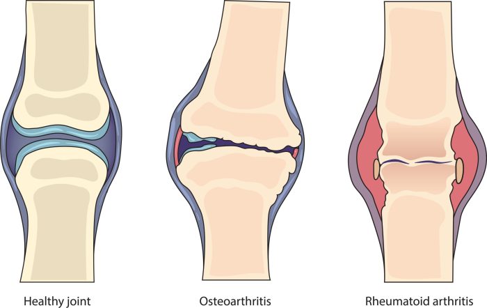
    A typical inflammatory response in RA involves a cascade of events. Imagine a healthy joint, with smooth cartilage surfaces and synovial fluid lubricating the joint. In RA, the immune system mistakenly attacks the joint lining (synovium). This triggers an inflammatory response, characterized by increased blood flow to the affected area. The synovium becomes inflamed and thickened, producing a thick fluid called pannus.

    This pannus erodes the cartilage, causing the joint to lose its smooth surface and leading to pain, stiffness, and swelling. Further, the inflammatory process involves the release of various inflammatory mediators, which contribute to the pain and damage within the joint.

    Types of Joint Inflammation

    Different types of arthritis affect joints in various ways. Understanding the characteristics of each type is vital for proper diagnosis and management.

    Type Description Rheumatoid Arthritis (RA) General Arthritis
    Synovitis Inflammation of the synovial membrane, the lining of the joint. A hallmark of RA, where the synovial membrane becomes inflamed and thickened. Can occur in various types of arthritis, including osteoarthritis and gout.
    Cartilage Damage Erosion and breakdown of cartilage, the protective tissue covering the ends of bones in joints. Progressive cartilage destruction is a key feature, leading to joint deformity and instability. Can be caused by wear and tear (osteoarthritis), crystal deposits (gout), or infections.
    Bone Erosion Breakdown of the bone tissue surrounding the joint. Bone erosion is often a result of the inflammatory process and pannus formation. Can be seen in various forms of arthritis, particularly those involving chronic inflammation.
    Joint Deformity Changes in the shape and structure of the joint. Deformities are common, with joints becoming misaligned and unstable, and often involving progressive loss of joint function. Various types of arthritis can lead to joint deformities, ranging from mild to severe.

    Typical Progression of Joint Damage in RA

    The progression of joint damage in RA is often gradual, but can be accelerated by factors such as lack of treatment or lifestyle choices. Visualize the joint as a building. Initially, there are cracks in the foundation (early-stage cartilage damage). Over time, these cracks widen, leading to structural instability (intermediate-stage cartilage and bone erosion). Finally, the building collapses (severe joint damage and deformity).

    This progression varies among individuals, influenced by factors such as the severity of the disease, age, and treatment response.

    Various Types of Arthritis

    A wide range of conditions fall under the umbrella term “arthritis.” Each type presents unique characteristics and management approaches.

    • Osteoarthritis (OA): This is the most common type of arthritis, often associated with aging and wear and tear on joints. The cartilage in the affected joints gradually wears down, leading to pain, stiffness, and reduced mobility. Imagine the smooth surface of a joint becoming rough, similar to a worn-out road. Osteoarthritis can develop in any joint, with weight-bearing joints (like knees and hips) being particularly susceptible.

      Symptoms usually worsen with activity and improve with rest. Examples include knee pain, hip pain, and hand pain.

    • Gout: This form of arthritis is caused by uric acid crystals accumulating in the joints, often forming painful tophi. The sudden onset of severe pain, redness, and swelling in a single joint is a hallmark of gout. This is often triggered by factors like high purine intake in the diet, such as consuming too much seafood or alcohol. Imagine tiny needles of uric acid crystals irritating the joint lining.

      Treatment focuses on managing the acute attack and preventing future episodes.

    • Psoriatic Arthritis: This type of arthritis is often associated with psoriasis, a skin condition characterized by red, scaly patches. It affects the joints, often with inflammation and swelling, particularly in the fingers and toes. The inflammatory process is often more aggressive in this condition, potentially leading to quicker joint damage than other types of arthritis. It can manifest with inflammation in different joints of the body, making it complex to diagnose and manage.

    • Juvenile Idiopathic Arthritis (JIA): This is a type of arthritis that develops in children and adolescents. It can affect many joints, leading to pain, stiffness, and swelling. It’s characterized by its onset in childhood, and it can lead to chronic joint damage if left untreated. The long-term effects of JIA can impact growth and development in children, emphasizing the importance of early diagnosis and treatment.

    Research and Future Directions

    Unraveling the complexities of rheumatoid arthritis (RA) and arthritis in general requires continuous research. Current studies aim to improve diagnostics, treatments, and ultimately, find cures or effective disease-modifying therapies. Understanding the underlying mechanisms driving these conditions is crucial for developing personalized approaches to care.Ongoing research endeavors are exploring novel avenues to address the multifaceted nature of these diseases, offering hope for better outcomes and improved quality of life for those affected.

    A critical examination of existing research and future directions is essential to identify potential breakthroughs and inform clinical practice.

    Current Research on Rheumatoid Arthritis

    Research into rheumatoid arthritis (RA) focuses on several key areas. Understanding the genetic predisposition to RA, identifying biomarkers for early detection, and developing more targeted therapies are high priorities. Immunological mechanisms driving inflammation and joint destruction are intensely scrutinized, aiming to develop therapies that modulate the immune response. The role of environmental factors in RA development is also under investigation.

    Ongoing Research on General Arthritis

    Research on general arthritis extends beyond RA, encompassing various forms like osteoarthritis (OA). Researchers are investigating the interplay between aging, genetics, and lifestyle factors in the development of OA. The development of non-invasive diagnostic tools and effective therapies tailored to different subtypes of arthritis are active areas of inquiry. The role of cartilage degradation and joint remodeling in OA is a subject of ongoing study.

    Comparison of Research Directions in RA and General Arthritis

    While both RA and general arthritis research share the common goal of improving patient outcomes, their specific research directions differ based on the unique characteristics of each disease. RA research often focuses on the autoimmune nature of the disease, targeting the immune system’s aberrant response. Research on general arthritis, particularly OA, often emphasizes the degenerative aspect, examining the breakdown of cartilage and joint structures.

    This leads to different research approaches and potential treatment strategies.

    Future Directions of Research in RA and General Arthritis

    Future research into RA and general arthritis will likely involve personalized medicine approaches, tailoring therapies to individual patients based on their genetic profiles and disease characteristics. Developing more precise diagnostic tools for early detection is another crucial area, allowing for earlier intervention and potentially more effective treatments. The integration of technology, such as advanced imaging techniques and bioinformatics, promises to accelerate research and improve patient care.

    Further research into the role of lifestyle factors in both conditions is also expected to contribute significantly to disease management.

    Summary of Current Research Findings

    Area of Research RA Findings General Arthritis Findings Notes
    Genetics Specific genes identified as contributing factors to RA susceptibility. Genetic predisposition to OA is increasingly understood, with various genes linked to risk. Further research needed to pinpoint specific genetic variations influencing disease progression.
    Biomarkers Biomarkers for early detection and disease activity are being developed. Biomarkers for OA severity and progression are being investigated. Validation and clinical application of biomarkers are essential for routine use.
    Immunology Understanding the aberrant immune response in RA is crucial for developing targeted therapies. Inflammation and the role of inflammatory cytokines in different forms of arthritis are under investigation. Targeted immunomodulatory therapies hold promise for RA, and similar approaches are being explored for other arthritis types.
    Environmental Factors Environmental triggers are being investigated to determine their impact on RA development. Lifestyle factors and environmental factors are studied to assess their influence on OA progression. Identifying environmental factors may lead to preventive strategies.

    Conclusion

    In conclusion, while both rheumatoid arthritis and general arthritis involve joint pain and inflammation, RA presents a more systemic and complex picture. This detailed comparison emphasizes the importance of accurate diagnosis and tailored treatment plans. Understanding the nuances of each condition is paramount for individuals seeking effective management strategies and improved quality of life.

  • Hyperglycemic Hyperosmolar Nonketotic Syndrome A Deep Dive

    Hyperglycemic Hyperosmolar Nonketotic Syndrome A Deep Dive

    Hyperglycemic hyperosmolar nonketotic syndrome (HHNS) is a serious complication of diabetes, characterized by extremely high blood sugar levels without the presence of ketones. This often occurs in individuals with type 2 diabetes, particularly those who aren’t managing their blood sugar well. Understanding the underlying mechanisms, symptoms, and management strategies is crucial for anyone at risk. This blog post will explore the intricacies of HHNS, from its pathophysiology to potential complications and preventative measures.

    HHNS typically develops gradually, unlike diabetic ketoacidosis (DKA), often stemming from a combination of factors like inadequate insulin, infections, or medications. The elevated blood sugar levels lead to a dangerous increase in the body’s osmotic pressure, causing dehydration and electrolyte imbalances. This complex condition requires immediate medical attention to prevent severe complications.

    Introduction to Hyperglycemic Hyperosmolar Nonketotic Syndrome (HHNS)

    Hyperglycemic hyperosmolar nonketotic syndrome (HHNS) is a serious, life-threatening complication of diabetes mellitus, primarily in type 2 diabetes. It’s characterized by extremely high blood sugar levels without the presence of ketones in the urine or blood. This distinct feature differentiates HHNS from diabetic ketoacidosis (DKA), another serious complication. Understanding the pathophysiology, presentation, and risk factors of HHNS is crucial for early diagnosis and effective management.HHNS develops when the body’s ability to regulate blood sugar is overwhelmed, often due to a combination of factors.

    The key mechanism involves a profound deficiency in insulin secretion and/or action, leading to uncontrolled hyperglycemia. This elevated blood glucose draws water out of cells, causing a dangerous shift in the body’s fluid balance and electrolyte levels. The resulting hyperosmolar state, characterized by high blood glucose levels and elevated serum osmolality, disrupts vital organ function.

    Hyperglycemic hyperosmolar nonketotic syndrome (HHNS) can be a serious complication of diabetes, requiring careful management. While focusing on blood sugar control is crucial, it’s also important to consider factors like the best time to take probiotics for overall gut health. Optimizing your probiotic intake, as detailed in this guide on best time to take probiotics , might indirectly support your body’s ability to manage blood sugar levels, contributing to better outcomes for HHNS.

    Ultimately, consistent management of diabetes and a healthy lifestyle are key to preventing HHNS.

    Pathophysiology of HHNS

    The pathophysiology of HHNS centers around the interplay of impaired insulin secretion and/or action, and insufficient fluid intake. Insufficient insulin action allows for a significant rise in blood glucose levels. This hyperglycemia leads to osmotic diuresis, where excess glucose is excreted in the urine, pulling water with it. The loss of water leads to dehydration and an increase in serum osmolality.

    The body attempts to compensate by producing more urine, further exacerbating the dehydration. This cycle perpetuates the hyperosmolar state, ultimately leading to impaired organ function.

    Typical Presentation of HHNS

    HHNS typically presents with a gradual onset of symptoms, often over several days to weeks. The symptoms are related to the severe dehydration and electrolyte imbalances. Patients may experience:

    • Extreme thirst (polydipsia): The body’s attempt to compensate for dehydration.
    • Increased urination (polyuria): Osmotic diuresis caused by high blood glucose levels.
    • Dry mouth and skin: Direct effects of dehydration.
    • Weakness, lethargy, and confusion: Due to the impact of hyperglycemia and dehydration on brain function.
    • Headache: A common symptom that may indicate an increase in intracranial pressure.
    • Vision problems: Possible consequence of the hyperosmolar state affecting the optic nerve.

    Risk Factors for HHNS

    Several factors can increase the risk of developing HHNS. These include:

    • Poorly controlled diabetes: This is the most significant risk factor, as uncontrolled blood sugar levels increase the likelihood of severe hyperglycemia.
    • Infections: Illnesses, such as pneumonia or urinary tract infections, can increase insulin needs, thereby disrupting blood glucose control.
    • Certain medications: Some medications, such as diuretics, steroids, and some antipsychotics, can impair glucose regulation.
    • Recent surgery or trauma: Stressful events can increase insulin requirements and impair blood glucose control.
    • Insufficient fluid intake: Dehydration can worsen hyperglycemia and contribute to the development of HHNS.
    • Advanced age: Older adults may have reduced ability to regulate blood glucose.

    Symptoms, Severity, and Potential Causes of HHNS

    Symptom Severity Potential Causes
    Extreme thirst Mild to severe, depending on the degree of dehydration Insufficient fluid intake, osmotic diuresis, hyperglycemia
    Increased urination Mild to severe, reflecting the osmotic diuresis Hyperglycemia, dehydration
    Weakness and lethargy Can range from mild to profound, indicative of organ dysfunction Dehydration, electrolyte imbalances, cerebral dysfunction
    Confusion and altered mental status Can progress to coma, indicating severe brain dysfunction Hyperosmolarity, electrolyte abnormalities, cerebral edema
    Dry mouth and skin Severe, reflecting the dehydration Osmotic diuresis, insufficient fluid intake

    Diagnosis and Evaluation of HHNS

    Hyperglycemic hyperosmolar nonketotic syndrome (HHNS) is a serious medical emergency requiring prompt diagnosis and management. Early recognition and intervention are crucial for minimizing complications and improving patient outcomes. Accurately assessing the patient and interpreting laboratory results are vital steps in establishing a diagnosis and developing an effective treatment plan.

    Diagnostic Criteria for HHNS

    The diagnosis of HHNS relies on a combination of clinical presentation and laboratory findings. Key diagnostic criteria include severe hyperglycemia, typically exceeding 600 mg/dL, coupled with a serum osmolality significantly elevated above normal ranges. Absence of ketoacidosis, a hallmark of diabetic ketoacidosis (DKA), is also a defining characteristic. The clinical picture often includes profound dehydration, altered mental status, and neurological dysfunction.

    Hyperglycemic hyperosmolar nonketotic syndrome (HHNS) is a serious complication of diabetes, often triggered by infections or poor blood sugar control. Managing blood sugar levels is key, and finding delicious, satisfying treats like those found in go to diabetic desserts can make a big difference in overall well-being. While enjoying these treats, it’s crucial to maintain a balanced diet and medication schedule to prevent HHNS from recurring.

    These factors, alongside the laboratory data, confirm the diagnosis.

    Laboratory Findings in HHNS

    Several key laboratory parameters support the diagnosis of HHNS. Blood glucose levels are usually markedly elevated, frequently exceeding 600 mg/dL. Serum osmolality is also significantly elevated, reflecting the increased concentration of solutes in the blood. Electrolyte imbalances, particularly hyponatremia (low sodium), hypokalemia (low potassium), and hypochloremia (low chloride), are common. These imbalances result from the osmotic diuresis caused by the high blood glucose levels.

    In addition, the serum bicarbonate levels are usually normal to slightly elevated. The presence of these findings, in combination with clinical symptoms, strongly suggests HHNS.

    Evaluation Procedures for Suspected HHNS

    Evaluating patients suspected of HHNS involves a comprehensive approach, including a thorough history and physical examination, alongside a series of laboratory tests. The history should focus on the patient’s medical history, including any underlying conditions, recent illness, medication use, and dietary habits. The physical examination should assess vital signs, including blood pressure, pulse, and respiratory rate. Assessing the patient’s mental status and neurological function is critical.

    Blood samples are collected for comprehensive laboratory analysis, including complete blood count (CBC), serum electrolytes, glucose, blood urea nitrogen (BUN), creatinine, and serum osmolality. These analyses help confirm the diagnosis and evaluate the severity of the condition.

    Dealing with hyperglycemic hyperosmolar nonketotic syndrome can be tough, especially when it comes to managing the emotional rollercoaster. It’s all too easy to feel overwhelmed, but finding healthy coping mechanisms, like those discussed in this insightful article on coping with emotions and eczema , can make a real difference. Ultimately, understanding how to navigate these feelings is key to managing the symptoms and overall well-being associated with hyperglycemic hyperosmolar nonketotic syndrome.

    Significance of Key Laboratory Values

    Blood glucose levels are crucial for diagnosing and monitoring HHNS. Elevated blood glucose levels, often exceeding 600 mg/dL, signify a severe hyperglycemic state. Serum osmolality reflects the concentration of solutes in the blood. Significant elevations indicate dehydration and the need for aggressive fluid resuscitation. Electrolyte imbalances, particularly hyponatremia, hypokalemia, and hypochloremia, are significant complications of HHNS.

    These imbalances need prompt correction to prevent further complications.

    Comparison of HHNS with Other Conditions

    | Feature | HHNS | DKA ||—|—|—|| Blood Glucose | Extremely high (>600 mg/dL) | High, but can be variable || Ketones | Absent | Present || Serum Osmolality | Extremely high | Usually normal or slightly elevated || Electrolytes | Often low (hyponatremia, hypokalemia) | Can be low (hyponatremia) || Mental Status | Often altered | Can be altered || Dehydration | Often profound | Can be present, but often less severe than HHNS |The table highlights the key differences between HHNS and DKA.

    While both conditions involve hyperglycemia, their underlying pathophysiology and laboratory findings differ significantly. This distinction is vital for accurate diagnosis and appropriate treatment.

    Interpreting Blood Test Results Related to HHNS

    Interpreting blood test results in the context of HHNS requires a thorough understanding of the patient’s clinical presentation and the specific laboratory findings. A combination of elevated blood glucose, significantly elevated serum osmolality, and electrolyte imbalances, particularly hyponatremia, provides strong evidence for HHNS. For example, a patient presenting with altered mental status, severe dehydration, and blood glucose exceeding 700 mg/dL, coupled with hyponatremia and elevated serum osmolality, strongly suggests HHNS.

    Careful evaluation of the complete picture is essential for accurate interpretation and appropriate management.

    Management and Treatment of HHNS

    Hyperglycemic hyperosmolar nonketotic syndrome

    Hyperglycemic hyperosmolar nonketotic syndrome (HHNS) necessitates prompt and aggressive management to restore hydration and electrolyte balance while addressing the underlying hyperglycemia. Early intervention is crucial to prevent severe complications and improve patient outcomes. Effective management focuses on correcting the fluid and electrolyte imbalances, reducing hyperglycemia, and supporting the patient’s overall stability.The cornerstone of HHNS management lies in the meticulous administration of intravenous fluids, electrolyte replacement, and insulin therapy.

    A coordinated approach, closely monitoring vital signs and laboratory values, is essential to prevent adverse reactions and ensure the patient’s safety throughout the treatment process.

    Intravenous Fluid Therapy

    Intravenous (IV) fluids are paramount in the initial management of HHNS. They rapidly rehydrate the patient, correct the hyperosmolar state, and improve tissue perfusion. The choice of fluid and the rate of administration are crucial for patient safety.

    • Types of IV Fluids: The initial fluid choice is typically 0.9% normal saline (NS) due to its isotonic nature and ability to quickly restore intravascular volume. Later, as the serum sodium level decreases, 0.45% normal saline or even dextrose-containing solutions may be necessary to avoid hyponatremia.
    • Administration Rates: The rate of fluid administration is carefully titrated based on the patient’s clinical status, including vital signs, urine output, and serum sodium levels. Initial rates might be 250-500 mL/hour, adjusted according to response. Monitoring for signs of fluid overload, such as pulmonary edema, is critical. Close monitoring of the patient’s condition is paramount throughout the process.

      The goal is to rehydrate the patient without causing complications.

    Electrolyte Correction

    Electrolyte imbalances are common in HHNS and require careful attention. Correcting these imbalances is essential to prevent further complications and ensure the effectiveness of the overall treatment plan.

    • Potassium: Hyperglycemia drives potassium into the cells, leading to hypokalemia. Frequent monitoring of potassium levels is crucial, and potassium replacement may be necessary, given the potential for cardiac arrhythmias if not addressed properly. Replacement is guided by the potassium level and clinical status. It is vital to avoid rapid potassium correction, as it can lead to potentially fatal complications.

    • Sodium: HHNS is characterized by hypernatremia. Correcting hypernatremia should be done cautiously, avoiding rapid shifts in serum sodium levels, as this can lead to cerebral edema. A gradual decrease in serum sodium is usually recommended to mitigate this risk. Slow correction minimizes risk.
    • Other Electrolytes: Other electrolytes like magnesium and phosphate may also be depleted, and their correction should be managed under close medical supervision.

    Insulin Therapy

    Insulin therapy is necessary to lower blood glucose levels and promote rehydration.

    • Rationale: Insulin facilitates the uptake of glucose into cells, reducing hyperglycemia and decreasing osmotic diuresis. This in turn helps restore fluid balance.
    • Approach: Low-dose insulin infusion is typically initiated. The goal is to gradually lower blood glucose levels to prevent hypoglycemia and to improve the overall patient response. A slow, steady reduction in blood glucose levels is preferred.

    Step-by-Step Procedure for IV Fluid Administration in HHNS

    1. Assessment: Thorough patient assessment, including vital signs, blood glucose levels, and electrolyte levels, is critical. This initial assessment establishes a baseline for subsequent interventions.
    2. Initial Fluid Bolus: Administer a rapid IV fluid bolus (e.g., 500 mL of 0.9% normal saline) to quickly restore intravascular volume.
    3. Maintenance Fluid Infusion: Initiate a maintenance IV fluid infusion (e.g., 0.9% normal saline) at a rate appropriate for the patient’s needs, closely monitoring vital signs and urine output. The goal is to maintain adequate hydration and to address the underlying fluid deficit.
    4. Electrolyte Monitoring and Replacement: Frequent electrolyte monitoring (including potassium) is crucial. Replacement of electrolytes is administered according to laboratory results and clinical judgment.
    5. Insulin Therapy: Initiate low-dose insulin infusion according to protocol and patient response.
    6. Close Monitoring: Continuously monitor vital signs, blood glucose, electrolytes, and urine output to adjust therapy as needed.

    Complications and Prognosis of HHNS

    Hyperglycemic hyperosmolar nonketotic syndrome

    Hyperglycemic hyperosmolar nonketotic syndrome (HHNS) is a serious medical condition requiring prompt and effective management. While often treatable, the potential for complications and long-term effects on patient well-being must be carefully considered. Understanding the potential complications, influencing factors, and prognosis is crucial for both patient care and healthcare provider decision-making.

    Potential Acute Complications

    HHNS can lead to a cascade of acute complications, primarily stemming from the severe dehydration and electrolyte imbalances. These imbalances can disrupt various organ systems, potentially causing significant harm. Acute kidney injury is a particularly concerning complication, often resulting from the reduced blood flow to the kidneys due to dehydration and the resulting decreased glomerular filtration rate. Other potential complications include cardiovascular issues, such as heart failure or arrhythmias, and neurological complications, such as seizures or coma.

    Long-Term Effects on Health and Well-being

    While prompt treatment can often mitigate the acute effects of HHNS, long-term consequences can still impact patients’ health and well-being. Patients may experience persistent cognitive impairment or other neurological deficits if the brain is significantly affected by the severe hyperglycemia and dehydration. Moreover, individuals may develop an increased susceptibility to future episodes of HHNS. Sustained hyperglycemia, a hallmark of this condition, can also negatively impact vascular health, increasing the risk of cardiovascular complications over time.

    Factors Influencing Prognosis

    Several factors influence the prognosis of HHNS, ranging from the severity of the initial presentation to the promptness and efficacy of treatment. The patient’s underlying health status, including pre-existing conditions like cardiovascular disease or renal impairment, can significantly impact the outcome. Age, nutritional status, and the presence of concomitant infections or other illnesses also play a crucial role in determining the prognosis.

    Additionally, timely access to appropriate medical care, including intensive monitoring and effective management strategies, is essential in improving the prognosis.

    Mortality Rates

    The mortality rate associated with HHNS is influenced by the severity of the condition and the timeliness of intervention. While the mortality rate is not as high as with diabetic ketoacidosis (DKA), it still presents a considerable risk. Studies have shown that patients with severe HHNS, particularly those with co-morbidities or delayed diagnosis, have higher mortality rates. It is important to note that the mortality rate is influenced by access to timely and appropriate medical care.

    Correlation Between Severity and Complications

    Severity of HHNS Potential Complications
    Mild Electrolyte imbalances, mild dehydration, possible confusion
    Moderate Moderate dehydration, electrolyte abnormalities, possible seizures, acute kidney injury
    Severe Severe dehydration, significant electrolyte disturbances, coma, acute kidney injury, cardiovascular complications, neurological deficits

    The table above provides a general overview. The specific complications and their severity can vary significantly depending on individual factors and the promptness of treatment. The most severe cases often require intensive care unit (ICU) management.

    Prevention and Education of HHNS

    Hyperglycemic hyperosmolar nonketotic syndrome (HHNS) is a serious but preventable complication of diabetes. Early detection and proactive management of risk factors are crucial in preventing HHNS. By understanding the triggers and implementing preventative measures, individuals with diabetes can significantly reduce their risk of developing this potentially life-threatening condition.

    Strategies for Preventing HHNS in Susceptible Individuals

    Effective prevention hinges on meticulous self-management and proactive engagement with healthcare providers. Individuals at high risk of HHNS development, such as those with poorly controlled diabetes, should prioritize consistent blood glucose monitoring, adherence to medication regimens, and prompt medical attention for any signs of illness. This proactive approach significantly minimizes the risk of hyperglycemia escalating into HHNS.

    Importance of Patient Education

    Patient education plays a pivotal role in empowering individuals with diabetes to actively participate in their care. Thorough education regarding blood glucose monitoring techniques, recognizing symptoms of rising blood sugar, and understanding the importance of lifestyle modifications is crucial. Comprehensive patient education programs can equip individuals with the knowledge and skills needed to effectively manage their condition and prevent complications like HHNS.

    Role of Healthcare Providers in Educating Patients

    Healthcare providers have a critical role in educating patients about HHNS prevention. This includes providing clear and concise information about the risks, triggers, and symptoms of HHNS. Regular follow-up appointments, personalized diabetes management plans, and ongoing support are essential components of patient education. Diabetes educators and registered dietitians can provide invaluable assistance in this process. Healthcare providers should tailor their educational approach to each patient’s individual needs and circumstances, ensuring clarity and understanding.

    Tips for Managing Risk Factors

    Several risk factors contribute to the development of HHNS. Managing these factors effectively can significantly reduce the likelihood of developing the syndrome. Strategies include meticulous adherence to medication regimens, consistent blood glucose monitoring, timely recognition and treatment of infections or illnesses, and proactive management of underlying medical conditions. Maintaining a healthy lifestyle, including regular exercise and a balanced diet, also plays a significant role in preventing HHNS.

    Prevention Strategies Flowchart

    Prevention Flowchart
    This flowchart visualizes the key steps in preventing HHNS. It begins with routine blood glucose monitoring, progressing through identifying and managing risk factors, and culminates in early intervention when signs of illness or hyperglycemia appear.

    Blood Glucose Monitoring Techniques, Hyperglycemic hyperosmolar nonketotic syndrome

    Effective blood glucose monitoring is fundamental to preventing HHNS. Regular monitoring allows individuals to detect rising glucose levels early, enabling timely interventions to prevent severe hyperglycemia. Different methods, such as finger-prick testing and continuous glucose monitors (CGMs), are available, and selecting the most suitable method is crucial. Proper technique and interpretation of results are essential to effective glucose management.

    Case Studies and Illustrations of HHNS: Hyperglycemic Hyperosmolar Nonketotic Syndrome

    Hyperglycemic hyperosmolar nonketotic syndrome (HHNS) is a serious medical emergency requiring prompt diagnosis and management. Understanding real-world case studies allows healthcare professionals to better recognize the spectrum of presentations and develop effective treatment strategies. This section delves into illustrative cases, highlighting different patient presentations, management approaches, and outcomes.

    Illustrative Case Studies of HHNS

    Various patient presentations characterize HHNS, from subtle symptoms to severe complications. Case studies offer invaluable insights into recognizing HHNS in diverse patient populations.

    • Case Study 1: A 75-year-old female with a history of type 2 diabetes and hypertension presented with progressively worsening polyuria, polydipsia, and altered mental status. Blood glucose levels were found to be extremely elevated (approximately 600 mg/dL). The patient exhibited a profound state of dehydration, with dry mucous membranes and decreased skin turgor. Management involved aggressive fluid resuscitation, insulin therapy, and electrolyte correction.

      The patient’s mental status improved gradually, and she was discharged after several days of intensive care.

    • Case Study 2: A 62-year-old male with type 2 diabetes, uncontrolled by medication, presented with profound weakness, lethargy, and confusion. Initial blood glucose was elevated to 550 mg/dL. The patient had a history of poor adherence to his diabetes management plan. Treatment involved correcting dehydration and administering insulin. The patient showed improvement in his mental status but required several days of monitoring due to a prolonged response to therapy.

    • Case Study 3: A 30-year-old female with type 1 diabetes, poorly managed, presented to the emergency room with a Glasgow Coma Scale of 10. Her blood glucose was 700 mg/dL. This case highlighted the need for rapid diagnosis and intervention. Prompt treatment, including aggressive fluid and electrolyte replacement, and insulin therapy, led to gradual improvement in the patient’s neurological status.

      The patient required prolonged hospitalization and close monitoring.

    Outcomes for Patients with HHNS

    Patient outcomes in HHNS vary depending on the severity of the presentation, promptness of diagnosis and treatment, and underlying comorbidities.

    • Favorable Outcomes: Patients with timely and appropriate treatment, generally respond well, demonstrating improvement in their mental status and overall condition. Complete recovery and discharge from the hospital are common in cases where the disease process is contained, and the underlying condition is adequately managed.
    • Moderate Outcomes: Some patients experience a prolonged recovery period with residual neurological symptoms. These cases may necessitate ongoing rehabilitation or therapy to address the lingering effects of the syndrome.
    • Unfavorable Outcomes: Severe cases of HHNS, especially those with delayed diagnosis or inadequate treatment, can result in severe complications such as seizures, coma, and even death. The outcomes depend on the patient’s overall health status, severity of the syndrome, and the speed and effectiveness of treatment.

    Physiological Changes During HHNS

    HHNS involves a complex cascade of physiological changes, primarily centered around severe hyperglycemia and dehydration.

    • Hyperglycemia: A profound increase in blood glucose levels leads to osmotic diuresis, resulting in significant fluid loss from the body. This diuresis exacerbates dehydration and further contributes to hyperosmolarity.
    • Dehydration: The significant fluid loss due to osmotic diuresis causes electrolyte imbalances and impaired cellular function. This dehydration is a major contributor to the patient’s clinical presentation.
    • Electrolyte Imbalances: Dehydration leads to changes in sodium, potassium, and other electrolytes. These imbalances further compromise cellular function and can contribute to neurological dysfunction.
    • Neurological Dysfunction: The combination of hyperglycemia, hyperosmolarity, and electrolyte imbalances can lead to a spectrum of neurological symptoms, ranging from confusion and lethargy to seizures and coma.

    Diagnosis of HHNS in a Case Study

    Accurate diagnosis of HHNS involves a combination of clinical assessment and laboratory findings.

    • Clinical Evaluation: The clinical picture, including symptoms such as polyuria, polydipsia, and altered mental status, is crucial in raising suspicion for HHNS.
    • Laboratory Findings: Key laboratory tests include serum glucose, serum osmolality, serum electrolytes, and blood urea nitrogen (BUN)/creatinine levels to evaluate the degree of dehydration.

    Final Wrap-Up

    In conclusion, hyperglycemic hyperosmolar nonketotic syndrome (HHNS) is a potentially life-threatening condition requiring prompt diagnosis and aggressive management. Understanding the risk factors, symptoms, and treatment protocols is vital for both patients and healthcare providers. Prevention through proper blood glucose control and lifestyle modifications plays a key role in mitigating the risk of developing HHNS. This discussion highlights the importance of proactive diabetes management to prevent this severe complication.

  • Identifying Insect Bites and Stings A Comprehensive Guide

    Identifying Insect Bites and Stings A Comprehensive Guide

    Identifying insect bites and stings is crucial for prompt and appropriate treatment. This guide delves into the differences between bites and stings, explores common symptoms, and details assessment procedures, first aid, prevention, and specific insect types. From recognizing the subtle variations in bite marks to understanding potential allergic reactions, this comprehensive resource will equip you with the knowledge to handle insect encounters safely and effectively.

    The guide will cover a range of topics from distinguishing a bee sting from a mosquito bite to assessing the severity of a reaction. We’ll also explore preventative measures to minimize the risk of future encounters, including practical tips for identifying and eliminating potential breeding grounds for insects around your home.

    Identifying Bites vs. Stings

    Insect bites and stings, while seemingly similar, have distinct characteristics. Understanding these differences is crucial for prompt and appropriate treatment. Misidentification can lead to unnecessary anxiety and potentially inappropriate medical interventions. This section will delve into the key features that differentiate insect bites from stings, highlighting common insects and their characteristic marks.

    Bites vs. Stings: Key Differences, Identifying insect bites and stings

    Insect bites and stings are distinct actions resulting in different marks on the skin. Bites are typically caused by insects that pierce the skin to feed on blood or tissue fluids. Stings, on the other hand, are the result of an insect injecting venom or a toxin into the skin.

    Physical Characteristics of Bites and Stings

    A bite typically manifests as a puncture wound or a small, flat area of swelling. The skin might be broken or appear slightly reddened around the puncture site. Stings often appear as a more localized, raised area, with a small, central puncture mark. The surrounding skin may be inflamed and swollen. The size and shape of the wound depend on the size and type of insect involved.

    Comparison of Bite and Sting Marks

    Characteristic Bite Sting
    Typical Location Often on exposed skin, like hands, arms, legs, and face. Can occur anywhere on the body, but often on exposed areas.
    Appearance Small puncture wound, flat swelling, redness, sometimes bleeding. Raised welt, small central puncture, swelling, redness, localized pain.
    Size Usually small, ranging from a few millimeters to a centimeter in diameter. Can vary in size depending on the insect, typically from a few millimeters to a few centimeters in diameter.
    Other Symptoms Possible itching, mild pain, localized swelling. Pain, itching, swelling, redness, possible allergic reactions (anaphylaxis in severe cases).

    Misidentification and Its Importance

    Accurate identification of bites and stings is crucial for proper treatment. A misdiagnosis might lead to ineffective or even harmful interventions. For instance, treating a sting as a bite might result in neglecting the need for anti-venom, potentially exacerbating the condition. Conversely, treating a bite with an anti-allergic medication might be inappropriate and unnecessary.

    Common Insects and Their Characteristics

    Various insects are known for biting or stinging. Mosquitoes, for example, are notorious for their blood-feeding habits, resulting in small, itchy bites. Fleas typically cause small, red, itchy spots. Bees and wasps, on the other hand, deliver a sting that often leaves a painful, raised welt. Fire ants leave a distinctive, painful sting, often resulting in a larger area of redness and swelling.

    Recognizing the Danger of Misidentification

    Accurate identification is essential for selecting the right treatment and preventing potential complications.

    The severity of the reaction can vary significantly depending on the insect and the individual’s sensitivity. Some bites or stings can cause localized pain, itching, and swelling, while others may trigger more serious allergic reactions. Accurate identification allows for a more targeted and effective treatment approach.

    Recognizing Symptoms

    Insect bites and stings, while often minor annoyances, can sometimes indicate more serious issues. Understanding the symptoms is crucial for prompt and appropriate action. Identifying the specific insect involved can help determine the appropriate treatment and potential risks.The symptoms of an insect bite or sting can vary widely, depending on the insect, the individual’s sensitivity, and the location of the affected area.

    Common symptoms include pain, itching, swelling, redness, and warmth at the site. These reactions are often localized, meaning they primarily affect the area directly where the insect contacted the skin. However, more severe reactions can occur, especially in individuals with allergies.

    Common Symptoms

    A variety of symptoms are commonly associated with insect bites and stings. These include localized pain, which can range from mild discomfort to intense throbbing. Itching is another frequent symptom, often accompanied by a desire to scratch, which can lead to further skin irritation. Swelling is a common reaction, characterized by a noticeable increase in the size of the affected area.

    Redness is often visible, resulting from increased blood flow to the area. Finally, warmth at the site is a physiological response to the insect’s venom or saliva.

    Severity Levels and Insect Types

    The severity of symptoms can vary considerably. Mild reactions are typically characterized by localized pain, itching, and slight swelling, often resolving within a few hours. These reactions are commonly associated with bites from insects like mosquitoes, fleas, and bed bugs. More severe reactions, involving significant swelling, intense pain, or difficulty breathing, may indicate a more serious reaction or an allergic response.

    In such cases, the insect may include wasps, bees, hornets, or fire ants. A table outlining the potential severity levels and corresponding insect types is presented below.

    Severity Level Symptoms Potential Insect Types
    Mild Localized pain, itching, slight swelling, resolves within hours Mosquitoes, fleas, bed bugs, some spider bites
    Moderate Significant swelling, increased pain, localized redness, warmth Wasps, bees, hornets, some spider bites
    Severe Difficulty breathing, swelling of the face or throat, hives, nausea, dizziness, rapid heartbeat, or loss of consciousness Wasps, bees, hornets, fire ants, some spider bites

    Allergic Reactions

    An allergic reaction to an insect bite or sting can range from a mild skin rash to a life-threatening condition called anaphylaxis. Anaphylaxis is a severe, potentially fatal allergic reaction that can cause a rapid drop in blood pressure, difficulty breathing, and swelling of the throat and airways. Immediate medical attention is crucial in such cases.

    Symptoms by Location and Potential Culprits

    The location of the bite or sting can sometimes provide clues about the potential insect culprit. A bite on the face, for example, might be associated with a stinging insect like a bee, whereas a bite on an extremity could be from a mosquito or flea. The following table Artikels possible insect culprits based on the location of the reaction.

    Location Potential Insect Culprits
    Face Bees, wasps, hornets, mosquitos
    Extremities (arms, legs) Mosquitoes, fleas, spiders, ants
    Neck Bees, wasps, hornets, mosquitos
    Trunk Bees, wasps, hornets, mosquitos, ants

    Assessing the Situation

    Understanding the severity of an insect bite or sting is crucial for appropriate treatment. A thorough assessment helps determine the necessary response, whether it’s a simple first-aid measure or a visit to a medical professional. This involves a careful evaluation of the bite/sting site, symptoms, and the overall situation.Assessing the severity involves a combination of visual inspection and symptom evaluation.

    Accurate documentation is also essential to aid in the evaluation and provide useful information for medical professionals if needed.

    Visual Inspection Procedures

    A crucial first step in assessing an insect bite or sting is a careful visual inspection. This includes noting the location, size, shape, and any visible characteristics of the affected area. Precise details aid in determining the type of insect involved and the potential severity of the reaction.

    • Location: Precisely noting the location on the body (e.g., right forearm, behind the knee) is vital. This helps in understanding potential allergies or reactions based on body parts, as well as identifying areas prone to secondary infections.
    • Size and Shape: Measuring the diameter of the bite or sting with a ruler or using comparative objects (e.g., a coin) provides crucial information. The shape (e.g., round, oval, linear) can also offer clues about the insect species. For example, a linear mark suggests a crawling insect, while a round mark may indicate a stinging insect.
    • Appearance: Observing the characteristics of the bite/sting is important. Look for swelling, redness, discoloration (e.g., bruising), pus, or any other unusual signs. A simple red bump may be a minor reaction, while significant swelling or blistering could indicate a more serious allergic response.

    Symptom Evaluation

    Beyond the visual inspection, evaluating symptoms is critical for determining the severity of the reaction. Pay close attention to any changes in the body’s response to the bite/sting.

    • Pain: Note the intensity of the pain, from mild discomfort to severe throbbing. Consider how long the pain has lasted and whether it’s getting worse.
    • Itching: Observe the level of itching, from mild to intense. Constant or increasing itching can be a sign of an allergic reaction.
    • Swelling: Assess the extent of swelling around the bite/sting. Rapid or significant swelling may indicate an allergic response and require immediate medical attention.
    • Other symptoms: Monitor for other symptoms such as hives, nausea, difficulty breathing, or dizziness. These can signal a severe allergic reaction requiring immediate medical assistance.

    Stinger Removal

    If a stinger is visible, it’s crucial to remove it safely and effectively. Improper removal can potentially worsen the situation.

    1. Use a clean, blunt object: A credit card, a butter knife, or a similar flat, blunt object can be used to gently scrape the stinger away from the skin. Avoid pinching or squeezing the stinger, as this can inject more venom.
    2. Avoid tweezers: Tweezers can crush the stinger and potentially inject more venom. If the stinger is deeply embedded, do not attempt to remove it. Instead, seek medical attention.
    3. Apply pressure: After removal, apply gentle pressure to the area to help stop any further bleeding.

    Documentation

    Accurate documentation is key for tracking the bite/sting’s progression and for future reference. This information can be crucial in case of a severe reaction or for identifying potential patterns.

    Category Information to Document
    Time Record the exact time the bite/sting occurred.
    Location Precisely describe the location of the bite/sting on the body.
    Appearance Describe the initial appearance of the bite/sting (e.g., size, shape, redness, swelling).
    Symptoms Record any symptoms experienced, including pain, itching, swelling, or other reactions.

    Accurate documentation is essential for monitoring the reaction and seeking appropriate medical care if needed.

    Learning to identify insect bites and stings is crucial, especially if you’re venturing outdoors. While activities like walking with a weighted vest walking with a weighted vest can be great for building strength, it’s important to be vigilant for any unusual reactions. Knowing the differences between a harmless bite and a more serious one can make all the difference in getting the right treatment, so pay close attention to the symptoms and seek medical advice if necessary.

    First Aid and Treatment

    Identifying insect bites and stings

    Dealing with insect bites and stings promptly and correctly is crucial for minimizing discomfort and preventing complications. Understanding the appropriate first aid measures can significantly influence the healing process and reduce the risk of allergic reactions. This section details essential steps for managing these situations effectively.

    Basic First Aid Measures

    Effective first aid for insect bites and stings begins with immediate actions. Cleaning the affected area thoroughly and applying soothing remedies are key initial steps. Proper management of pain and swelling can accelerate healing and prevent further complications.

    • Cleaning the Area: Gently wash the affected area with mild soap and water. Avoid harsh scrubbing, which can irritate the skin further. Use clean hands or a clean cloth to avoid spreading bacteria.
    • Soothing the Area: Applying a cool compress can help reduce pain and swelling. A cold compress can also help minimize inflammation and discomfort. A cold pack wrapped in a clean cloth is ideal.
    • Elevating the Affected Area: If possible, elevate the affected area above the heart. This helps to reduce swelling by promoting the drainage of excess fluid. This is especially important for bites or stings on extremities.

    Applying Cold Compresses

    Applying cold compresses effectively can significantly reduce pain and swelling associated with insect bites and stings. The duration and application method are important for maximizing the benefits.

    • Cold Compress Application: Use a clean cloth or paper towel to wrap an ice pack or cold, wet washcloth. Apply the compress to the affected area for 10-15 minutes at a time, with breaks in between to avoid skin damage. Avoid applying ice directly to the skin, as it can cause frostbite.
    • Frequency of Application: Apply cold compresses several times a day as needed, especially during the initial stages of the reaction, to help reduce pain and swelling. Adjust the frequency based on the severity of the reaction and the individual’s response.

    Other Remedies for Pain Relief

    Various remedies, both over-the-counter and home-based, can help alleviate the pain and itching associated with insect bites and stings. Always consult with a healthcare professional if symptoms worsen or if there are concerns about an allergic reaction.

    • Over-the-Counter Medications: Antihistamines, such as diphenhydramine (Benadryl), can help relieve itching. Pain relievers like ibuprofen or acetaminophen can reduce pain and swelling. Follow the dosage instructions carefully.
    • Home Remedies: Some people find relief from applying a paste of baking soda and water to the affected area. Alternatively, a mixture of apple cider vinegar and water can also be used to reduce itching. It’s crucial to remember that these are not always guaranteed remedies and may not work for everyone. It’s important to consult a doctor if the symptoms don’t subside.

    Seeking Immediate Medical Attention

    Certain situations require immediate medical attention for insect bites and stings. Recognizing these situations is crucial for preventing serious complications.

    • Severe Reactions: Signs of a severe allergic reaction, including difficulty breathing, swelling of the face, throat, or tongue, hives, or dizziness, require immediate medical attention. This is an emergency, and immediate medical intervention is essential. Anaphylaxis is a life-threatening reaction that requires immediate medical intervention.
    • Suspected Allergies: If you have a known allergy to insect bites or stings, seek medical attention immediately after any incident. Prompt treatment can be critical for preventing a serious reaction.
    • Infection: If the bite or sting becomes infected (redness, swelling, pus, or excessive pain), seek medical attention promptly. An infection can quickly escalate and require antibiotics or other medical treatments.

    Preventing Future Bites and Stings: Identifying Insect Bites And Stings

    Identifying insect bites and stings

    Staying safe from insect bites and stings is largely about understanding their habits and taking proactive steps to avoid encounters. By recognizing the conditions that attract insects and adjusting your environment, you can significantly reduce your risk of experiencing these unpleasant encounters. A proactive approach to prevention is key to enjoying outdoor activities and maintaining a comfortable living space.Effective prevention involves a combination of smart choices about where you spend time, what you wear, and how you maintain your surroundings.

    This proactive approach not only minimizes the risk of bites and stings but also contributes to a healthier environment for everyone.

    Insect Repellent Use

    Using insect repellent is a crucial aspect of preventing bites and stings. Choosing a repellent containing DEET, picaridin, or oil of lemon eucalyptus can offer effective protection. Follow the product instructions carefully, ensuring proper application and reapplication intervals. It’s essential to note that some repellents are more effective against certain types of insects than others. For example, DEET is often more effective against mosquitoes, while picaridin may be a better choice for those with sensitive skin.

    Always apply repellent to exposed skin, avoiding the eyes and mouth.

    Clothing Choices

    Proper clothing choices play a significant role in preventing bites and stings. Wearing long sleeves and pants, especially in areas known for insect activity, can significantly reduce the chances of being bitten or stung. Light-colored clothing can also be beneficial as darker colors can sometimes attract insects more readily. Consider using permethrin-treated clothing, which can provide added protection against mosquitoes, ticks, and other biting insects.

    Make sure the clothing is properly fitted and comfortable, allowing for freedom of movement while offering the best protection.

    Identifying and Eliminating Breeding Grounds

    Identifying and eliminating potential breeding grounds for insects is vital in preventing future bites and stings. Standing water, such as stagnant pools, puddles, or flowerpots with accumulated water, can be ideal breeding grounds for mosquitoes and other insects. Regularly emptying and cleaning containers that collect water will significantly reduce the insect population in your immediate surroundings. Inspect your yard for any areas where water might collect, and take steps to eliminate these potential breeding grounds.

    Avoiding Areas Known for Insect Activity

    Knowing where insects are most active is crucial in preventing bites and stings. Certain times of day, such as dawn and dusk, are prime times for mosquito activity. Similarly, specific locations like wooded areas, wetlands, or areas with dense vegetation may have higher concentrations of biting insects. When possible, avoid these areas during peak activity times to minimize your exposure to insects.

    Furthermore, understanding the typical flight patterns of insects in your area can inform your decisions on where to avoid. For instance, some insects are more prevalent near bodies of water, while others favor shaded or humid environments.

    Figuring out if a bite or sting is from an insect can be tricky, but careful observation is key. Sometimes, the reaction isn’t just from the bite itself, but from the body’s response to the venom or allergen. This can be similar to some side effects of consuming too much protein, like digestive issues , or even allergic reactions.

    So, paying attention to the swelling, itching, and any other unusual symptoms is crucial in determining the severity and cause of the insect bite or sting.

    Proper Hygiene

    Maintaining good hygiene practices can also help prevent insect bites and stings. Regularly washing your skin, especially after spending time outdoors, can help remove any lingering insect attractants. Sweaty skin can also attract certain insects. Avoiding perfumes and strong scents can also deter insects, as their natural olfactory senses are highly sensitive. These practices contribute to a less appealing environment for insects.

    Additionally, be mindful of where you store food and garbage, as these can attract insects to your home.

    Learning to identify insect bites and stings is crucial for quick treatment, especially if you’re dealing with underlying health conditions like heart failure. Understanding the signs of a reaction is key, and for those managing heart failure, it’s especially important to be mindful of self-care techniques, like those outlined in this comprehensive guide on heart failure self care.

    Even seemingly minor insect bites can sometimes trigger a more significant reaction, so careful observation and prompt action are always best practices.

    Specific Insect Types

    Knowing the type of insect involved in a bite or sting is crucial for accurate diagnosis and appropriate treatment. Different insects have distinct characteristics and produce varying symptoms. This section delves into the features of common biting and stinging insects, enabling better identification and response.

    Common Biting Insects

    Understanding the characteristics of biting insects aids in recognizing potential threats. Identifying the insect involved is a critical first step in determining the appropriate course of action.

    • Mosquitoes: These small, flying insects are easily recognizable by their slender bodies, long legs, and characteristic buzzing sound. Females are known for their blood-feeding habits, while males typically feed on nectar. Mosquito bites typically manifest as small, itchy welts or bumps, often appearing in clusters. Certain species of mosquitoes are known vectors for diseases like malaria, dengue fever, and Zika virus, which are important considerations in the treatment and management of symptoms.

    • Bed Bugs: These small, flat insects are reddish-brown in color and feed on human blood. Bed bugs are nocturnal and typically hide in bedding, mattresses, and furniture crevices. Their bites often appear as itchy, red welts, sometimes in a linear pattern. Identifying bed bugs is critical in controlling infestations, which may require professional intervention.
    • Fleas: Fleas are tiny, wingless insects that infest animals and humans. They are typically dark-colored and characterized by their ability to jump. Fleas feed on blood and cause intense itching, leading to skin irritation and sometimes secondary infections. Regular checks for fleas on pets and thorough cleaning are important to prevent infestation.

    Common Stinging Insects

    Identifying stinging insects is essential to prevent severe reactions. Recognizing the specific insect is key to managing the potential consequences of a sting.

    • Bees: Bees are typically fuzzy, with yellow and black stripes. They are generally docile unless provoked. Bee stings typically result in a painful, localized swelling, redness, and itching. A bee sting may leave a stinger embedded in the skin; removing it promptly is important. Allergies to bee venom can cause severe reactions, necessitating immediate medical attention.

    • Wasps: Wasps are characterized by their slender bodies, often with thin waists, and varying colors, frequently yellow and black stripes. Wasps are often more aggressive than bees. Wasps stings produce similar symptoms to bee stings, but the severity can vary depending on the individual’s sensitivity. Aggressive wasp behavior often necessitates careful avoidance and appropriate precautions.
    • Hornets: Hornets are larger than wasps and have similar color patterns. They build paper nests and are known for their aggressive nature. Hornet stings typically cause significant pain and swelling, with the potential for more severe reactions than those from bees or wasps. Distinguishing hornets from other stinging insects is important for appropriate responses.

    Comparison of Insect Characteristics

    This table provides a concise overview of the characteristics of common biting and stinging insects, aiding in identification.

    Insect Type Appearance Feeding Habits Sting/Bite Characteristics
    Mosquito Slender body, long legs Blood-feeding (female) Itchy welts, often in clusters
    Bed Bug Flat, reddish-brown Blood-feeding Itchy welts, sometimes in a linear pattern
    Fleas Tiny, wingless, dark-colored Blood-feeding Intense itching, skin irritation
    Bee Fuzzy, yellow and black stripes Nectar and pollen Painful, localized swelling, redness, itching
    Wasp Slender body, often with a thin waist Nectar, fruit, insects Similar to bee stings, varying in severity
    Hornet Larger than wasps, similar color patterns Nectar, fruit, insects Significant pain, swelling, potential for severe reactions

    Images and Visual Aids

    Knowing what to look for is crucial in identifying insect bites and stings. Visual aids can help tremendously in determining the type of insect involved and the appropriate course of action. Accurate identification can prevent unnecessary panic and ensure the most effective treatment.Visual recognition is key in these situations. Detailed descriptions of bite/sting marks and insect appearances allow for faster, more precise identification, which can then inform appropriate treatment.

    The use of clear images further enhances this process, especially in situations where an individual might be uncertain about the insect involved.

    Common Bite/Sting Marks

    Visual recognition of bite/sting marks is a crucial first step. Different insects leave unique patterns, aiding in diagnosis. A simple, red bump may be a mosquito bite, while a larger, more swollen area could be a spider bite. Understanding these distinctions is critical in determining the proper course of action.

    • Mosquito Bites: Typically small, red bumps, often with a tiny puncture mark in the center. A slightly raised area with redness may surround the puncture. Multiple bites are common, appearing as a cluster of small red bumps.
    • Bee Stings: A small, round, slightly raised area with a noticeable puncture wound in the center. The area surrounding the sting is often red and swollen. The bee’s stinger, a small, dark object, may remain embedded in the skin.
    • Spider Bites: Can vary significantly depending on the species. Some spiders cause a small, localized area of redness and swelling, while others might create a more severe reaction with significant swelling, pain, and possible blistering. Look for distinct patterns or marks that might indicate the species. For example, a black widow bite may show a characteristic red hourglass shape.

    • Ant Bites: Often a small, itchy bump, possibly with a raised center. The bite site might be red or have a wheal, a slightly elevated, pale-centered area. Ant bites can be painful and cause swelling, particularly in sensitive individuals.

    Insect Species for Visual Identification

    Identifying the insect is paramount in determining the appropriate first aid. Visual characteristics are key for quick identification. Knowing what insect caused the bite or sting helps in understanding the potential severity of the reaction and how to proceed with treatment.

    • Mosquitoes: Small, slender insects with long legs and feathery antennae. Females have a distinctive proboscis (mouthpart) for feeding. Often, but not always, light-colored or dark-colored, depending on species and location.
    • Bees: Fuzzy-bodied insects with yellow and black stripes or solid color patterns, depending on the species. They have two pairs of wings and typically a noticeable stinger. Note the size and shape for accurate identification.
    • Spiders: Vary widely in size and appearance. They have eight legs and often a distinct body structure. Important features include the shape and size of the body, legs, and any markings. Be mindful of patterns or markings on their abdomen, as these can aid in identification.
    • Ants: Small insects with segmented bodies and usually six legs. They often have distinctive heads and antennae. Pay attention to the size, color, and shape of the ant’s body and antennae for proper identification.

    First Aid Image Examples

    Proper first aid is essential for managing insect bites and stings. Images are invaluable in demonstrating the appropriate steps, ensuring safety. Visual aids show the correct approach to minimize harm and promote healing.

    • Cleaning the wound: Use a clean cloth or gauze pad soaked in cool water or saline solution to gently clean the bite or sting site. Avoid harsh scrubbing. Images should demonstrate gentle, circular motions.
    • Applying a cold compress: Use a clean cloth or ice pack wrapped in a thin towel to reduce swelling and pain. Images should illustrate proper application and duration to prevent skin damage from direct ice contact.
    • Using an antihistamine: If itching or swelling is severe, use an over-the-counter antihistamine cream or tablet. Images should show proper application and dosage.
    • Seeking medical attention: If the reaction is severe or persistent, or if there are signs of infection, seek medical attention immediately. Images should show the signs of a severe reaction that require immediate medical care.

    Insect Bite/Sting Characteristics Table

    This table provides a visual guide to help identify common insect bites and stings.

    Insect Type Bite/Sting Mark Appearance Insect Appearance
    Mosquito Small red bump, possible puncture mark Slender body, long legs, feathery antennae
    Bee Small, round raised area, puncture wound, surrounding redness Fuzzy body, yellow and black stripes
    Spider (e.g., Black Widow) Variable, localized redness, swelling, possible blistering, distinctive pattern Eight legs, distinct body structure, markings (e.g., hourglass)
    Ant Small itchy bump, raised center, possible redness or wheal Segmented body, six legs, distinctive head and antennae

    Final Conclusion

    In conclusion, recognizing and responding to insect bites and stings requires careful observation, a thorough understanding of potential symptoms, and knowledge of appropriate first aid measures. This comprehensive guide provides a framework for identifying the type of insect involved, assessing the severity of the reaction, and implementing effective treatment strategies. Remember, prompt action and accurate identification are key to minimizing discomfort and potential complications.

    By arming yourself with the information presented here, you can better navigate insect encounters and ensure your well-being.

  • Why Do I Keep Getting Sick? The Truth

    Why Do I Keep Getting Sick? The Truth

    Why do I keep getting sick? This persistent illness can be frustrating, and understanding the underlying causes is key to regaining your well-being. This post delves into potential reasons, from lifestyle choices to underlying health conditions, environmental factors, and nutritional deficiencies. We’ll explore how each element impacts your immune system, offering insights and solutions for a healthier you.

    The path to understanding your frequent illnesses often involves a multifaceted approach. From checking for potential underlying conditions to assessing lifestyle habits and stress levels, a thorough evaluation is needed. This post aims to help you uncover the root causes and empowers you to take proactive steps toward better health.

    Underlying Health Conditions

    Frequent illness can sometimes be a symptom of an underlying health condition, rather than simply a consequence of lifestyle or environmental factors. Understanding potential medical issues can lead to earlier diagnosis and appropriate treatment, improving overall well-being. Identifying these conditions requires a careful evaluation by a healthcare professional.Many conditions can cause frequent infections or illness symptoms that mimic the common cold or flu.

    These conditions often involve weakened immune systems or chronic inflammation, making individuals more susceptible to various pathogens. A thorough medical evaluation is crucial to determine the root cause of persistent illness.

    Potential Underlying Health Conditions

    A range of underlying health conditions can contribute to frequent illness. These conditions may affect the immune system’s ability to fight off infections or cause chronic inflammation, making individuals more vulnerable to pathogens.

    • Chronic Fatigue Syndrome (CFS): CFS is characterized by persistent fatigue, often accompanied by other symptoms such as muscle pain, headaches, and cognitive difficulties. These symptoms can easily mimic frequent illnesses, making diagnosis challenging. The exact cause of CFS remains unknown, but it’s believed to involve a combination of genetic and environmental factors.
    • Autoimmune Diseases: Autoimmune diseases, such as lupus or rheumatoid arthritis, involve the immune system mistakenly attacking the body’s own tissues. This chronic inflammation can lead to frequent infections and fatigue. Symptoms can vary widely, but fever, joint pain, and skin rashes are common indicators.
    • Hypothyroidism: Hypothyroidism, a condition where the thyroid gland doesn’t produce enough hormones, can lead to a weakened immune response. Symptoms often include fatigue, weakness, and increased susceptibility to infections. These symptoms can be subtle and easily overlooked, leading to misdiagnosis.
    • Vitamin Deficiencies: Certain vitamin deficiencies, particularly vitamin D, can impact the immune system’s function. Low vitamin D levels have been linked to increased susceptibility to infections. Other deficiencies, such as vitamin C or zinc, also contribute to compromised immunity. Diet and supplementation can play a significant role in correcting these deficiencies.
    • Weakened Immune System (e.g., HIV/AIDS): Conditions that weaken the immune system, such as HIV/AIDS, make individuals more susceptible to opportunistic infections. This can result in frequent illnesses that are unusual or severe.

    Comparing Symptoms and Potential Causes

    The following table provides a comparison of symptoms, potential causes, and further information regarding potential underlying health conditions that may contribute to frequent illness.

    Condition Symptoms Potential Causes Further Information
    Chronic Fatigue Syndrome (CFS) Persistent fatigue, muscle pain, headaches, cognitive difficulties, sleep disturbances, tender lymph nodes Unknown, likely a combination of genetic and environmental factors Diagnosis often involves ruling out other conditions with similar symptoms. Treatment focuses on symptom management.
    Autoimmune Diseases (e.g., Lupus, Rheumatoid Arthritis) Joint pain, swelling, fever, fatigue, skin rashes, organ inflammation, hair loss Genetic predisposition, environmental triggers, and immune system dysfunction Diagnosis typically involves blood tests, imaging scans, and physical examination. Treatment focuses on managing symptoms and slowing disease progression.
    Hypothyroidism Fatigue, weakness, cold intolerance, weight gain, constipation, dry skin, hair loss, slow heart rate Insufficient production of thyroid hormones Diagnosis involves blood tests to measure thyroid hormone levels. Treatment typically involves hormone replacement therapy.
    Vitamin Deficiencies (e.g., Vitamin D, C, Zinc) Fatigue, weakness, frequent infections, slow wound healing, bone pain, muscle cramps Inadequate intake of essential vitamins from diet or poor absorption Diagnosis involves blood tests to assess vitamin levels. Treatment may include dietary changes and/or supplements.
    Weakened Immune System (e.g., HIV/AIDS) Frequent, severe, or unusual infections, fever, weight loss, fatigue, swollen lymph nodes Infections or conditions that damage the immune system Diagnosis involves blood tests and other assessments to identify the specific condition. Treatment focuses on managing the underlying condition and preventing infections.

    Lifestyle Factors

    Why do i keep getting sick

    Beyond underlying health conditions, our daily choices significantly impact our immune system’s ability to fight off illnesses. Poor lifestyle habits can weaken the body’s defenses, making us more susceptible to infections. Conversely, adopting healthy habits can strengthen our immune response and improve overall well-being. Understanding these factors is crucial for taking proactive steps towards a healthier, more resilient immune system.

    Nutrition

    A balanced diet is paramount for immune function. Essential nutrients, such as vitamins, minerals, and antioxidants, are crucial for supporting immune cell activity. A diet lacking in these vital components can impair the immune system’s ability to respond effectively to pathogens. Insufficient intake of fruits, vegetables, and lean proteins can contribute to immune deficiency, increasing the risk of illness.

    Conversely, a diet rich in fruits, vegetables, and whole grains provides the necessary nutrients for a robust immune response. Proper hydration is also essential for transporting immune cells and nutrients throughout the body.

    Sleep

    Adequate sleep is critical for immune system function. During sleep, the body repairs and regenerates, including immune cells. Chronic sleep deprivation weakens the immune system, making individuals more vulnerable to infections. Research consistently demonstrates a link between insufficient sleep and increased susceptibility to illnesses. Aiming for 7-9 hours of quality sleep each night is crucial for maintaining a healthy immune response.

    Stress Management

    Chronic stress negatively impacts immune function. Stress hormones, like cortisol, can suppress the activity of immune cells, making the body less capable of fighting off infections. Prolonged stress can lead to a weakened immune system, increasing susceptibility to illness. Conversely, managing stress effectively through techniques like exercise, meditation, or spending time in nature can support a healthy immune response.

    Finding healthy ways to cope with stress is essential for maintaining a strong immune system.

    Exercise

    Regular physical activity is vital for immune function. Exercise helps improve blood circulation, delivering essential nutrients and immune cells to tissues throughout the body. Moderate-intensity exercise, such as brisk walking or cycling, can boost immune function. Conversely, a sedentary lifestyle can impair immune response, making individuals more susceptible to infections. Incorporating regular exercise into daily routines strengthens the immune system and contributes to overall health.

    I’ve been feeling under the weather lately, and I’m starting to wonder why I keep getting sick. Could it be stress? Maybe I’m not taking care of myself as well as I should. Recent research suggests that underlying issues like narcissistic personality disorder (NPD) might play a role in overall health. Understanding NPD traits, causes, and potential treatments could offer valuable insight ( narcissistic personality disorder npd traits causes treatment ).

    Perhaps examining my coping mechanisms and emotional well-being will help me figure out why I keep getting sick.

    Smoking and Alcohol Consumption

    Smoking and excessive alcohol consumption are detrimental to immune function. Smoking damages the respiratory system, reducing the body’s ability to fight off infections. Alcohol impairs the function of immune cells, hindering the body’s ability to respond to pathogens. These detrimental habits compromise the immune system, making individuals more prone to various illnesses. Conversely, avoiding smoking and limiting alcohol intake are essential for maintaining a healthy immune response.

    Healthy vs. Unhealthy Lifestyle Choices

    Lifestyle Choice Impact on Immune System Healthy/Unhealthy Explanation
    Balanced Diet Supports immune cell activity Healthy Provides essential nutrients for a robust immune response.
    Chronic Sleep Deprivation Weakens immune system, increases vulnerability to infections Unhealthy Disrupts immune cell regeneration and function.
    Chronic Stress Suppresses immune cell activity, increasing susceptibility to illness Unhealthy Elevated stress hormones impair immune response.
    Regular Exercise Improves blood circulation, boosting immune function Healthy Delivers nutrients and immune cells to tissues effectively.
    Smoking Damages respiratory system, reduces infection-fighting ability Unhealthy Compromises the body’s defense mechanisms.
    Excessive Alcohol Consumption Impairs immune cell function, hinders pathogen response Unhealthy Disrupts immune system’s ability to combat infections.

    Nutritional Deficiencies

    A crucial aspect of maintaining a robust immune system is ensuring adequate intake of essential nutrients. Nutritional deficiencies can significantly compromise the body’s ability to fight off infections and illnesses. Understanding which nutrients are vital for immune function and how deficiencies manifest is key to preventative health.

    Common Nutritional Deficiencies and Their Impact

    Several vitamins and minerals are essential for the proper functioning of immune cells. Deficiencies in these crucial nutrients can lead to a weakened immune response, making individuals more susceptible to infections. Common deficiencies include vitamin C, zinc, vitamin D, and various B vitamins.

    Foods Rich in Immune-Boosting Nutrients

    A balanced diet rich in fruits, vegetables, lean proteins, and whole grains provides the necessary nutrients for a strong immune system. Including a variety of colorful fruits and vegetables is particularly important as they offer a wide range of vitamins and minerals. Lean proteins are crucial for building and repairing tissues, while whole grains provide complex carbohydrates for sustained energy.

    I’ve been feeling under the weather lately, and I’m starting to wonder why I keep getting sick. Could it be my diet? Recent research into the Bernstein Diabetes Diet, a low-carbohydrate approach, bernsteins diabetes diet overview , suggests a connection between blood sugar control and immunity. Maybe tweaking my eating habits could be the key to fighting off these pesky colds and flus.

    It’s worth looking into!

    Potential Nutrient Deficiencies and Symptoms

    Recognizing the symptoms of nutritional deficiencies can help individuals address potential issues early. Symptoms can vary depending on the specific nutrient deficiency, but common indicators include fatigue, frequent infections, slow wound healing, and weakened immune response.

    Nutrient Deficiency Symptoms Food Sources Impact on Immune Function
    Vitamin C Fatigue, frequent colds, bleeding gums, impaired wound healing Citrus fruits, berries, peppers, broccoli Crucial for collagen production, supporting immune cell function.
    Zinc Impaired wound healing, loss of appetite, slow growth, delayed sexual maturation, impaired immune response Oysters, beef, poultry, beans, nuts Essential for immune cell development and function.
    Vitamin D Muscle weakness, bone pain, increased risk of infections Fatty fish (salmon, tuna), egg yolks, fortified foods Plays a role in regulating immune cell activity and preventing infections.
    Iron Fatigue, weakness, shortness of breath, pale skin Red meat, poultry, beans, spinach Important for oxygen transport to immune cells, and crucial for immune function.
    B Vitamins (e.g., B6, B12) Fatigue, anemia, neurological problems Meat, poultry, fish, eggs, leafy green vegetables Support the production of immune cells and their function.

    Environmental Factors

    Environmental factors play a significant role in influencing our susceptibility to illness. Beyond the obvious dangers of pathogens, our surroundings can directly impact our immune system’s ability to fight off infection. This influence extends from the air we breathe to the stress levels we experience, highlighting the interconnectedness of our health and our environment.Our immune system is constantly adapting to the environment.

    Exposure to pollutants, chronic stress, and insufficient sleep can disrupt this delicate balance, leaving us more vulnerable to illness. Understanding these environmental stressors and their effects is crucial in developing strategies to boost our immune defenses and maintain optimal health.

    Pollution’s Impact on Immune Function

    Air pollution, a pervasive environmental concern, is directly linked to a weakened immune response. Particulate matter and other pollutants can trigger inflammatory responses in the body, which, if chronic, can compromise the effectiveness of the immune system. Studies have shown a correlation between exposure to high levels of air pollution and an increased risk of respiratory infections and other illnesses.

    This is particularly concerning for vulnerable populations, such as children and the elderly.

    Stress and the Immune System

    Chronic stress, whether stemming from work, relationships, or other life pressures, has a profound impact on the immune system. The body’s response to stress involves the release of hormones like cortisol, which can suppress the activity of immune cells. This suppression can lead to a decreased ability to fight off infections and heal wounds. Prolonged periods of stress can make individuals more susceptible to a range of illnesses, including colds, flu, and even more serious conditions.

    Sleep Deprivation and Immune Response

    Adequate sleep is essential for a healthy immune system. During sleep, the body repairs and strengthens its defenses. Chronic sleep deprivation disrupts this process, reducing the production of immune cells and impairing their function. This makes individuals more susceptible to infections and slower to recover from illness. The importance of getting enough sleep cannot be overstated in maintaining a robust immune response.

    Environmental Stressors and Their Effects

    Numerous environmental factors can negatively impact the immune system. Exposure to certain chemicals, heavy metals, and toxins can suppress immune function. Furthermore, noise pollution and electromagnetic fields have been linked to stress responses that can compromise the immune system’s effectiveness. These factors, when combined, can create a complex web of negative influences on our health.

    • Exposure to mold and mildew can trigger allergic reactions and weaken the immune system. This can lead to respiratory issues, skin problems, and a higher susceptibility to infections.
    • Extreme temperatures, both heat and cold, can put a strain on the body, potentially suppressing immune function. This effect is amplified in vulnerable populations.
    • Noise pollution can trigger stress responses, leading to the release of stress hormones, which in turn can weaken the immune system.

    Table: Environmental Factors and Risk of Illness

    Environmental Factor Impact on Immune System Mitigation Strategies Explanation
    Air pollution (e.g., particulate matter) Triggers inflammation, reduces immune cell activity Improving air quality, using air purifiers, limiting outdoor time during high-pollution periods. Exposure to high levels of air pollution has been linked to increased respiratory infections and other illnesses.
    Chronic stress Suppresses immune cell activity, reduces ability to fight infection Stress management techniques (yoga, meditation, mindfulness), time management, seeking support. Prolonged stress can make individuals more susceptible to a variety of illnesses.
    Sleep deprivation Reduces immune cell production, impairs immune function Establishing a regular sleep schedule, creating a relaxing bedtime routine, addressing underlying sleep disorders. Chronic sleep deprivation compromises the body’s ability to repair and strengthen its immune defenses.
    Exposure to mold/mildew Triggers allergic reactions, weakens immune system Identifying and removing sources of mold/mildew, improving ventilation. Exposure to mold can lead to respiratory issues and other health problems.

    Hygiene Practices: Why Do I Keep Getting Sick

    Maintaining good hygiene is a cornerstone of preventing illness. Consistent adherence to these practices significantly reduces the risk of infection and promotes overall well-being. Simple, everyday actions can dramatically impact your health. The chain of infection is broken when proper hygiene is implemented.Understanding how hygiene impacts the spread of germs is crucial. Germs, including bacteria and viruses, can be easily transmitted through direct contact, contaminated surfaces, or airborne particles.

    Proper handwashing, along with other hygienic practices, plays a critical role in interrupting this transmission. By reducing the number of germs present, we significantly lower the likelihood of contracting an infection.

    Handwashing Effectiveness

    Thorough handwashing is arguably the most effective preventive measure against the spread of many illnesses. Frequent and proper handwashing significantly reduces the transmission of pathogens. Soap and water are the most effective means for removing germs from hands, particularly when visibly soiled. Washing hands with soap and water for at least 20 seconds is essential. This allows for sufficient contact time to dislodge and remove germs from the skin.

    Using antimicrobial soaps can offer some additional benefit but may not be significantly more effective than regular soap.

    Other Preventive Measures

    Beyond handwashing, other hygienic practices contribute to disease prevention. These practices are critical in minimizing the risk of infection. Proper food handling techniques, such as avoiding cross-contamination and maintaining safe temperatures, are vital. Cleanliness of living spaces and regularly disinfecting frequently touched surfaces are also important aspects of infection control.

    Hygiene Practices and Effectiveness

    Hygiene Practice Effectiveness Mechanism Further Information
    Handwashing (with soap and water) High Removes germs from hands, interrupting the transmission chain. Wash hands for at least 20 seconds, especially after using the restroom, handling food, or being in public areas.
    Food Handling Practices Moderate to High Prevents cross-contamination, ensuring food safety and preventing contamination of surfaces. Separate raw and cooked foods, use clean utensils, and maintain safe food temperatures to avoid bacterial growth.
    Surface Disinfection Moderate Reduces the number of germs on surfaces, limiting their spread. Regularly disinfect frequently touched surfaces like doorknobs, countertops, and light switches.
    Coughing and Sneezing Etiquette High Reduces the spread of airborne pathogens by containing respiratory droplets. Cover your mouth and nose with a tissue or your elbow when coughing or sneezing. Dispose of used tissues properly.
    Personal Hygiene Moderate to High Maintaining personal cleanliness, including showering or bathing regularly, reduces the amount of germs on the body. Shower or bathe daily and keep clothing clean.

    Stress Management Techniques

    Stress, a ubiquitous part of modern life, plays a surprisingly significant role in our susceptibility to illness. Chronic stress can negatively impact our immune system’s ability to fight off pathogens, leaving us more vulnerable to infections and hindering our recovery. Understanding how stress influences our immune response and implementing effective stress management strategies is crucial for overall well-being.Stress weakens the immune system by disrupting the delicate balance of immune cell activity.

    Cortisol, the primary stress hormone, suppresses the activity of lymphocytes, crucial white blood cells that combat infection. This suppression can result in a compromised immune response, making individuals more prone to catching and recovering from illnesses more slowly. A well-managed stress response, conversely, can enhance the immune system’s ability to function optimally.

    Stress’s Impact on Immune Response

    The body’s immune system is a complex network of cells and proteins designed to defend against foreign invaders like bacteria and viruses. Stress disrupts this intricate network by altering the production and function of immune cells. This disruption can lead to reduced antibody production, slower inflammation responses, and a decrease in the effectiveness of vaccines. This compromised immune function manifests as an increased susceptibility to infections, longer recovery times, and a greater likelihood of developing chronic diseases.

    Effective Stress Management Techniques, Why do i keep getting sick

    Stress management techniques are vital tools in maintaining a robust immune system. These techniques aim to reduce the production of stress hormones, promote relaxation, and enhance overall well-being. By incorporating these strategies into daily routines, individuals can create a protective shield against the detrimental effects of stress on the immune system.

    Relaxation Techniques and Their Benefits

    Implementing relaxation techniques can positively influence immune function. These techniques help to reduce stress hormones and promote a state of calm, thereby bolstering the immune system’s ability to fight off infections. A relaxed state allows the immune system to function more effectively, improving the body’s response to pathogens.

    I’ve been feeling under the weather lately, and wondering why I keep getting sick. Recent blood work revealed high neutrophils and low lymphocytes, which, according to this resource on what does high neutrophils low lymphocytes mean , might indicate a possible immune response issue. This could definitely be contributing to my frequent illnesses. It’s definitely something I’ll need to discuss with my doctor.

    • Deep Breathing Exercises: Deep, slow breaths engage the parasympathetic nervous system, promoting relaxation and reducing the release of stress hormones. These exercises can calm the mind and body, leading to a more balanced immune response. For example, practicing deep breathing for 10 minutes daily can noticeably reduce stress levels and improve overall immune function.
    • Mindfulness Meditation: Mindfulness meditation encourages present-moment awareness, reducing rumination on stressful thoughts and promoting a sense of calm. This practice can reduce cortisol levels and enhance the immune system’s ability to fight off infections. Studies have shown that regular mindfulness meditation can improve immune cell function and reduce inflammation markers in the body.
    • Progressive Muscle Relaxation: This technique involves systematically tensing and relaxing different muscle groups, releasing physical tension and promoting relaxation. By reducing physical tension, progressive muscle relaxation can also alleviate stress and reduce the body’s stress response, thus indirectly improving immune function.
    • Yoga and Tai Chi: These practices combine physical postures, breathing techniques, and meditation, promoting both physical and mental well-being. Yoga and Tai Chi can lower stress hormones, improve blood flow, and increase immune cell activity. Regular practice can create a sense of calm and reduce stress-induced inflammation, positively impacting immune response.
    • Spending Time in Nature: Exposure to natural environments has been shown to reduce stress levels and improve mood. This can result in a reduction in stress hormones, leading to an enhanced immune response. A simple walk in a park or spending time in a garden can provide significant stress relief and boost immune function.

    Medical History & Medications

    Why do i keep getting sick

    Understanding your medical history and the medications you’re taking is crucial in pinpointing potential factors contributing to frequent illness. Pre-existing conditions and certain medications can significantly impact your immune system’s ability to fight off infections. This section delves into the intricate relationship between your health background, medications, and susceptibility to illness.Pre-existing conditions, such as autoimmune diseases, chronic illnesses, and weakened immune systems (like those resulting from organ transplants), often alter the body’s natural defenses, making individuals more prone to infections.

    Medications, particularly those suppressing the immune response, can further diminish the body’s ability to combat pathogens. This interplay between underlying health issues and medications needs careful consideration when evaluating illness patterns.

    Impact of Pre-existing Conditions

    Pre-existing conditions can directly affect the immune system’s function. Autoimmune diseases, for example, cause the body’s immune system to attack its own tissues, leading to chronic inflammation and reduced immune response. Chronic conditions like diabetes and heart disease can also compromise the immune system’s efficiency. Individuals with these conditions often experience a slower recovery from infections, and their immune systems may not respond as effectively to vaccines.

    Impact of Medications

    Certain medications, particularly immunosuppressants, are designed to dampen the immune response. These drugs are crucial in managing autoimmune diseases and preventing organ rejection after transplantation, but they can also leave patients vulnerable to infections. Examples include corticosteroids, chemotherapy drugs, and some biologics. The duration and dosage of these medications are carefully managed to balance the benefits of treatment with the risks of immune suppression.

    Correlation Between Medical History and Illness Frequency

    A detailed medical history is vital in understanding the patterns of illness. The frequency and severity of infections can often be linked to specific conditions or medications.

    Medical History Impact on Immune System Medications Explanation
    Autoimmune diseases (e.g., lupus, rheumatoid arthritis) Weakened immune response, increased inflammation Immunosuppressants, corticosteroids These conditions and medications directly suppress immune function, increasing susceptibility to infections.
    Chronic illnesses (e.g., diabetes, heart disease) Compromised immune function, delayed healing Various medications These conditions can indirectly impact the immune system, often through effects on blood sugar regulation or overall health.
    Organ transplant Significantly weakened immune system Immunosuppressants Immunosuppressants are essential after transplantation to prevent organ rejection, but they dramatically reduce the ability to fight off infections.
    HIV/AIDS Severely compromised immune system Antiretroviral therapy HIV/AIDS directly attacks the immune system, while antiretroviral therapy can impact immune function through side effects.

    Importance of Open Communication

    Honest communication with your healthcare provider about your complete medical history and all medications is paramount. This information allows for a comprehensive evaluation of your health status and helps your doctor tailor treatment strategies to minimize the risk of illness and optimize your well-being.

    Last Word

    So, why do you keep getting sick? The answer likely isn’t singular. By understanding the complex interplay of health conditions, lifestyle factors, nutrition, environmental influences, hygiene practices, stress levels, and medical history, you can gain valuable insights and implement changes for a healthier, stronger immune system. Remember, proactive steps toward better health are crucial. This comprehensive guide offers a starting point to understanding and improving your well-being.

  • Proton Pump Inhibitors What They Do & How They Work

    Proton Pump Inhibitors What They Do & How They Work

    Proton pump inhibitors what they do and how they work – Proton pump inhibitors (PPIs) what they do and how they work are crucial for understanding their impact on digestion and overall health. These medications, commonly prescribed for conditions like heartburn and ulcers, exert their effects by targeting the proton pumps in the stomach lining. By inhibiting these pumps, they reduce the production of stomach acid, which can alleviate symptoms and promote healing.

    This in-depth exploration delves into the mechanism of action, pharmacological effects, clinical applications, potential risks, and even alternative treatments.

    Understanding the intricate process of how PPIs function is essential for informed decision-making about their use. This article will provide a comprehensive overview, allowing you to better understand the complexities and nuances of these often-prescribed drugs. We’ll explore the science behind their effectiveness, potential downsides, and considerations for use.

    Introduction to Proton Pump Inhibitors (PPIs): Proton Pump Inhibitors What They Do And How They Work

    Proton pump inhibitors, or PPIs, are a class of medications that play a crucial role in managing conditions involving excess stomach acid production. They work by specifically targeting and inhibiting the proton pumps in the stomach lining, reducing the amount of acid produced. This reduction in acidity is beneficial for various health issues. Understanding how PPIs function is key to appreciating their use and potential side effects.

    Primary Function

    PPIs primarily reduce the production of stomach acid. This decreased acidity is vital in treating conditions where excessive stomach acid is a contributing factor. This includes heartburn, acid reflux, and various forms of peptic ulcers. By lowering stomach acidity, PPIs create a more neutral environment, alleviating symptoms and promoting healing in affected areas.

    Mechanism of Action

    PPIs work by irreversibly inhibiting the H+/K+-ATPase enzyme, commonly known as the proton pump. This enzyme is responsible for transporting hydrogen ions (H+) into the stomach lumen, thereby generating the acidic environment needed for digestion. By inhibiting this pump, PPIs effectively block the production of stomach acid. This process occurs in the parietal cells of the stomach lining.

    The irreversible nature of the inhibition is crucial for their sustained effect.

    Types of PPIs

    Different PPIs have varying chemical structures and slight differences in their potency and absorption rates. This leads to different durations of action and typical uses. The following table summarizes some of the most commonly prescribed PPIs.

    PPI Name Simplified Chemical Structure (Note: Not to scale and highly simplified representation) Typical Use
    Omeprazole

    A bicyclic aromatic compound with a substituted imidazole ring.

    Simplified diagram of Omeprazole's chemical structure.

    Treatment of gastroesophageal reflux disease (GERD), peptic ulcers, and Zollinger-Ellison syndrome.
    Lansoprazole

    A substituted benzimidazole derivative. Its structure is similar to omeprazole but with a different substituent.

    Simplified diagram of Lansoprazole's chemical structure.

    Treatment of GERD, peptic ulcers, and other conditions associated with excess stomach acid.
    Pantoprazole

    A substituted benzimidazole derivative with a different substituent pattern compared to omeprazole and lansoprazole.

    Simplified diagram of Pantoprazole's chemical structure.

    Treatment of GERD, peptic ulcers, and other conditions involving acid hypersecretion.
    Rabeprazole

    A substituted benzimidazole derivative with a different substituent pattern.

    Simplified diagram of Rabeprazole's chemical structure.

    Treatment of GERD, peptic ulcers, and Zollinger-Ellison syndrome.
    Esomeprazole

    A chiral derivative of omeprazole, featuring a different spatial arrangement of atoms.

    Simplified diagram of Esomeprazole's chemical structure.

    Treatment of GERD, peptic ulcers, and other conditions associated with acid hypersecretion.

    Note: The simplified chemical structures are for illustrative purposes only and do not reflect the precise complexity of the molecules. Consult a medical professional for accurate information and guidance on the use of these medications.

    Mechanism of Action in Detail

    Proton pump inhibitors (PPIs) are a class of drugs that effectively reduce stomach acid production. Their precise mechanism of action is crucial to understanding their therapeutic benefits and potential side effects. This detailed look at the molecular processes involved provides a deeper understanding of how PPIs achieve their goal.

    The Role of H+/K+ ATPase

    The stomach lining contains specialized cells called parietal cells, which secrete hydrochloric acid (HCl) into the stomach lumen. This process is essential for digestion, but excessive acid can lead to various issues. The key player in this acid secretion is the H+/K+ ATPase enzyme, a proton pump that actively transports hydrogen ions (H+) into the stomach. This pumping action, driven by the hydrolysis of ATP, creates the acidic environment necessary for proper digestion.

    The enzyme sits within the cell membrane, strategically positioned to pump H+ against its concentration gradient, making it crucial for maintaining stomach acidity.

    PPI Binding and Inactivation

    PPIs are designed to inhibit the H+/K+ ATPase enzyme, effectively blocking the acid production process. Their action involves a multi-step process that ultimately leads to the inactivation of the proton pump.

    Proton pump inhibitors, like omeprazole, work by reducing stomach acid production. This is helpful for various conditions, but it’s important to remember that protecting your gut isn’t just about medication; vaccinations, like the menactra vaccine for meningococcal disease , are crucial for overall health. Ultimately, a balanced approach to health, including medication and preventive measures, is key for well-being and managing digestive issues effectively.

    1. PPI Binding: PPIs are weak bases that accumulate in the acidic environment of the stomach. They specifically bind to the cysteine residues within the H+/K+ ATPase enzyme, particularly the active site. This binding is crucial as it disrupts the enzyme’s structure and function.
    2. Proton Pump Inactivation: Once bound, PPIs undergo a chemical transformation. This transformation, often involving a protonation step, causes a conformational change in the enzyme. This altered shape prevents the enzyme from functioning properly. The enzyme can no longer catalyze the crucial transport of H+ ions, effectively shutting down acid production.

    Molecular Interactions

    Different PPIs exhibit slight variations in their interaction with the H+/K+ ATPase enzyme at a molecular level. These differences affect their potency and duration of action. For instance, some PPIs may bind more tightly than others, leading to a more prolonged inhibition of acid production. The specific chemical structure of the PPI determines its affinity for the enzyme and its effectiveness.

    Stages of PPI Action and Impact on Acid Production

    Stage PPI Action Impact on Acid Production
    Binding PPI molecules bind to specific sites on the H+/K+ ATPase enzyme. Reduces the enzyme’s ability to transport H+ ions.
    Protonation PPIs undergo protonation, changing their chemical structure. Causes a conformational change in the enzyme, rendering it inactive.
    Inactivation The altered enzyme structure prevents further H+ transport. Significantly reduces or completely stops acid secretion.

    Pharmacological Effects

    Proton pump inhibitors what they do and how they work

    Proton pump inhibitors (PPIs) dramatically alter stomach acidity, impacting various digestive processes and nutrient absorption. Understanding these effects is crucial for comprehending both the benefits and potential risks associated with PPI use. This section delves into the profound influence of PPIs on stomach acidity, digestion, nutrient absorption, and common prescribing scenarios.

    Impact on Stomach Acidity

    PPIs effectively reduce stomach acid production by inhibiting the proton pump in parietal cells. This inhibition leads to a significant decrease in gastric acidity, often resulting in near-neutral stomach pH. This profound change in acidity has far-reaching consequences for the digestive system.

    Effects on Digestion

    Reduced stomach acidity alters the environment for digestive enzymes and processes. The acidic environment of the stomach plays a critical role in initial food breakdown, protein denaturation, and the activation of pepsin, a crucial enzyme for protein digestion. Decreased acidity can impact the efficiency of these early steps in digestion, potentially affecting the absorption of nutrients further down the digestive tract.

    For example, a less acidic environment can affect the digestion of proteins, leading to incomplete breakdown and potentially affecting nutrient absorption.

    Effects on Nutrient Absorption

    Reduced stomach acidity can impact the absorption of several nutrients. Vitamin B12 absorption, for example, relies on the intrinsic factor secreted by the stomach lining. Low stomach acidity can impair the release and binding of intrinsic factor, hindering the absorption of vitamin B12. Calcium absorption is also affected, as the acidic environment helps in the breakdown of dietary calcium compounds, making them more readily absorbed.

    Proton pump inhibitors, like omeprazole, reduce stomach acid production by blocking the pumps in the stomach lining. This can be crucial for various digestive issues, but did you know that a tailored exercise program for spinal stenosis, like the one outlined in this guide exercise program for spinal stenosis , can also significantly impact overall health and well-being?

    Understanding how these inhibitors work is important for managing related conditions and optimizing overall health.

    Iron absorption, although not as directly dependent on stomach acidity as B12, can also be indirectly impacted by a reduced acidic environment.

    Proton pump inhibitors, like omeprazole, work by reducing stomach acid production. This is great for conditions like heartburn, but it’s important to consider potential interactions. For example, if you’re taking ADHD medication, understanding how magnesium carbonate might react with it is crucial. Learning about magnesium carbonate reaction with adhd medication can help you manage potential side effects and ensure optimal treatment.

    Ultimately, understanding how these different medications work together is key to safe and effective health management.

    Conditions Requiring PPI Use

    PPIs are frequently prescribed for a range of conditions that involve excessive stomach acid production or gastric reflux. Gastroesophageal reflux disease (GERD) is a common indication, where the acidic stomach contents reflux into the esophagus, causing heartburn and other symptoms. Peptic ulcers, caused by the erosion of the stomach lining, are another frequent reason for PPI use. Zollinger-Ellison syndrome, a rare condition characterized by excessive stomach acid production, is also treated with PPIs.

    Potential Side Effects of PPI Use

    Potential Side Effect Description
    Gastrointestinal Infections Increased risk of Clostridium difficile infection, a serious bacterial infection of the colon, particularly with long-term PPI use.
    Vitamin and Mineral Deficiencies Long-term PPI use can potentially lead to deficiencies in vitamin B12, vitamin D, calcium, and magnesium due to reduced absorption.
    Bone Loss Decreased calcium absorption can contribute to bone loss, potentially increasing the risk of osteoporosis, especially with long-term PPI use.
    Headache Some individuals experience headaches as a side effect of PPI therapy.
    Pneumonia Long-term use is linked to a slightly increased risk of community-acquired pneumonia, particularly in individuals with other underlying conditions.
    Kidney Problems Some studies suggest a potential link between long-term PPI use and a slightly increased risk of kidney problems, though more research is needed to confirm this.

    Note: This table provides a summary of potential side effects. Individual experiences may vary. Always consult with a healthcare professional for personalized advice.

    Clinical Applications

    Proton pump inhibitors (PPIs) have revolutionized the treatment of various gastrointestinal conditions. Their ability to significantly reduce stomach acid production makes them a cornerstone in managing a range of ailments. This section delves into the specific medical conditions where PPIs are frequently prescribed, their role in treating ulcers and GERD, and the considerations surrounding long-term use.

    Conditions Requiring PPI Prescription

    PPIs are prescribed for a variety of conditions where excessive stomach acid is a contributing factor. These include peptic ulcers, both gastric and duodenal, caused by Helicobacter pylori infection or non-steroidal anti-inflammatory drug (NSAID) use. Gastroesophageal reflux disease (GERD) and its complications, such as esophageal inflammation (esophagitis), are also frequently treated with PPIs. Furthermore, Zollinger-Ellison syndrome, a condition characterized by excessive stomach acid production, often necessitates PPI therapy.

    Finally, certain types of esophageal disorders, including Barrett’s esophagus, can benefit from PPI use.

    Ulcers and GERD Treatment

    PPIs are highly effective in treating both peptic ulcers and GERD. In peptic ulcers, they neutralize the acidic environment that facilitates ulcer formation, allowing the ulcer to heal. The healing process is often accelerated compared to conventional treatments, and in the case of ulcers caused by H. pylori, PPIs are often used in combination with antibiotics. In GERD, PPIs reduce the amount of stomach acid that refluxes into the esophagus, alleviating heartburn and other symptoms.

    This reduction in acid exposure allows the esophageal lining to heal and prevents further damage.

    Preventing Complications

    The prevention of complications is a crucial aspect of PPI use. In GERD, PPIs can prevent the development of Barrett’s esophagus, a precancerous condition. Furthermore, in chronic peptic ulcer disease, PPIs can prevent recurrent ulcers and associated complications.

    Long-Term PPI Use: Benefits and Drawbacks

    While PPIs are highly effective for managing acute conditions, long-term use warrants careful consideration. The primary benefit of long-term use is the prevention of recurrent symptoms and complications. However, potential drawbacks include the risk of developing Clostridium difficile infection (C. diff), vitamin B12 deficiency, and increased risk of fractures. Individual patient needs and potential risks must be weighed carefully against the benefits when considering long-term PPI use.

    Dosage Regimens, Proton pump inhibitors what they do and how they work

    Dosage regimens for PPIs vary depending on the specific condition and individual patient response. A common dosage for treating ulcers or GERD is 20 mg once daily, taken in the morning. Higher doses may be necessary for more severe conditions, and the duration of treatment is determined by the physician. Specific dosing instructions should always be followed as directed by a healthcare professional.

    Comparative Effectiveness of PPIs

    PPI Effectiveness in Treating Ulcers Effectiveness in Treating GERD Potential Side Effects
    Omeprazole High High Headache, diarrhea, abdominal pain
    Lansoprazole High High Headache, nausea, abdominal pain
    Pantoprazole High High Headache, diarrhea, abdominal pain
    Rabeprazole High High Headache, nausea, abdominal pain
    Esomeprazole High High Headache, diarrhea, abdominal pain

    Note: This table provides a general comparison. Effectiveness and potential side effects may vary depending on individual patient characteristics and other factors. Always consult with a healthcare professional for personalized advice.

    Potential Risks and Side Effects

    Proton pump inhibitors (PPIs) are powerful medications, effectively reducing stomach acid production. While beneficial for numerous conditions, prolonged use can lead to a range of potential risks and side effects. Understanding these risks is crucial for responsible prescribing and patient management.Long-term use of PPIs, even at recommended dosages, can disrupt the delicate balance of the gut microbiome, potentially increasing the risk of certain infections and other complications.

    This disruption is a significant concern and highlights the importance of careful consideration regarding duration of use.

    Long-Term Consequences of PPI Use

    Prolonged PPI use can lead to a range of potential long-term consequences. These consequences often stem from the profound impact on gastric acid production and the subsequent changes in the digestive system. The suppression of stomach acid can disrupt the normal functioning of the digestive tract, affecting nutrient absorption and potentially leading to various complications.

    • Increased Risk of Infections: Reduced stomach acid weakens the body’s natural defense against certain bacterial and fungal infections, potentially leading to increased susceptibility to Clostridium difficile infections, pneumonia, and certain types of enteric infections.
    • Vitamin and Mineral Deficiencies: Reduced acid production can impact the absorption of essential nutrients like vitamin B12, iron, and calcium. Prolonged PPI use can thus contribute to deficiencies in these crucial vitamins and minerals.
    • Bone Loss: Reduced absorption of calcium, a crucial component of bone health, can increase the risk of osteoporosis and fractures, especially in long-term users.
    • Increased Risk of Cardiovascular Events: Some studies suggest a potential link between long-term PPI use and an increased risk of cardiovascular events, such as heart attack or stroke. However, more research is needed to definitively establish a causal relationship.

    Mechanisms Behind Documented Side Effects

    The mechanisms behind various side effects, including those related to bacterial overgrowth, are complex and often involve multiple interacting factors. Understanding these mechanisms is vital for predicting and managing potential complications.

    • Bacterial Overgrowth: The reduction in stomach acid allows for the proliferation of certain bacteria in the upper gastrointestinal tract. This bacterial overgrowth can lead to symptoms such as diarrhea, abdominal discomfort, and bloating. The overgrowth can also trigger nutrient malabsorption and contribute to the aforementioned deficiencies.
    • Reduced Stomach Acid Production and its Implications: The suppression of stomach acid by PPIs can disrupt the normal balance of the gut microbiome, making it more susceptible to opportunistic infections. This reduction also impacts the digestion of food, affecting nutrient absorption and potentially leading to other digestive issues.

    Conditions Where PPI Use Might Be Contraindicated

    Certain conditions may render PPI use contraindicated. A careful assessment of patient history and current health status is essential before prescribing PPIs.

    • Active or Recent History of Infections: If a patient has active or recent infections, PPIs may exacerbate the situation by reducing the body’s ability to fight off these infections.
    • History of Vitamin Deficiencies: Individuals with a history of vitamin or mineral deficiencies should be carefully monitored during PPI therapy to ensure proper nutrient intake and prevent further complications.
    • Underlying Liver Disease: PPIs can impact liver function, so caution is advised for patients with underlying liver conditions.

    Comparison of Side Effects Among Different PPIs

    The following table provides a comparison of the incidence and severity of various side effects among different PPIs. Note that this is not an exhaustive list and individual experiences may vary.

    PPI Incidence of Side Effects (approximate percentage) Severity of Side Effects (mild/moderate/severe)
    Omeprazole 1-5% Mild to moderate
    Lansoprazole 2-6% Mild to moderate
    Pantoprazole 1-4% Mild to moderate
    Rabeprazole 1-3% Mild to moderate
    Esomeprazole 1-5% Mild to moderate

    Interactions with Other Medications

    Proton pump inhibitors (PPIs) are powerful medications, but their effectiveness can be significantly altered when taken alongside other drugs. Understanding these interactions is crucial for optimizing treatment outcomes and minimizing potential side effects. This section delves into the common drug interactions associated with PPIs, highlighting the mechanisms and potential consequences.

    Common Medications Interacting with PPIs

    Several medications can interact with PPIs, impacting their absorption, metabolism, or both. These interactions can lead to either increased or decreased efficacy of the interacting drugs, potentially causing complications. Understanding these interactions is vital for safe and effective medication management.

    Nature of PPI Interactions

    PPI interactions can manifest in various ways. Some medications may experience altered absorption rates when taken concurrently with PPIs. For example, if a medication relies on the acidic environment of the stomach for proper absorption, a PPI can neutralize this environment, potentially reducing the drug’s absorption. Conversely, some medications may have their metabolism influenced by PPIs, leading to either higher or lower blood levels of the medication.

    This can affect the drug’s efficacy or potentially result in toxicity.

    Impact on Efficacy

    The altered absorption or metabolism of interacting medications can significantly affect their effectiveness. If a medication’s absorption is reduced by a PPI, it may not reach the target site in sufficient quantities to produce its intended effect. Conversely, if the metabolism is altered, the medication’s breakdown may be slowed, leading to elevated blood levels and potentially harmful side effects.

    This highlights the importance of considering PPI interactions when prescribing or taking multiple medications.

    Medications to Avoid or Use Cautiously with PPIs

    Certain medications should be avoided or used with caution in conjunction with PPIs due to the potential for adverse interactions. These include, but are not limited to, certain drugs that are affected by gastric acidity, or have a narrow therapeutic index.

    Table of Potential Drug Interactions with PPIs

    Medication Class Specific Medications Potential Interaction Impact on PPI/Other Medication
    Antibiotics (e.g., ketoconazole, itraconazole, clarithromycin, erythromycin) Ketoconazole, Itraconazole, Clarithromycin, Erythromycin Increased absorption of PPI Potentially increased PPI blood levels
    Clopidogrel Clopidogrel Decreased absorption of clopidogrel Decreased effectiveness of clopidogrel
    Calcium channel blockers Verapamil, Diltiazem Increased absorption of PPI Potentially increased PPI blood levels
    Warfarin Warfarin Altered metabolism of warfarin Potential for increased or decreased warfarin blood levels, requiring close monitoring
    Oral anticoagulants Warfarin, Dabigatran, Apixaban, Rivaroxaban Altered metabolism of oral anticoagulants Potential for increased or decreased anticoagulant blood levels, requiring close monitoring

    Important Note: This table is not exhaustive and should not be used as a substitute for professional medical advice. Always consult with your doctor or pharmacist before taking PPIs with other medications to assess potential interactions and ensure safe medication management.

    Alternatives and Complementary Treatments

    Proton pump inhibitors (PPIs) are powerful medications, but they’re not without potential downsides. Many individuals seek alternative or complementary approaches to managing conditions that might lead to PPI use. These approaches can range from lifestyle adjustments to natural remedies, offering potential benefits alongside or instead of conventional pharmaceutical interventions. It’s crucial to understand that these alternatives often lack the rigorous scientific backing of PPIs, and their efficacy can vary significantly.This section explores various alternative and complementary approaches to managing conditions often treated with PPIs.

    It examines the potential role of lifestyle modifications, highlights some natural remedies, and presents a comparison of their efficacy and safety profiles. Remember that consulting with a healthcare professional is always recommended before making any changes to your treatment plan, especially when considering natural remedies or supplements.

    Lifestyle Modifications

    Adopting a healthier lifestyle can significantly reduce the need for long-term PPI use. Dietary changes, stress management techniques, and regular exercise are key components. A balanced diet rich in fruits, vegetables, and whole grains can help maintain a healthy gut microbiome, which plays a crucial role in digestion. Stress reduction techniques, such as yoga, meditation, or deep breathing exercises, can also be beneficial in alleviating symptoms.

    Regular physical activity contributes to overall well-being and may help regulate digestion.

    Natural Remedies and Supplements

    Several natural remedies and supplements are often touted as alternatives or adjuncts to PPI therapy. These include herbal extracts like licorice root, chamomile, and slippery elm, as well as probiotics. However, the evidence supporting their effectiveness in managing PPI-related conditions is often limited or inconsistent. Furthermore, some natural remedies may interact with medications, potentially leading to adverse effects.

    It’s essential to approach these with caution and under the guidance of a healthcare professional.

    Comparison of Alternative Treatments

    Treatment Efficacy (Evidence Level) Safety Profile (Evidence Level) Potential Interactions
    Licorice Root Limited evidence, some studies show potential benefits for some individuals. Generally safe, but can interact with certain medications. May cause high blood pressure in some. Can interact with blood pressure medications, diuretics, and corticosteroids.
    Chamomile Limited evidence for digestive issues; mostly for calming effects. Generally safe for oral consumption, but may cause allergic reactions in some. Potential interactions with certain medications, such as anticoagulants, are possible.
    Slippery Elm Limited evidence, potentially helpful for soothing the lining of the digestive tract. Generally considered safe, but may interact with certain medications. Potential interactions with medications that affect blood sugar and blood pressure.
    Probiotics Some evidence suggests a potential role in maintaining a healthy gut microbiome. Generally considered safe, but may cause mild digestive discomfort in some. Potential interactions with certain antibiotics are possible.
    Dietary Changes Strong evidence for reducing symptoms and potentially reducing the need for PPIs. Generally safe, but careful consideration of individual dietary needs is important. No significant interactions with medications.
    Stress Management Techniques Evidence suggests that stress management can improve overall well-being and potentially alleviate some digestive symptoms. Generally safe and beneficial for overall health. No significant interactions with medications.

    Note: The evidence level for each treatment is based on the quality and quantity of available research. The table is not exhaustive and does not represent all possible alternative treatments. Consult with a healthcare professional for personalized advice.

    Future Research and Development

    Proton pump inhibitors what they do and how they work

    Proton pump inhibitors (PPIs) have revolutionized the treatment of acid-related disorders, but ongoing research aims to refine their use, minimize potential risks, and explore alternative approaches. The quest for improved safety profiles and potentially better-tolerated medications is a driving force behind many of these efforts.The current understanding of PPI mechanisms and their effects on the human body continues to evolve, leading to a deeper understanding of potential side effects and long-term consequences.

    Researchers are diligently seeking to pinpoint the specific molecular pathways involved in PPI-related complications, paving the way for the development of more targeted therapies.

    Improving PPI Safety and Efficacy

    Researchers are actively investigating ways to reduce the potential side effects associated with long-term PPI use. One area of focus is the development of PPIs with shorter half-lives, potentially minimizing the duration of exposure to the drug and its potential consequences. Another avenue of exploration involves creating formulations that release the active ingredient more gradually, thereby maintaining consistent acid-suppressing effects while lowering peak concentrations and minimizing potential adverse reactions.

    Alternative Therapies for Acid-Related Disorders

    Emerging research is exploring alternative therapeutic strategies for managing acid-related conditions. Studies are investigating the potential of novel drugs targeting different components of the acid-producing system in the stomach. This includes exploring the modulation of other enzymes and receptors involved in gastric acid secretion. This approach may lead to treatments with fewer side effects and a broader range of applicability.

    Targeting Specific Patient Populations

    There’s a growing recognition of the need for personalized PPI therapy. Future research will likely involve studies that investigate the genetic and environmental factors that influence individual responses to PPIs. This personalized approach aims to optimize treatment effectiveness and reduce the risk of adverse events by tailoring the medication to specific patient characteristics.

    Key Areas of Ongoing Research

    Research Area Description
    Reduced side effect profiles Developing PPIs with shorter half-lives, more controlled release mechanisms, or altered chemical structures to minimize long-term adverse effects.
    Alternative therapies Investigating novel drugs and approaches that target different components of gastric acid secretion, potentially offering safer and more effective alternatives to PPIs.
    Personalized medicine Exploring the genetic and environmental factors influencing individual responses to PPIs to tailor treatment strategies and optimize efficacy while minimizing risk.
    Mechanism of PPI-related complications Unraveling the precise molecular pathways and interactions that lead to side effects to develop more targeted therapies.

    Final Wrap-Up

    In conclusion, proton pump inhibitors (PPIs) are a critical aspect of managing various digestive issues, but their long-term use warrants careful consideration. This comprehensive look at PPIs, from their mechanism of action to clinical applications, highlights their efficacy while emphasizing the importance of understanding potential risks and alternatives. Hopefully, this information empowers you to make well-informed choices regarding your health and treatment options.

  • High Heeled Shoes Bad for the Body?

    High Heeled Shoes Bad for the Body?

    High heeled shoes bad for the body? This exploration delves into the often-overlooked impact of these fashionable footwear choices on our physical well-being. From altering foot structure to affecting knee alignment and potentially causing injuries, we’ll examine the science behind the style. Understanding the potential consequences can empower us to make informed decisions about our footwear choices.

    This comprehensive look at high heels examines the structural impact on feet, knees, and lower backs, exploring the potential for injuries, and providing alternatives for those seeking healthier footwear options. We’ll also touch upon cultural and societal influences, and individual variations in how high heels affect different people.

    Impact on Foot Structure

    High heels, while fashionable, can have a detrimental impact on the delicate structure of the foot. Their unnatural elevation forces the body to adapt in ways that can lead to pain and long-term problems. Understanding how high heels alter the foot’s alignment is crucial for anyone considering wearing them regularly or for extended periods.High heels dramatically alter the foot’s natural alignment, shifting the weight distribution and stressing various anatomical components.

    This unnatural positioning can lead to a cascade of problems, from immediate discomfort to chronic conditions. The consequences are particularly pronounced with increased heel height.

    Alteration of Foot Arch

    The human foot is designed with an arch to absorb shock and provide stability during movement. High heels, however, compress the arch, flattening it over time. This loss of arch support leads to a reduction in the foot’s shock absorption capacity, increasing the risk of injuries like plantar fasciitis and metatarsalgia. The pressure on the plantar fascia, the thick band of tissue along the bottom of the foot, can become inflamed, causing significant pain.

    Impact on Metatarsals

    The metatarsals are the long bones in the midfoot. High heels force the weight of the body onto the balls of the feet, placing immense stress on the metatarsals. This can lead to pain, calluses, and even fractures, particularly in higher heel heights. The unnatural shift in weight distribution can also contribute to the development of bunions.

    For example, a woman wearing 6-inch heels may experience a significant increase in pressure on the metatarsal heads, potentially leading to acute pain and discomfort.

    Influence on Ankle Joints

    High heels also affect the ankle joints, altering their natural range of motion and increasing stress on the ligaments and tendons. The unnatural angle created by the heel forces the ankle into a position of dorsiflexion, or pulling the toes upwards. This can strain the Achilles tendon and cause ankle instability. The ankle’s position is further affected by the height of the heel.

    High heels, while stylish, can wreak havoc on your body, causing everything from foot pain to back problems. But did you know that some of those issues might be linked to increased stomach acid? Understanding the connection between poor posture from high heels and digestive issues like high stomach acid symptoms is crucial for overall well-being.

    Ultimately, prioritizing foot health and proper posture is key to avoiding the many negative consequences of wearing high heels too frequently.

    Examples of Heel Types and Effects

    Different types of high heels have varying degrees of impact on foot structure. Stiletto heels, known for their sharp pointed toes and high elevation, are particularly detrimental due to the extreme shift in weight distribution and restriction of the foot’s natural movement. Wedge heels, while often perceived as more stable, still exert significant pressure on the forefoot, potentially causing issues like metatarsalgia.

    Platform heels, though appearing stable, still exert pressure on the metatarsals and can alter the ankle’s natural alignment.

    Long-Term Consequences

    Chronic wear of high heels can result in a variety of long-term foot health problems. These include, but are not limited to, plantar fasciitis, metatarsalgia, bunions, hammertoe, and Achilles tendinitis. The repetitive stress on the foot can also lead to arthritis, impacting mobility and overall quality of life.

    Comparison of Foot Alignment in Different Heel Heights

    Heel Height Arch Alignment Metatarsal Stress Ankle Angle
    2 inches Slightly altered, arch support maintained Moderate stress on metatarsals Minimal alteration of ankle angle
    4 inches Significant arch flattening Increased stress on metatarsals, potential for pain Increased dorsiflexion of the ankle
    6 inches Severely flattened arch High stress on metatarsals, high risk of pain and injury Significant dorsiflexion, instability

    Effects on the Knees and Lower Back

    High heels, while often stylish, can significantly impact the biomechanics of our bodies, particularly affecting the knees and lower back. Understanding these effects is crucial for making informed decisions about footwear choices and potentially mitigating long-term health issues. The altered weight distribution and posture changes associated with high heels can contribute to pain and discomfort.High heels dramatically alter the body’s natural alignment.

    This shift in weight distribution is not just cosmetic; it directly influences the way our joints work and the stress placed on our muscles and ligaments. The consequences for the knees and lower back can range from mild discomfort to chronic pain. This section delves into how high heels affect the biomechanics of these crucial areas.

    High Heels and Knee Biomechanics

    High heels force the knees into a hyperextended position. This unnatural bending can put undue stress on the ligaments and tendons surrounding the knee joint, potentially leading to pain, inflammation, and even long-term damage. The increased pressure on the patella (kneecap) can also contribute to patellofemoral pain syndrome. For example, individuals wearing high heels may experience a grinding sensation or pain behind the kneecap during or after wearing them for prolonged periods.

    Impact on Lower Back Posture and Alignment

    High heels alter the natural curve of the spine, shifting the body’s center of gravity. This forward lean and shift in posture can strain the muscles in the lower back, leading to pain and discomfort. The muscles in the lower back are designed to support the body’s weight in a neutral position. High heels disrupt this natural alignment, forcing the back muscles to work harder to maintain balance.

    High Heels and Back Pain Development

    The alteration of the spine’s natural curve, caused by high heels, is a significant contributor to back pain. This misalignment can lead to muscle imbalances and chronic pain. For instance, prolonged wear of high heels can result in a noticeable increase in lower back pain and stiffness. Furthermore, the unnatural forward lean required to maintain balance while wearing high heels can exacerbate pre-existing back conditions or contribute to the development of new ones.

    Altered Body Weight Distribution

    High heels redistribute the body’s weight, shifting the emphasis to the balls of the feet. This shift forces the hips and pelvis forward, increasing the stress on the lower back and knees. This redistribution of weight can lead to compensatory movements, further impacting the biomechanics of the body.

    Comparison of Knee Angle and Lower Back Curvature in Different Heel Heights, High heeled shoes bad for the body

    Heel Height Knee Angle Lower Back Curvature
    2 inches Slightly increased knee flexion Normal, slightly increased lumbar lordosis
    4 inches Significant increase in knee flexion and hyperextension Increased lumbar lordosis, potentially leading to a more pronounced swayback posture
    6 inches Further increase in knee hyperextension, with significant strain on knee ligaments and tendons Significant increase in lumbar lordosis, with a greater forward lean and increased risk of lower back pain

    The table above provides a general overview. Individual experiences may vary based on factors like body type, gait, and the specific design of the heel. The table highlights how progressively higher heels lead to more pronounced alterations in posture and joint angles.

    Potential for Injuries

    High heels, while fashionable, can significantly impact foot health. Beyond the structural changes they induce, high heels increase the risk of various foot and lower limb injuries. Understanding these risks is crucial for making informed choices about footwear and preventing potential problems.High heel-related injuries often stem from the unnatural positions and stresses they impose on the feet, ankles, knees, and lower back.

    These injuries can range from minor discomfort to severe conditions requiring extensive treatment. Recognizing the common injuries and their risk factors can empower individuals to take proactive steps to safeguard their foot health.

    High heels, while stylish, can wreak havoc on your body, causing foot pain and posture problems. Think about how important gut health is for overall well-being, and how issues like constipation can affect your entire system. Sometimes, simple remedies like warm prune juice and butter can help with constipation, as discussed in this article on warm prune juice and butter constipation.

    Ultimately, prioritizing your body’s comfort and health, even with fashion choices like shoes, is key to long-term well-being.

    Common Foot Injuries Associated with High Heel Use

    High heels can lead to a variety of foot injuries. Understanding these injuries and their causes is vital for prevention and treatment. Common problems include: bunions, hammertoes, plantar fasciitis, and heel spurs. Each condition has unique characteristics and treatment approaches.

    Bunions

    Bunions are bony protrusions that develop on the joint at the base of the big toe. They often result from the pressure and friction exerted on the big toe joint by the narrow toe box of high heels. This pressure forces the joint to deviate, leading to the formation of the bunion. Pain, swelling, and redness are common symptoms.

    Early intervention is key to managing bunions. Conservative treatments, such as padding and orthotics, can alleviate discomfort. In more severe cases, surgery may be necessary.

    Hammertoes

    Hammertoes occur when the toe joints bend abnormally, often in a claw-like position. The tight fit of high heels, particularly those with pointed toes, can contribute to this condition. Prolonged wearing of high heels can restrict toe movement, increasing the risk of hammertoes. Symptoms include pain, swelling, and difficulty with movement. Appropriate footwear, orthotics, and stretching exercises can help manage hammertoe symptoms.

    In severe cases, surgery might be required to correct the toe’s alignment.

    Plantar Fasciitis

    Plantar fasciitis is an inflammation of the plantar fascia, a thick band of tissue that runs along the bottom of the foot. High heels can contribute to plantar fasciitis due to the increased tension and strain they place on the plantar fascia. The constant pressure and stretching from high heels can irritate the fascia, leading to inflammation, pain, and stiffness, particularly in the heel and arch areas.

    Rest, ice, and stretching exercises are often effective in alleviating symptoms. Custom orthotics and physical therapy can provide additional support and rehabilitation.

    Heel Spurs

    Heel spurs are bony growths that develop on the heel bone. They are often associated with plantar fasciitis. High heels can contribute to heel spurs by placing excessive stress on the plantar fascia. The constant tension and strain can cause inflammation and calcium deposits to form, resulting in the development of a heel spur. Conservative treatments like rest, ice, and stretching can help manage symptoms.

    In severe cases, injections or surgery might be necessary.

    Risk Factors Contributing to High Heel-Related Injuries

    Several factors increase the risk of developing high heel-related injuries. These include the height of the heel, the frequency of wear, the type of footwear, and individual foot anatomy. The higher the heel, the greater the pressure on the forefoot and the increased stress on the ankle and knee. Individuals who wear high heels frequently are at higher risk than those who wear them occasionally.

    The type of toe box and material of the shoe can also influence the risk of injury. Individuals with pre-existing foot conditions or biomechanical abnormalities are also more susceptible.

    Treatment Options for High Heel-Related Injuries

    Treatment options for high heel-related injuries vary depending on the specific condition and severity. Conservative measures, such as rest, ice, and physical therapy, are often the first line of defense. Custom orthotics can provide support and alleviate pressure on affected areas. In some cases, medications, such as anti-inflammatory drugs, might be necessary to manage pain and inflammation.

    Surgery may be considered for severe cases that do not respond to conservative treatments.

    Incidence of Injuries in High Heel Wearers

    Studies have shown a correlation between high heel use and an increased incidence of certain foot injuries, such as bunions, hammertoes, plantar fasciitis, and heel spurs. Individuals who frequently wear high heels are more likely to experience these conditions compared to those who do not. However, individual susceptibility to injury can vary based on factors like foot anatomy, gait, and the type of high heel worn.

    The frequency and duration of high heel use also significantly impact the risk of developing these conditions.

    Alternative Footwear Options

    High heeled shoes bad for the body

    High heels, while stylish, often come at a cost to our foot health. Fortunately, there are many excellent alternatives that prioritize comfort and support, allowing us to maintain good posture and overall well-being without sacrificing style. Choosing the right footwear can significantly impact our daily lives and long-term health.Understanding the importance of proper footwear goes beyond just aesthetics; it plays a crucial role in maintaining the health of our feet, knees, and back.

    By selecting shoes that provide adequate support and alignment, we can prevent discomfort, pain, and potential injuries. This involves considering not only the style but also the structure and materials of the shoe, ensuring it accommodates our foot’s natural contours.

    High heels, while stylish, can wreak havoc on your body, causing everything from foot pain to back problems. It’s a similar story with ultraprocessed food, which, according to this study on the subject ultraprocessed food will increase your chance of an early death , significantly increases your risk of an early demise. Ultimately, prioritizing your health through balanced choices, like ditching those stilettos for some comfy flats, is key to living a longer, healthier life.

    Healthier Alternatives to High Heels

    A variety of footwear options provide excellent support and comfort without sacrificing style. These alternatives range from flats and low heels to supportive sneakers and ankle boots. The key is selecting footwear that properly aligns the foot and supports the arches, reducing strain on the knees and lower back.

    Examples of Supportive Footwear

    A wide range of footwear options can support natural foot alignment. These include:

    • Flat shoes: These shoes offer the most support for the feet, knees, and lower back because they distribute weight evenly across the foot, promoting proper posture. Examples include ballet flats, loafers, and slip-ons. They are generally more comfortable for prolonged wear compared to high heels.
    • Low-heeled shoes: Shoes with low heels, typically 1-2 inches, provide a balance between support and style. They can be more comfortable than high heels, reducing pressure on the feet and the impact on the knees and lower back. Examples include heeled loafers, wedges, and some ankle boots.
    • Supportive sneakers: Sneakers designed with support features, like cushioned soles and arch support, can be comfortable and functional for everyday use. They are particularly beneficial for individuals who are on their feet frequently. They also provide good shock absorption.
    • Ankle boots with proper support: Many ankle boots offer substantial support for the ankles and feet, especially those made with cushioned insoles or structured soles. Look for features that align with your foot type and support your arches.

    Importance of Proper Footwear for Overall Body Health

    Proper footwear directly impacts our posture, gait, and overall well-being. Shoes that support the natural alignment of the feet can reduce strain on the knees, hips, and lower back. This leads to improved balance and stability, which is especially important for individuals prone to falls. It can also contribute to reduced pain and discomfort, enhancing our ability to participate in daily activities.

    Benefits of Flat Shoes or Low-Heeled Shoes

    Flat and low-heeled shoes offer numerous advantages over high heels. They are typically more comfortable for extended periods of wear, reducing pressure points on the feet. The reduced heel height also decreases the impact on the knees and lower back, minimizing the risk of injury. These types of shoes provide better support for the arches of the feet, promoting a healthier gait and posture.

    Different Types of Supportive Footwear

    Type of Footwear Description Benefits
    Ballet flats Elegant, low-profile shoes without heels. Excellent support, comfortable for prolonged wear.
    Loafers Slip-on shoes with a simple design. Comfortable and supportive, suitable for various occasions.
    Wedges Shoes with a platform heel. Good support and comfort, provides a touch of style.
    Supportive sneakers Sneakers with cushioned soles and arch support. Excellent for everyday use, shock absorption.
    Ankle boots with support Boots that provide good support for the ankles and feet. Suitable for various weather conditions, supportive design.

    Cultural and Societal Influences

    High heeled shoes bad for the body

    High heels, a seemingly simple piece of footwear, have a complex history deeply intertwined with cultural and societal norms. Beyond their practical function, high heels have become a powerful symbol, influencing fashion trends, body image ideals, and even power dynamics across various societies. This exploration delves into the pressures that drive high heel use, tracing their historical trajectory and contrasting perceptions worldwide.The allure of high heels extends far beyond mere aesthetics.

    They are often associated with sophistication, femininity, and even a certain level of power. Understanding these cultural and societal influences is crucial to comprehending the enduring popularity of high heels, despite their inherent health risks.

    Cultural Pressures Encouraging High Heel Use

    Cultural expectations significantly shape individuals’ choices regarding footwear. In many societies, high heels are associated with specific social events, professional settings, or personal expressions of femininity. These perceptions often dictate the types of shoes considered appropriate or desirable. For instance, the appropriateness of wearing high heels in a workplace setting can vary dramatically depending on the cultural norms and the specific profession.

    Historical Context of High Heels and Their Role in Fashion

    High heels have a rich history, evolving from practical necessities to powerful fashion statements. Early forms, often worn by men, were intended to enhance status and mobility. Over time, high heels became increasingly associated with women’s fashion, symbolizing elegance and social standing. The evolution of high heels reflects changing societal values and fashion trends. The design and height of high heels have continuously adapted to match the changing aesthetics of the era.

    Comparing Perceptions of High Heels Across Cultures

    The perception of high heels varies significantly across different cultures. In some societies, high heels are embraced as a symbol of femininity and beauty, whereas in others, they may be seen as impractical or even ostentatious. This difference in perception is rooted in cultural values and social norms. For example, in Western cultures, high heels are often associated with power and sophistication, while in some Eastern cultures, traditional footwear may prioritize comfort and practicality.

    Marketing Strategies for Promoting High Heels

    The fashion industry employs various marketing strategies to promote high heels, appealing to different demographics and societal pressures. High heel advertisements often feature models and celebrities, showcasing the shoes in glamorous settings and linking them to desirable lifestyles. This creates an association between high heels and social status, desirability, and femininity. Marketing campaigns often emphasize the shoes’ ability to enhance the wearer’s physical appearance and confidence, aligning with cultural beauty standards.

    The media, fashion magazines, and social media platforms contribute significantly to shaping the image of high heels as a symbol of style and desirability.

    Impact of Societal Beauty Standards

    Societal beauty standards play a crucial role in shaping the demand for high heels. The emphasis on slender figures and elegant postures contributes to the desire to achieve an idealized image, often associated with wearing high heels. The marketing strategies often highlight how high heels can create an illusion of longer legs and a more slender silhouette.

    Individual Variations: High Heeled Shoes Bad For The Body

    High heels, while a fashion staple for many, can have vastly different impacts on different individuals. This isn’t just about personal preference; underlying anatomical variations play a significant role in how our bodies respond to the altered biomechanics of wearing high heels. Understanding these individual differences is crucial for assessing the risks and making informed decisions about high heel use.Individual foot types and body structures significantly influence the impact of high heels.

    A person with a naturally high arch will experience different pressures and stresses compared to someone with flat feet. This is due to the varying distribution of weight on the foot, which in turn affects the strain on the ankle, knee, and hip joints. Moreover, leg length discrepancies can lead to compensatory mechanisms that exacerbate the negative effects of high heels.

    Foot Arch Types

    Foot arch types are a key factor in how the foot absorbs impact when wearing high heels. Individuals with high arches often experience more pressure on the ball of the foot and the front of the heel, potentially leading to metatarsalgia (pain in the ball of the foot). Conversely, individuals with flat feet may experience excessive pronation (inward rolling of the foot), which can put additional strain on the ankles and knees.

    These differences necessitate a tailored approach to heel selection and usage. A proper understanding of one’s foot arch type allows for the selection of heels that better distribute weight and reduce stress on specific areas.

    Leg Length Discrepancies

    Leg length discrepancies can lead to compensatory postural changes, potentially increasing the risk of injuries. If one leg is shorter than the other, the body may adopt a tilted posture to compensate, which can lead to imbalances in the hips, knees, and ankles. This imbalance can further exacerbate the stress on joints and soft tissues when wearing high heels.

    Proper footwear and potentially orthotics might help mitigate these effects. Consideration should be given to the potential impact of leg length discrepancies on the alignment of the body when wearing heels.

    Other Anatomical Factors

    Other anatomical factors, such as the position of the kneecap and the flexibility of the ankle joints, can influence how a person experiences high heels. Individuals with naturally stiff ankles or knees might experience greater discomfort and increased risk of injury compared to those with more flexible joints. Individuals with a naturally more flexible spine may be less prone to back pain from heel use.

    These variations need to be considered in conjunction with foot arch type and leg length to get a more comprehensive picture of the individual’s susceptibility to injury from high heel use.

    Susceptibility to Injuries

    Several factors can increase a person’s susceptibility to injuries when wearing high heels. Pre-existing foot conditions, such as plantar fasciitis or bunions, can be exacerbated by the increased pressure and stress placed on the foot by high heels. A history of ankle sprains or knee problems may also increase the risk of re-injury with high heel use. Individuals with poor posture or weak core muscles are also more vulnerable to heel-related injuries.

    Advice for Specific Foot Conditions

    Individuals with specific foot conditions, such as plantar fasciitis, bunions, or hammertoes, should be cautious when wearing high heels. In some cases, high heels may be completely contraindicated. Consulting with a podiatrist or physical therapist is recommended for personalized advice on appropriate footwear choices and potential modifications to reduce the risk of injury. Individuals with pre-existing conditions should carefully consider the suitability of high heels based on the severity of the condition and their level of pain.

    Individual Reactions

    Individual reactions to high heel use vary significantly. Some people may experience minimal discomfort, while others may suffer from significant pain and injuries. Factors influencing these reactions include the height of the heel, the duration of wear, the type of activity, and the individual’s anatomical characteristics. It is important to be aware of one’s own body’s response to high heels and adjust accordingly to minimize the risk of injury.

    Different individuals have different pain thresholds and tolerances for the stress placed on their body by high heels.

    Last Recap

    In conclusion, while high heels can offer a powerful aesthetic statement, their impact on our bodies shouldn’t be underestimated. From foot structure to back pain, injuries, and cultural influences, this discussion highlights the need for mindful choices about footwear. Ultimately, prioritizing comfort and supporting our bodies with appropriate footwear can lead to better overall health and well-being. Consider the long-term consequences and explore the wide array of supportive alternatives available.

  • Stoma Problems Recognizing the Signs

    Stoma Problems Recognizing the Signs

    Signs of a problem with your stoma can range from mild discomfort to serious complications. Understanding these signs is crucial for managing your stoma effectively and maintaining your well-being. This guide delves into potential issues, their severity, and the importance of seeking timely medical attention.

    Identifying the specific signs and symptoms associated with your type of stoma (ileostomy, colostomy, or urostomy) is key. The table below provides a general overview of typical outputs and potential problems. However, individual experiences can vary, so it’s essential to pay attention to any changes in your stoma’s function or appearance.

    Identifying Possible Issues

    Living with a stoma requires vigilance and understanding of potential problems. Knowing the possible signs of a malfunction allows for prompt intervention, ensuring optimal health and well-being. Early detection and treatment can prevent minor issues from escalating into more serious complications.

    Potential Stoma Malfunctions

    Stoma complications can range from minor discomfort to serious medical emergencies. Identifying the specific symptoms associated with each type of stoma is crucial for timely intervention. Understanding the potential problems and their associated symptoms empowers individuals to seek prompt medical attention if necessary.

    Types of Stoma Complications

    A variety of issues can affect a stoma, impacting its function and overall health. Some common complications include blockages, infections, leaks, and skin irritation. These problems can vary in severity and may require different treatment approaches. Recognizing the specific symptoms associated with each complication is vital for timely management.

    Symptoms Based on Stoma Type

    The symptoms of a stoma malfunction can differ depending on the type of stoma. An ileostomy, colostomy, and urostomy each have unique characteristics in terms of output and potential complications. Knowing the typical output for each type aids in recognizing deviations that may signal a problem.

    Hey everyone! Sometimes, a change in your stoma output can signal a problem. Things like unusual color changes, increased output, or a lot of bleeding could indicate an issue. Understanding your body is key, and sometimes, it’s similar to figuring out if you have allergies. For example, if you’re experiencing unusual reactions after eating certain foods, checking out how do you know if you have allergies can be helpful.

    It’s always a good idea to contact your healthcare provider if you notice any of these signs.

    Ileostomy Symptoms

    Ileostomy output is typically liquid or semi-liquid. Changes in the consistency of the output, such as increased or decreased frequency, or the presence of blood, mucus, or excessive gas, can be signs of a problem. Blockages, leaks, and infections are potential issues associated with an ileostomy. The severity of symptoms can range from mild discomfort to significant pain.

    Colostomy Symptoms

    Colostomy output is typically solid or semi-solid. Changes in the consistency, color, or frequency of the output, as well as the presence of blood or mucus, may indicate a problem. Blockages, leaks, infections, and odor issues are possible concerns. Symptoms can range from mild discomfort to more serious conditions like perforation.

    Urostomy Symptoms

    Urostomy output is urine. Changes in the color or clarity of the urine, increased or decreased frequency, or the presence of blood or clots can signal a problem. Blockages, leaks, and infections are potential complications. The severity of symptoms can vary, but it’s important to monitor for any significant changes.

    Sometimes, a change in your stoma’s appearance can signal a problem. For example, a sudden change in colour or a noticeable amount of bleeding might indicate an underlying issue. While we’re on the topic of potential health concerns, did you know that stress can sometimes lead to hair loss? Learning more about the connection between stress and hair loss could be beneficial for overall health management.

    does stress cause hair loss Regardless of the cause, it’s always best to keep a close eye on your stoma for any unusual signs, and if you notice anything concerning, consult your healthcare provider promptly.

    Common Warning Signs

    Several warning signs can indicate a stoma issue. Changes in output consistency or color, pain or discomfort around the stoma, unusual odor, and signs of infection are all potential indicators. Early recognition of these warning signs is crucial for preventing complications.

    Severity of Stoma Problems

    The severity of stoma problems can vary greatly. Mild discomfort, such as skin irritation, may be manageable with adjustments to the appliance or diet. However, more severe problems, such as blockages or infections, can require immediate medical attention. Some complications can be life-threatening if not addressed promptly.

    Table of Stoma Types and Outputs

    Stoma Type Typical Output Potential Problems
    Ileostomy Liquid or semi-liquid Blockage, leakage, infection
    Colostomy Solid or semi-solid Blockage, leakage, infection, odor issues
    Urostomy Urine Blockage, leakage, infection

    Assessing the Severity of Signs

    Understanding the severity of your stoma-related symptoms is crucial for prompt and effective management. Ignoring or downplaying issues can lead to complications, while overreacting can cause unnecessary anxiety. This section provides a framework for evaluating the seriousness of signs and symptoms, emphasizing the importance of documentation and seeking timely medical advice.

    Symptom Severity Scale

    A consistent method for assessing stoma issues is vital. A standardized scale aids in determining the urgency and nature of the problem. A subjective scale, ranging from mild to severe, can be used. The scale will help you determine the appropriate course of action. Examples of such symptoms are pain, redness, or leakage.

    Symptoms that are sudden, intense, or accompanied by other concerning signs warrant immediate medical attention.

    Determining Minor vs. Immediate Attention

    Determining whether a stoma problem is minor or requires immediate medical attention involves careful consideration of the symptom’s characteristics and progression. A minor issue might involve slight redness or a minor leak, while a severe issue might involve intense pain, significant leakage, or signs of infection. The table below Artikels common stoma complications and their corresponding severity levels.

    Symptom Description Severity Level Action
    Pain Sharp, constant pain around stoma, accompanied by nausea or fever. High Seek immediate medical help.
    Redness Localized redness around stoma, extending beyond the stoma, accompanied by warmth, swelling, or pus. Moderate Monitor closely. Contact your healthcare provider if symptoms worsen or persist for more than 24 hours. Check for signs of infection.
    Leakage Significant leakage around stoma, despite adjusting the appliance. Leakage causing skin irritation, or accompanied by a foul odor. High Seek immediate medical help. Change the appliance immediately, and monitor for signs of infection.
    Bleeding Persistent bleeding from the stoma. High Seek immediate medical help.
    Fever Temperature above 100.4°F (38°C) High Seek immediate medical help.
    Swelling Significant swelling around the stoma. Moderate Monitor closely. Contact your healthcare provider if symptoms worsen or persist for more than 24 hours.

    Importance of Symptom Documentation

    Thorough documentation of stoma-related symptoms is crucial for effective communication with your healthcare provider. Record the type of symptom, its intensity (e.g., mild, moderate, severe), location, and when it started. Note any associated symptoms, such as pain, fever, or nausea. Also, note the frequency and duration of the symptom. Detailed records aid in identifying patterns and trends, helping your healthcare provider diagnose and treat any potential issues effectively.

    Role of Time in Assessing Severity

    The time frame in which symptoms manifest and progress significantly impacts the severity assessment. Sudden onset of intense pain or severe leakage demands immediate attention. A gradual worsening of symptoms, such as increasing redness or persistent leakage, also warrants prompt medical attention. A consistent pattern over time, such as recurring pain or leakage at the same time each day, might indicate a particular issue that needs addressed.

    Ever noticed some unusual changes around your stoma? That could be a sign of a problem. While focusing on a healthy diet is key, it’s also important to be mindful of ultra-processed foods to avoid for weight loss, as they can sometimes lead to digestive issues. Things like sugary cereals and highly processed snacks can wreak havoc on your system, impacting your overall health, including potential stoma complications.

    So, keeping a close eye on what you eat, like checking out this helpful guide on ultra processed foods to avoid for weight loss , can make a big difference in how your stoma functions. Regular monitoring of your stoma output and consistency is vital for early detection of any issues.

    By monitoring the progression of symptoms, you and your healthcare provider can determine the appropriate course of action.

    Understanding the Impact on Daily Life

    Signs of a problem with your stoma

    Living with a stoma can significantly alter daily routines and impact various aspects of life. It’s crucial to recognize the full scope of these changes to effectively manage the condition and maintain a good quality of life. This section will explore the practical and emotional challenges associated with stoma issues and highlight strategies for navigating them.Stoma problems, ranging from minor discomfort to serious complications, can dramatically affect a person’s daily activities.

    The impact extends beyond physical discomfort, affecting emotional well-being and social interactions. Understanding these impacts is key to developing effective coping mechanisms and support systems.

    Impact on Daily Activities

    Stoma issues can disrupt daily routines in numerous ways. Eating, working, and social interactions can all be affected by pain, leakage, or infection. A change in bowel habits, for example, can necessitate adjustments to meal planning and eating schedules. Working from home or having more flexible work arrangements can become necessary to accommodate stoma care needs.

    Impact on Mental Well-being

    Stoma problems can cause significant emotional distress. Anxiety, depression, and low self-esteem are common responses to the physical and practical challenges of managing a stoma. The stigma associated with the condition can also contribute to feelings of isolation and embarrassment. Acknowledging and addressing these emotional responses is crucial for overall well-being.

    Importance of Self-Care Strategies

    Effective self-care strategies are vital for managing stoma issues and maintaining a positive outlook. These strategies encompass physical, emotional, and social aspects of well-being. Maintaining a healthy diet, engaging in regular exercise, and practicing relaxation techniques are essential components of self-care. Seeking support from healthcare professionals, support groups, and loved ones is also crucial for emotional well-being.

    Maintaining a Sense of Normalcy

    It’s important to remember that living with a stoma doesn’t have to define a person’s life. Maintaining a sense of normalcy is possible through proactive management and a positive mindset. Finding healthy coping mechanisms, like engaging in hobbies or spending time with loved ones, can help to mitigate the emotional impact of stoma issues. Building a strong support network can also provide emotional and practical assistance.

    Table of Daily Life Impacts

    Stoma Issue Impact on Daily Life
    Pain Difficulty with movement, eating, and social activities, potentially impacting work productivity. Pain can also lead to sleep disturbances and reduced overall well-being.
    Leakage Anxiety, embarrassment, hygiene concerns, and the need for frequent changes to the appliance. Leakage can disrupt work and social activities and create practical challenges in managing daily tasks.
    Infection Discomfort, fever, possible hospitalization, and potential delays in daily routines. Infection can significantly impact work productivity, social interactions, and overall well-being.

    Seeking Professional Help

    Navigating stoma care can be a journey filled with both challenges and triumphs. Understanding the importance of professional support is crucial for maintaining optimal health and well-being. A dedicated healthcare team can provide personalized guidance, addressing specific needs and concerns related to your stoma.Seeking professional help is not a sign of weakness but rather a proactive step towards managing your stoma effectively and confidently.

    Early intervention and ongoing support are key to preventing complications and ensuring a higher quality of life.

    Importance of Consulting a Healthcare Professional

    Early and consistent communication with a healthcare professional is essential for managing any stoma-related issues. A healthcare provider can assess the specific needs of your stoma, tailor a personalized care plan, and provide guidance on adapting to changes in your daily life. Regular check-ups allow for early detection of potential problems and facilitate prompt intervention, minimizing complications and maximizing comfort.

    Questions to Ask a Doctor About Stoma Issues

    Effective communication is paramount when discussing stoma concerns with your healthcare provider. A prepared list of questions will help you get the most out of your appointments and ensure your concerns are addressed. This proactive approach helps ensure you receive the support and care you need.

    • What are the potential causes of my current stoma symptoms?
    • What are the possible long-term effects of my stoma?
    • How can I best manage my stoma care at home?
    • What adjustments can I make to my diet to improve my stoma health?
    • Are there any support groups or resources available for people with stomas?
    • How can I ensure proper wound healing around my stoma?
    • What are the signs of potential complications, and what should I do if I notice them?

    Communicating Effectively with a Healthcare Provider

    Clear and detailed communication is crucial for effective healthcare. When discussing your stoma issues, provide specific and detailed information about your symptoms. This includes the duration, frequency, and intensity of the symptoms, as well as any recent changes in your stoma’s appearance or function.

    • Be prepared with specific examples to help your doctor understand the nuances of your situation.
    • Keep a detailed journal of your stoma-related experiences, noting the date, time, and details of each event.
    • Bring a list of your current medications and supplements.
    • Take clear photos of any changes in your stoma or surrounding skin.

    Importance of Accurate and Detailed Descriptions of Symptoms

    Precise descriptions of your stoma-related symptoms allow for accurate diagnosis and treatment planning. This includes details about the location, severity, duration, and frequency of discomfort or changes in stoma function. Accurate descriptions empower your healthcare provider to make informed decisions regarding your care.

    Strategies for Managing Stoma Care at Home

    Effective stoma care extends beyond the doctor’s office. Adopting proactive strategies for home care contributes to optimal stoma health and well-being. Following a consistent routine and diligently maintaining your stoma will contribute to a more comfortable and manageable lifestyle.

    • Regularly inspect your stoma and surrounding skin for any signs of irritation or infection.
    • Maintain a well-stocked supply of stoma appliances and supplies.
    • Ensure proper hygiene and cleanliness of the stoma area.
    • Follow your healthcare provider’s instructions for cleaning and changing your stoma appliance.

    Healthcare Professionals Who Can Provide Support, Signs of a problem with your stoma

    A multidisciplinary approach to stoma care can offer comprehensive support. A network of healthcare professionals can address diverse aspects of your stoma care needs.

    • Gastroenterologist: Specializes in digestive health and can address underlying digestive issues that may affect your stoma.
    • General surgeon: Responsible for surgical procedures related to the stoma.
    • Stoma nurse specialist: Provides specialized expertise in stoma care and can offer valuable guidance and support.
    • Registered dietitian: Can help create a diet plan to ensure optimal nutritional intake for your stoma health.
    • Psychologist/counselor: Can offer emotional support and coping strategies to manage the emotional impact of living with a stoma.

    Managing Stoma Problems at Home

    Taking charge of your stoma care at home is crucial for maintaining comfort, preventing complications, and enjoying a fulfilling life. This involves proactive steps in appliance management, hygiene, nutrition, and seeking support. A well-structured routine will significantly reduce stress and empower you to live independently.Understanding the specific needs of your stoma and tailoring your care routine is essential.

    This involves careful attention to details, from appliance changes to odor management. The key is consistency and a proactive approach to maintaining a healthy stoma and overall well-being.

    Changing Stoma Appliances

    A consistent schedule for changing your stoma appliance is important for preventing skin irritation and maintaining a secure seal. Proper technique minimizes discomfort and ensures a reliable barrier against leakage.

    • Preparation: Gather all necessary supplies, including a new appliance, adhesive remover, skin barrier wipes, and a clean cloth or paper towels.
    • Cleaning: Carefully remove the old appliance, ensuring you remove all residue and adhesive. Gently clean the stoma area with a mild cleanser and warm water, patting dry with a clean cloth.
    • Measuring and Applying: Measure the stoma accurately, ensuring a precise fit for the new appliance. Apply the new appliance according to the manufacturer’s instructions. Ensure the appliance is properly secured and forms a snug seal around the stoma.
    • Post-Application: Check for any leakage or discomfort. If you experience leakage, carefully remove the appliance and repeat the process. Monitor the stoma area for any signs of irritation or skin breakdown. Regular checks are crucial.

    Maintaining a Clean and Healthy Stoma Area

    Maintaining a clean and healthy stoma area is paramount for preventing infections and promoting healing. This requires consistent care and attention to detail.

    • Daily Cleansing: Clean the stoma area daily with mild soap and warm water. Gently pat the area dry to avoid irritation. Avoid harsh scrubbing or harsh chemicals.
    • Inspecting for Issues: Regularly examine the stoma and surrounding skin for any signs of redness, irritation, or breakdown. Note any changes and discuss them with your healthcare professional.
    • Skin Barrier Creams: Using skin barrier creams or ointments can prevent skin irritation and maintain a healthy barrier. Apply these products as directed by your healthcare provider.
    • Avoiding Irritants: Be mindful of potential irritants that could affect the stoma area, such as certain fabrics, clothing, or soaps. Identify any personal triggers.

    Proper Nutrition for Managing Stoma Problems

    Maintaining a balanced diet is essential for overall health and well-being, especially when managing a stoma. Proper nutrition supports healing and minimizes potential complications.

    • Hydration: Drink plenty of fluids to prevent dehydration. Adjust your fluid intake as needed to accommodate changes in your stoma function. Consult your doctor for specific recommendations.
    • Fiber Intake: A balanced intake of fiber is crucial to prevent constipation and maintain bowel regularity. However, individual fiber requirements may vary; consult with a registered dietitian.
    • Protein Intake: Adequate protein intake is important for wound healing and overall health. Consult with a registered dietitian for specific recommendations.
    • Balanced Diet: A balanced diet rich in fruits, vegetables, and whole grains is beneficial for overall health and may help manage potential complications. A dietitian can provide tailored dietary advice.

    Support Groups in Managing Stoma-Related Issues

    Connecting with others who understand your experience can provide invaluable support and practical advice. Support groups offer a safe space to share experiences and receive encouragement.

    • Emotional Support: Sharing experiences and challenges with others who understand can provide significant emotional support.
    • Practical Advice: Support groups can offer practical advice and coping strategies from individuals who have firsthand experience with stoma care.
    • Building Community: Building a community with shared experiences creates a sense of belonging and reduces feelings of isolation.
    • Finding Resources: Support groups can often provide information on resources, such as support groups, local clinics, and medical professionals.

    Managing Stoma Odor

    Proper stoma care, including appliance selection and diet, can help significantly minimize odor.

    • Diet: Identify foods that may contribute to odor and adjust your diet accordingly. Consult with a registered dietitian for dietary recommendations.
    • Stoma Care: Ensure that you are using appropriate stoma appliances and changing them regularly to minimize leakage.
    • Odor-Reducing Products: Certain odor-reducing products, such as specialized pouches or sprays, can be beneficial.
    • Monitoring: Monitor your stoma output and identify any potential issues that may be contributing to the odor. Note changes in your diet or routine.

    Managing Potential Complications

    Monitoring for and addressing complications promptly is essential.

    • Skin Irritation: Early detection of skin irritation and appropriate treatment can prevent complications.
    • Leakage: Addressing leakage promptly and adjusting appliance size or type can prevent further problems.
    • Infection: Prompt medical attention for any signs of infection is crucial. Maintaining proper hygiene is important to minimize the risk of infection.
    • Other Issues: Monitor for any other issues and consult with your healthcare professional for guidance.

    Final Conclusion: Signs Of A Problem With Your Stoma

    Signs of a problem with your stoma

    Managing stoma problems involves proactive monitoring, understanding the severity of symptoms, and seeking professional help when needed. This guide empowers you to recognize potential issues, assess their impact on your daily life, and take appropriate steps toward effective management. Remember, early detection and prompt action are vital for maintaining your health and quality of life.

  • What are Squamous Cells A Comprehensive Guide

    What are Squamous Cells A Comprehensive Guide

    What are squamous cells? These flat, scale-like cells play crucial roles throughout the body, from lining our respiratory tracts to protecting our skin. Understanding their diverse functions, locations, and potential abnormalities is key to grasping their significance in health and disease.

    This in-depth look at squamous cells will cover their defining characteristics, highlighting their structural features and contrasting them with other cell types. We’ll explore their diverse locations in the body, examining their specific roles in various tissues and organs. Furthermore, we’ll delve into their formation, development, and significance in both health and disease. This includes an exploration of squamous cell abnormalities, associated diseases, and the crucial role they play in clinical diagnostics.

    Definition and Characteristics

    Squamous cells, characterized by their flattened, scale-like appearance, are a fundamental component of various tissues throughout the body. Understanding their diverse forms and functions is crucial for comprehending human physiology and disease processes. These cells play critical roles in protection, secretion, and absorption, depending on their location and specialization.The shape and structure of squamous cells directly relate to their function.

    Their thin, flat morphology allows for efficient diffusion of substances across their surfaces. This is particularly important in areas where gas exchange or absorption is paramount. This adaptability in structure contributes to their widespread distribution in the body.

    Types of Squamous Cells

    Squamous cells exhibit variations in their structure and function, categorized into keratinized and non-keratinized types. These differences stem from the presence or absence of keratin, a tough protein that provides a protective barrier. Keratinization is a crucial process in areas subjected to high wear and tear.

    • Keratinized Squamous Cells: These cells are found in areas exposed to the external environment, such as the epidermis of the skin. The presence of keratin provides a robust barrier against pathogens and mechanical stress. This layer of keratinized cells acts as a shield, protecting the underlying tissues from dehydration, abrasion, and infection. Examples include the skin of the palms and soles.

    • Non-keratinized Squamous Cells: These cells line internal surfaces, such as the lining of the mouth, esophagus, and vagina. The absence of keratin contributes to a moist, flexible surface that facilitates various functions, including protection from friction and allowing for ease of movement within the internal environment. These cells maintain a moist environment crucial for various physiological processes.

    Structural Features

    The defining characteristic of squamous cells is their flattened, scale-like shape. This structure is optimized for various functions, particularly in areas where diffusion is critical. The thinness allows for rapid movement of molecules across the cell layer.

    • Shape and Size: Squamous cells are typically thin and flat, resembling irregular, polygonal scales. Their size varies depending on their location and function.
    • Nucleus: The nucleus is typically centrally located and flattened in accordance with the shape of the cell.
    • Cytoplasm: The cytoplasm is thin and sparse, with a minimal amount of organelles. This streamlined structure allows for efficient movement of molecules.

    Comparison with Other Cell Types

    Squamous cells differ significantly from other cell types in terms of shape, structure, and function. Their flattened morphology and thin cytoplasm are distinct characteristics that set them apart from cuboidal and columnar cells. This adaptability allows them to perform a range of functions in different parts of the body.

    Cell Type Description Location Function
    Squamous Epithelial Cells (Keratinized) Flattened, scale-like cells with keratin Epidermis of skin (palms, soles) Protection against dehydration, abrasion, and infection
    Squamous Epithelial Cells (Non-keratinized) Flattened, scale-like cells without keratin Lining of mouth, esophagus, vagina Protection from friction, maintaining a moist environment
    Cuboidal Epithelial Cells Cube-shaped cells Glands, kidney tubules Secretion and absorption
    Columnar Epithelial Cells Column-shaped cells Intestinal lining Absorption and secretion

    Location and Function

    What are squamous cells

    Squamous cells, those flat, scale-like cells, are a crucial part of many bodily systems. Their thin, flattened shape allows for efficient diffusion of substances across their surface. This feature is vital for various functions, from gas exchange in the lungs to absorption in the intestines. Understanding their location and specific roles within different tissues provides a deeper appreciation for their importance in maintaining overall health.

    Locations of Squamous Cells

    Squamous cells are not found in isolation but rather form the lining of various surfaces throughout the body. Their prevalence in these locations highlights their vital role in covering and protecting underlying structures.

    • Respiratory System: In the lungs, squamous cells, also known as type I alveolar cells, form the walls of the alveoli, the tiny air sacs where gas exchange occurs. This location is critical for oxygen uptake and carbon dioxide release, essential for respiration. The thinness of these cells facilitates the rapid diffusion of gases across their surface.
    • Integumentary System: The epidermis, the outermost layer of skin, is primarily composed of squamous cells. This layer acts as a protective barrier against pathogens, dehydration, and physical trauma. The constant turnover of these cells is crucial for maintaining skin integrity.
    • Cardiovascular System: Endothelium, the lining of blood vessels, is composed of squamous cells. These cells regulate blood flow, prevent blood clotting, and facilitate the exchange of nutrients and waste products between blood and surrounding tissues. The smooth surface of the endothelium minimizes friction, promoting efficient blood circulation.
    • Digestive System: Parts of the digestive tract, including the mouth, esophagus, and vagina, contain squamous cells. These cells provide a protective barrier against the harsh conditions within the digestive system, preventing damage and facilitating absorption of nutrients.

    Functions of Squamous Cells

    The varied functions of squamous cells directly correlate with their location. Their primary function is to facilitate the diffusion of substances, whether gases, nutrients, or waste products.

    • Gas Exchange: In the alveoli of the lungs, squamous cells enable the rapid diffusion of oxygen from the air into the bloodstream and the diffusion of carbon dioxide from the bloodstream into the air. This efficient exchange is critical for respiration.
    • Protection: The squamous cells of the epidermis form a protective barrier against pathogens, preventing infection and protecting underlying tissues from environmental damage. This is essential for overall health and well-being.
    • Absorption and Secretion: In the digestive system, squamous cells in the lining of the mouth and esophagus protect these delicate tissues from the harsh environment and facilitate the absorption of nutrients. In some locations, squamous cells also play a role in secretion of mucus.
    • Blood Vessel Regulation: Squamous cells lining blood vessels (endothelium) regulate blood flow, prevent blood clotting, and facilitate the exchange of essential nutrients and waste products. These functions are vital for maintaining circulatory health.

    Importance of Squamous Cell Health

    Maintaining the health of squamous cells is crucial for optimal bodily function. Any disruption to these cells, such as damage from infection or trauma, can negatively impact the delicate balance of these processes. This can lead to serious consequences for health, ranging from respiratory problems to skin infections to cardiovascular issues. Healthy squamous cells are essential for overall well-being.

    Squamous cells are a type of flat, scale-like cell that line various parts of your body, including your mouth. Knowing what these cells look like is important, as they often play a crucial role in healing. If you happen to get a cut inside your mouth, proper care is essential. Following the advice in this guide on how to treat a cut inside your mouth can speed up the healing process and help prevent complications.

    Ultimately, understanding squamous cells is key to maintaining overall oral health.

    Location Function
    Alveoli (Lungs) Gas exchange (oxygen and carbon dioxide)
    Epidermis (Skin) Protection against pathogens and environmental factors
    Endothelium (Blood Vessels) Blood flow regulation, nutrient/waste exchange
    Digestive Tract (Mouth, Esophagus, Vagina) Protection, absorption (in some areas)

    Formation and Development: What Are Squamous Cells

    What are squamous cells

    Squamous cells, those flat, scale-like cells, don’t just appear fully formed. Their development is a fascinating process, influenced by various factors and progressing through distinct stages. Understanding this process is crucial for comprehending their role in tissue health and disease. This journey from precursor cells to mature squamous cells is key to maintaining the integrity of surfaces throughout the body.The formation and development of squamous cells are tightly regulated.

    This intricate process ensures that the cells are appropriately differentiated and functional. Factors like genetic instructions, local environment, and external signals all play a part in guiding the cells through their developmental path. A precise sequence of events and cellular changes determines the final form and function of the squamous cells.

    Stages of Squamous Cell Differentiation

    The transformation of precursor cells into mature squamous cells is a multi-step process. Each stage involves specific changes in cell shape, structure, and function. These changes reflect the increasing specialization of the cells.

    • Proliferation: In this initial stage, precursor cells undergo rapid division and multiplication. This increase in cell number is essential to replenish existing cells and support tissue growth. The environment plays a significant role in stimulating this rapid cell growth, ensuring proper cell replenishment.
    • Differentiation: As the cells progress, they begin to acquire specialized characteristics. This involves changes in cell morphology, with the cells becoming increasingly flattened and acquiring their characteristic squamous shape. Specific genes are activated and deactivated, guiding the cells down a particular developmental pathway.
    • Maturation: During this stage, the cells reach their full functional capacity. Their structural components, like keratin filaments, become more prominent, contributing to the cell’s protective properties. The production of specific proteins, vital for cell function and interactions with surrounding tissues, increases.

    Factors Influencing Squamous Cell Growth and Maturation

    Several factors influence the growth and maturation of squamous cells. These include both intrinsic factors, inherent to the cells themselves, and extrinsic factors, originating from the surrounding environment.

    • Genetic factors: The cell’s genetic makeup dictates the sequence of events and the characteristics of the developing squamous cells. Variations in the genetic code can affect the timing and efficiency of these developmental stages, potentially leading to abnormalities.
    • Growth factors: These signaling molecules stimulate cell proliferation and differentiation. The presence and concentration of growth factors in the microenvironment significantly impact the rate and direction of squamous cell development. For example, epidermal growth factor (EGF) is known to promote squamous cell growth in the skin.
    • Hormones: Hormonal influences can also play a significant role in squamous cell development. For instance, hormones can affect the rate of cell division and the production of specific proteins in the cells.

    Visual Representation of Squamous Cell Development

    Stage Description
    Proliferation Precursor cells rapidly divide and multiply, increasing cell numbers.
    Differentiation Cells acquire specialized characteristics, becoming flattened and adopting the squamous shape. Cellular organelles adapt to the new function.
    Maturation Cells achieve their full functional potential. Keratin production increases, enhancing protective qualities.

    Significance in Health and Disease

    Squamous cells, crucial components of our skin and lining of various organs, play a vital role in maintaining the integrity and function of these tissues. Their flat, scale-like structure provides a protective barrier against external insults and pathogens. However, abnormalities in squamous cells can lead to serious health concerns. Understanding these abnormalities and the associated diseases is critical for effective diagnosis and treatment.Squamous cell abnormalities represent a significant area of concern in medicine.

    These deviations from the normal structure and function of squamous cells can manifest as precancerous lesions or, in more advanced stages, as invasive cancers. The causes and risk factors behind these abnormalities are varied, but often include genetic predisposition, environmental exposures, and chronic irritations. Recognizing these factors is essential for prevention and early intervention.

    Squamous Cell Abnormalities

    Squamous cell abnormalities encompass a spectrum of conditions, ranging from benign lesions to cancerous growths. Identifying the specific type of abnormality is crucial for appropriate treatment and prognosis. These abnormalities can be characterized by changes in cell shape, size, and arrangement, often visible under a microscope.

    Causes and Risk Factors

    Numerous factors contribute to the development of squamous cell disorders. Genetic predispositions, such as certain inherited syndromes, can increase the risk. Environmental exposures, including prolonged sun exposure, certain chemical agents, and infections, also play a significant role. Chronic irritations, such as those caused by smoking, chewing tobacco, or chronic skin conditions, can further increase the risk. In some cases, the exact cause remains unknown.

    Examples of Squamous Cell Diseases

    Squamous cell disorders range from relatively benign conditions to life-threatening cancers. Understanding these conditions is critical for early detection and treatment.

    Table of Squamous Cell Diseases, What are squamous cells

    Disease Symptoms Causes Treatment Options
    Actinic Keratosis Rough, scaly patches on sun-exposed skin; may be slightly raised or discolored. Prolonged sun exposure, cumulative UV radiation, aging. Cryotherapy, topical medications (5-fluorouracil, imiquimod), surgical excision.
    Squamous Cell Carcinoma (SCC) Scaly, red, or flesh-colored lesions that may ulcerate, bleed, or crust over; may be firm or hard. Prolonged sun exposure, smoking, chewing tobacco, chronic skin irritation, certain genetic syndromes. Surgical excision, radiation therapy, chemotherapy (in advanced cases), targeted therapies.
    Bowen’s Disease Red, scaly patch that appears on skin; may be itchy or painful. Exact cause unknown, but possibly related to chronic irritation or viral infections. Topical medications, phototherapy, surgical excision.

    Microscopy and Imaging

    Peeking into the microscopic world of squamous cells reveals a wealth of information crucial for understanding their structure and behavior, both in health and disease. Different microscopy techniques offer unique perspectives, enabling us to visualize cellular details that are otherwise invisible to the naked eye. This detailed exploration will illuminate how these techniques provide invaluable insights into squamous cell morphology and their role in various clinical scenarios.Advanced imaging techniques play a vital role in the diagnostic and monitoring processes for squamous cell conditions.

    Analyzing images obtained through microscopy allows clinicians to identify specific features, detect abnormalities, and assess the progression of the disease. These techniques provide a precise picture of cellular structure, facilitating early diagnosis and targeted treatment strategies.

    Appearance of Squamous Cells Under Different Microscopy Techniques

    Visualizing squamous cells under various microscopy techniques provides a comprehensive understanding of their morphology. Light microscopy, with its relatively simple setup and affordability, is a fundamental tool for initial examination. High-quality light microscopy images can clearly reveal the flattened, scale-like appearance of squamous cells, along with their characteristic nuclei. Electron microscopy, including transmission electron microscopy (TEM) and scanning electron microscopy (SEM), offers significantly higher resolution.

    TEM images allow visualization of internal cellular structures like the nucleus, cytoplasm, and organelles, revealing details about cell function. SEM provides a three-dimensional perspective of the cell surface, showcasing surface features and textures.

    Use of Imaging Techniques in Diagnosis and Monitoring

    Imaging techniques are indispensable tools in the diagnosis and monitoring of squamous cell conditions. Microscopy images, particularly those obtained from biopsies or cytological samples, are crucial for identifying squamous cell carcinoma (SCC). The presence of abnormal cell shapes, sizes, and nuclear features can be indicative of malignant transformation. Furthermore, monitoring the progression of the disease, response to treatment, and recurrence can be assessed through serial microscopy images.

    This allows for adjustments in treatment plans and facilitates proactive intervention.

    Key Features Observed in Microscopy Images of Squamous Cells

    Microscopy images of squamous cells showcase a range of key features. The characteristic flattened shape and scale-like appearance are readily apparent. The presence and size of the nucleus, often centrally located, are also important indicators. Variations in these features, such as increased nuclear size, abnormal nuclear shape, or loss of cellular organization, can be indicative of squamous cell abnormalities.

    The cytoplasm surrounding the nucleus may exhibit different staining characteristics or cellular structures. In malignant squamous cells, these features are often significantly altered.

    Microscopy Techniques and Their Application in Studying Squamous Cells

    The following table summarizes various microscopy techniques and their applications in studying squamous cells:

    Microscopy Technique Application in Studying Squamous Cells
    Light Microscopy Initial examination; identification of general morphology, cell size, and shape; screening for abnormalities.
    Transmission Electron Microscopy (TEM) Detailed visualization of internal cellular structures, including the nucleus, cytoplasm, and organelles; identification of structural alterations associated with disease.
    Scanning Electron Microscopy (SEM) Three-dimensional visualization of the cell surface; examination of surface features, textures, and the presence of abnormalities or markers of infection.
    Fluorescence Microscopy Visualization of specific cellular components or markers using fluorescent dyes; identification of specific proteins, DNA, or other molecules relevant to the study of squamous cells.
    Confocal Microscopy Three-dimensional imaging of cells; acquisition of high-resolution images of thick specimens.

    Clinical Relevance

    Squamous cells, a ubiquitous component of various tissues, play a crucial role in clinical diagnostics. Their presence, morphology, and abnormalities can provide valuable insights into the health status of an individual and the potential presence of disease. Understanding the clinical relevance of squamous cells extends beyond basic identification to encompass the intricate relationship between their characteristics and a wide spectrum of pathological conditions.Detailed analysis of squamous cells is often integral to diagnostic pathways.

    Techniques such as cytology and histology, which examine the cellular components and tissue structures, rely heavily on the characteristics of squamous cells. The ability to identify and interpret squamous cell variations is essential for accurately assessing potential health concerns.

    Importance in Diagnostic Procedures

    The analysis of squamous cells is instrumental in various medical procedures. From routine pap smears to specialized biopsies, squamous cell evaluation provides critical information for early detection and management of diseases. This is particularly crucial in the context of cancers, where squamous cell abnormalities can be early indicators of malignant transformation.

    Squamous cells are flat, scale-like cells found throughout the body, playing a crucial role in lining various organs. Understanding these cells is vital in various medical contexts, including medical underwriting processes, which assess an applicant’s health risk before insurance coverage. This process, explained further in this insightful guide on what is medical underwriting , often looks at factors related to squamous cell health, like any potential abnormalities or conditions.

    In short, knowing what squamous cells are is fundamental to understanding medical underwriting and overall health assessment.

    Role in Disease Diagnosis

    Squamous cells play a pivotal role in the diagnosis of various diseases. Their presence, distribution, and cellular features can provide valuable clues about the underlying pathology. For instance, the presence of abnormal squamous cells in respiratory samples can suggest infections or malignancies. Similarly, the identification of atypical squamous cells in cervical smears is a significant indicator for the early detection of cervical cancer.

    Examples of Squamous Cell Abnormalities and Associated Diseases

    Several squamous cell abnormalities are strongly linked to specific diseases. One prominent example is the presence of dysplastic squamous cells in cervical tissue, which is a significant precursor to cervical cancer. Similarly, the identification of squamous cell carcinoma in situ (SCCIS) in the skin or oral cavity can indicate a high-risk of developing invasive squamous cell carcinoma. Other diseases that may involve squamous cell abnormalities include lung cancer, oral cancer, and various skin conditions.

    Clinical Applications of Squamous Cell Analysis

    Clinical Application Testing Method Results Interpretation
    Cervical Cancer Screening Pap smear Identification of abnormal squamous cells (e.g., dyskaryosis) suggests the possibility of precancerous or cancerous changes, requiring further investigation.
    Lung Cancer Diagnosis Bronchoscopy with cytology Presence of squamous cells with atypical features in sputum or bronchoalveolar lavage fluid can suggest lung cancer or other respiratory diseases.
    Oral Cancer Screening Oral cytology Identification of abnormal squamous cells in oral mucosa biopsies can indicate precancerous lesions or oral cancer.
    Skin Cancer Diagnosis Skin biopsy Presence of atypical squamous cells in skin biopsies may suggest skin cancer, requiring further evaluation and potentially biopsy.
    Vaginal/Vulvar Cancer Screening Cytology or biopsy Presence of abnormal squamous cells in vaginal or vulvar samples may suggest precancerous lesions or cancer.

    Related Cell Types

    Squamous cells, with their flattened, scale-like shapes, are a common cell type found throughout the body. Understanding their relationship to other cell types provides crucial insight into their function and role in maintaining health. This exploration delves into the similarities and differences between squamous cells and related cell types, highlighting the cellular mechanisms that distinguish them and the functional implications of these distinctions.The comparison of squamous cells with other cell types emphasizes the intricate interplay of cellular structures and functions within the body.

    The differences in morphology and function directly impact the roles these cells play in various tissues and organs, contributing to the overall homeostasis of the organism.

    Squamous cells are a type of flat, scale-like cell found throughout the body, including the cervix. Sometimes, changes in these cells can lead to unusual discharge, like brown discharge before your period. Understanding these changes can help you better interpret your body’s signals, and if you’re concerned, consulting a healthcare professional like the ones at brown discharge before period is always a good idea.

    Learning more about these cells and their role in reproductive health can be incredibly helpful.

    Comparison of Squamous Cells with Related Cell Types

    This comparison Artikels the key characteristics distinguishing squamous cells from their related counterparts, emphasizing the importance of understanding these differences for accurate diagnosis and treatment.

    Feature Squamous Cells Cuboidal Cells Columnar Cells Transitional Cells
    Shape Flattened, scale-like Cube-shaped Column-shaped Variable, depending on the degree of stretching
    Nucleus Flattened, centrally located Round, centrally located Oval, located basally Variable, depending on the degree of stretching
    Location Epithelial linings, alveoli, endothelium Glandular tissue, kidney tubules Gastrointestinal tract, respiratory tract Urinary bladder, ureters
    Function Protection, diffusion, filtration Secretion, absorption Secretion, absorption, protection Distensibility, protection

    Cellular Mechanisms Distinguishing Squamous Cells

    The unique characteristics of squamous cells arise from specific cellular mechanisms. Their thin, flattened shape maximizes surface area for diffusion and filtration. This is critical in areas like the lungs where oxygen uptake and carbon dioxide release are paramount. The structural simplicity also facilitates rapid cell turnover, crucial for tissue repair.

    Functional Implications of Differences

    The differences in shape, location, and function between squamous cells and other cell types have significant implications. For instance, the flattened structure of squamous cells in the alveoli of the lungs is essential for efficient gas exchange. In contrast, the columnar cells lining the intestines have a larger surface area and specialized structures (microvilli) for maximizing nutrient absorption.

    Understanding these differences is fundamental to comprehending the intricate workings of various organ systems.

    Illustrations and Visualizations

    Squamous cells, crucial components of various tissues, are best understood through visual representations. Illustrations and microscopic images provide critical insights into their structure, shape, and arrangement within different contexts. These visualizations bridge the gap between abstract descriptions and concrete understanding, enhancing comprehension of their roles in health and disease.

    Illustration of a Squamous Cell

    A squamous cell is typically depicted as a thin, flattened cell, resembling a fried egg. The cell’s nucleus is centrally located and oval or slightly irregular in shape. The cytoplasm surrounding the nucleus is thin and translucent, exhibiting minimal cellular organelles. A simple drawing of this flattened structure can be easily envisioned, with the nucleus prominently featured within the cell’s flattened body.

    Microscopic Structure of a Squamous Cell

    Under a microscope, a squamous cell displays a characteristically thin, flattened morphology. The cell’s nucleus, usually centrally located, is a prominent feature. Its chromatin appears finely dispersed within the nucleus, reflecting active cellular processes. The cytoplasm, being extremely thin, may not be easily distinguished from the surrounding extracellular matrix. The absence of prominent organelles, apart from the nucleus, further accentuates the cell’s flattened shape.

    Appearance of Squamous Cells in Various Tissues

    The appearance of squamous cells varies depending on the tissue in which they reside. In the epidermis (outer layer of skin), they form a continuous layer, protecting underlying tissues. In the lining of the respiratory tract, squamous cells are often found in the transitional zones between different tissue types, displaying slight variations in their shape and size depending on the specific location within the tract.

    In the lining of the esophagus, squamous cells form a protective barrier against harsh environmental conditions. This varied appearance reflects the diverse functions of squamous cells in different tissues.

    Table of Squamous Cell Illustrations and Descriptions

    Tissue Context Illustration Description
    Epidermis (Skin) A flat, mosaic-like arrangement of cells forming a continuous sheet Tightly packed, thin cells forming a protective barrier. The nucleus is relatively flat and centrally positioned, and the cytoplasm is extremely thin.
    Lining of the Respiratory Tract Cells varying in shape and size, transitioning between stratified and simple squamous epithelia Cells transitioning between cuboidal and squamous shapes, reflecting the dynamic nature of respiratory tissue. Nuclei vary in size and shape, and cytoplasm is visible but less extensive.
    Lining of the Esophagus Layered squamous cells A stratified squamous epithelium, with multiple layers of cells. The cells near the surface are flat, while those deeper are more cuboidal. This layered arrangement provides protection against the harsh environment.
    Inner lining of blood vessels (Endothelium) A single layer of thin cells lining the blood vessel lumen A single layer of squamous cells forming a smooth surface that facilitates blood flow. The nucleus is centrally located and the cytoplasm is very thin.

    Wrap-Up

    In conclusion, squamous cells are essential components of our bodies, performing a multitude of vital functions. From their fundamental structure to their complex roles in health and disease, their significance is undeniable. This exploration has provided a comprehensive overview of these cells, from their formation and development to their clinical relevance in diagnostics and disease understanding. Hopefully, this has given you a deeper appreciation for the intricate role squamous cells play in the overall health and function of the human body.

  • Magnesium L-Threonate vs Glycinate A Deep Dive

    Magnesium L-Threonate vs Glycinate A Deep Dive

    Magnesium l threonate vs magnesium glycinate – Magnesium L-Threonate vs Magnesium Glycinate: Unveiling the best magnesium supplement for your needs. Choosing the right magnesium form can significantly impact your health and well-being. This comprehensive guide explores the nuances of two popular magnesium supplements: Magnesium L-Threonate and Magnesium Glycinate. We’ll delve into their chemical structures, absorption rates, potential benefits, and potential side effects to help you make an informed decision.

    This exploration begins with a look at magnesium’s vital role in the human body, outlining its various functions and why it’s crucial for overall health. We’ll then proceed to discuss the different types of magnesium supplements available, highlighting the importance of choosing the right form for your individual needs. The comparison will extend to exploring factors like absorption, bioavailability, and potential side effects.

    Understanding these key factors is essential to effectively harness the benefits of magnesium supplementation.

    Introduction to Magnesium Supplements

    Magnesium is a crucial mineral that plays a vital role in numerous bodily functions. It’s involved in over 300 enzymatic reactions, supporting muscle and nerve function, blood sugar control, and bone health, among other things. A deficiency can lead to various health issues, ranging from fatigue and muscle cramps to more serious conditions. Supplementing with magnesium can help address deficiencies and potentially improve overall well-being.Different forms of magnesium supplements exist, each with varying absorption rates and potential effects.

    Understanding these differences is key to choosing the right form for your specific needs. A personalized approach, guided by a healthcare professional, is often the best strategy to ensure safety and effectiveness.

    Different Forms of Magnesium Supplements

    Magnesium is available in various forms, each with distinct characteristics impacting its absorption and bioavailability. These differences are important to consider when selecting a supplement.

    Ever wondered about magnesium L-threonate versus magnesium glycinate? While both are magnesium supplements, they differ in their absorption and potential benefits. Learning about the various types of skin lesions, their causes, and treatments, like those found on types of skin lesion pictures causes and treatment , can be surprisingly relevant. Ultimately, the best choice between magnesium L-threonate and magnesium glycinate depends on individual needs and goals, considering factors like potential skin sensitivities and overall health.

    More research into these supplements is always a good idea.

    • Magnesium Oxide: This is a common, inexpensive form, but its absorption is often poor. It’s frequently used as a laxative, rather than a primary nutritional supplement.
    • Magnesium Citrate: Known for its relatively good absorption rate, magnesium citrate can have a laxative effect in some individuals. This effect can be beneficial for those experiencing constipation, but should be monitored.
    • Magnesium Chloride: This form is highly soluble and is often touted for its high absorption rate. However, it may cause digestive discomfort in some people due to its concentrated nature.
    • Magnesium Glycinate: This form combines magnesium with the amino acid glycine. Glycine’s role is to enhance absorption and reduce potential digestive upset.
    • Magnesium L-Threonate: This form is designed for enhanced brain function. It’s believed to cross the blood-brain barrier more effectively than other forms, potentially supporting cognitive health.

    Choosing the Right Magnesium Form

    The best magnesium form depends on individual needs and tolerance. Factors such as existing health conditions, digestive sensitivities, and specific goals should be considered. For instance, someone with constipation might benefit from magnesium citrate, while someone focused on brain health might prefer magnesium L-threonate.

    Comparison of Common Magnesium Forms

    The table below highlights key characteristics of common magnesium forms, including absorption, bioavailability, and potential side effects. This comparison can aid in making informed decisions about supplementation.

    Magnesium Form Absorption Bioavailability Potential Side Effects
    Magnesium Oxide Low Low Laxative effects, potential digestive discomfort
    Magnesium Citrate Moderate to High Moderate to High Laxative effects, potential digestive discomfort (especially high doses)
    Magnesium Chloride High High Digestive discomfort, potential skin irritation (if high doses are used)
    Magnesium Glycinate High High Generally well-tolerated, but potential for mild digestive discomfort in some individuals
    Magnesium L-Threonate High High Generally well-tolerated, but potential for mild digestive discomfort in some individuals. Less likely to cause laxative effects.

    Magnesium L-Threonate

    Magnesium L-threonate is a relatively new form of magnesium that has gained popularity for its potential benefits, particularly in relation to brain health and overall well-being. Its unique chemical structure and superior absorption characteristics set it apart from other magnesium supplements, making it a compelling choice for those seeking targeted magnesium support.

    Chemical Structure and Absorption

    Magnesium L-threonate is a chelated form of magnesium, meaning it’s bonded to another molecule. In this case, the molecule is L-threonine, an amino acid. This unique chemical structure plays a crucial role in its absorption and bioavailability. The chelation process shields the magnesium from stomach acid, leading to better absorption and less digestive upset compared to some other forms of magnesium.

    Mechanisms of Absorption and Bioavailability

    Magnesium L-threonate demonstrates superior absorption rates compared to other forms like magnesium oxide or magnesium sulfate. This enhanced absorption is due to its ability to more easily pass through cell membranes. It’s readily absorbed in the small intestine, minimizing the potential for gastrointestinal issues. Its high bioavailability means a larger percentage of the ingested magnesium is effectively utilized by the body.

    Absorption Rate Comparison

    Magnesium L-threonate generally exhibits a faster and more efficient absorption rate than magnesium glycinate. This is because the threonate molecule aids in the rapid transport of magnesium across cell membranes. While glycinate is also a well-absorbed form, the specific chemical structure of threonate may provide a slight edge in terms of speed and efficiency. However, individual responses can vary.

    Potential Benefits for Specific Conditions

    Magnesium L-threonate has shown promise in several areas of health, especially concerning the central nervous system. Preliminary research suggests potential benefits for brain health, including improved cognitive function, reduced stress, and enhanced mood regulation. It may also support sleep quality and energy levels.

    Table of Potential Benefits and Applications

    Potential Benefit Application
    Improved Cognitive Function Enhanced focus, memory, and mental clarity
    Reduced Stress and Anxiety Support emotional well-being and stress management
    Enhanced Mood Regulation Potential to improve mood stability and reduce depressive symptoms (needs further research)
    Improved Sleep Quality Supporting relaxation and promoting deeper sleep
    Increased Energy Levels Potentially supporting energy production and combating fatigue
    Support for Brain Health Addressing cognitive decline and promoting overall brain function

    Magnesium Glycinate

    Magnesium l threonate vs magnesium glycinate

    Magnesium glycinate is a magnesium supplement that combines magnesium with the amino acid glycine. This combination offers a unique approach to magnesium supplementation, potentially enhancing absorption and utilization by the body. Understanding its chemical structure, absorption mechanisms, and potential benefits is key to evaluating its suitability for individual needs.Magnesium glycinate is a chelate, meaning that the magnesium ion is bonded to the glycine molecule.

    This bonding process creates a stable complex that is more readily absorbed and utilized by the body compared to some other forms of magnesium. This enhanced absorption is often a key factor in the perceived benefits of magnesium glycinate supplements.

    Chemical Structure of Magnesium Glycinate

    Magnesium glycinate is formed when a magnesium ion bonds with glycine, an amino acid. The specific chemical structure involves the magnesium ion interacting with the carboxyl group of glycine, forming a stable complex. This chelation process is important for enhancing the bioavailability of magnesium.

    Mechanisms of Absorption and Bioavailability

    Magnesium glycinate’s enhanced bioavailability is primarily attributed to its chelated form. The chelation process shields the magnesium from harsh stomach acids, preventing degradation and improving its absorption rate. The glycine component is also believed to promote magnesium transport across cell membranes, leading to higher intracellular magnesium levels.

    Absorption Rate Comparison to Magnesium L-Threonate

    Direct comparisons of absorption rates between magnesium glycinate and magnesium L-threonate are often difficult to definitively state, as absorption studies can vary based on methodology. While both forms are generally well-absorbed, the specific mechanisms and kinetics of absorption may differ slightly. Magnesium L-threonate is known for its rapid absorption, whereas magnesium glycinate’s absorption might be slightly slower, but often leads to a more sustained release of magnesium into the body.

    Potential Benefits for Specific Conditions

    Magnesium glycinate has shown potential benefits for various conditions. It’s often used to support stress reduction and relaxation, as glycine has a calming effect on the central nervous system. Some studies suggest that magnesium glycinate may be beneficial for improving sleep quality and addressing anxiety. However, more research is needed to definitively establish these benefits in specific populations.

    While exploring magnesium L-threonate versus magnesium glycinate, I stumbled upon a fascinating question about vitamin D supplements and their potential effect on skin tone. Wondering if those supplements might affect your skin’s pigmentation? Check out this article on does vitamin d supplement make skin darker. Ultimately, the best choice between magnesium L-threonate and magnesium glycinate will depend on individual needs and preferences, but understanding these nuances can be helpful in achieving optimal health.

    Potential Side Effects and Precautions

    • Gastrointestinal Issues: Some individuals may experience mild gastrointestinal discomfort, such as nausea or diarrhea, particularly with higher doses. Starting with a lower dose and gradually increasing it may help mitigate these effects.
    • Interactions with Medications: Magnesium glycinate may interact with certain medications, such as those that affect blood pressure or kidney function. Consult a healthcare professional before using magnesium glycinate if you are taking other medications.
    • Individual Sensitivity: As with any supplement, individual sensitivities may exist. Pay attention to your body’s response to the supplement and adjust the dosage as needed. If any adverse effects occur, discontinue use and consult a healthcare professional.
    Potential Side Effect Precautions
    Mild gastrointestinal upset (e.g., nausea, diarrhea) Start with a lower dose and gradually increase; consider taking with food.
    Interactions with certain medications Consult a healthcare professional before use if taking other medications.
    Individual sensitivity Monitor your body’s response; adjust dosage as needed. Discontinue use and consult a healthcare professional if adverse effects occur.

    Comparison of Magnesium L-Threonate and Magnesium Glycinate

    Magnesium supplements are becoming increasingly popular for their potential health benefits, but choosing the right form can be challenging. Two common types are magnesium L-threonate and magnesium glycinate, each with unique characteristics affecting absorption, bioavailability, and potential effects on the body. This comparison will explore their differences, potential advantages and disadvantages, and how they might impact individual responses.Magnesium absorption and bioavailability are crucial factors when selecting a supplement.

    The body’s ability to utilize the magnesium effectively determines its overall impact on health. Different magnesium forms vary in how readily the body absorbs and utilizes them.

    Absorption and Bioavailability

    Magnesium L-threonate is often cited for its superior absorption and bioavailability compared to other forms, including magnesium glycinate. This is due to its unique chemical structure, which allows it to cross the blood-brain barrier more efficiently. Magnesium glycinate, while generally well-absorbed, may not achieve the same level of bioavailability as L-threonate in some individuals. The differences in absorption and bioavailability might affect the effectiveness of these supplements for specific purposes.

    Potential Advantages and Disadvantages

    Magnesium L-threonate is often promoted for its potential to support brain health, potentially reducing anxiety and improving cognitive function. However, it might not be as effective for other magnesium-related needs like muscle relaxation or general magnesium support. Magnesium glycinate, on the other hand, might be a better choice for individuals seeking muscle relaxation or general magnesium support due to its ability to potentially support muscle function and relaxation.

    It also might be better tolerated by some individuals who experience gastrointestinal discomfort with other magnesium forms.

    Effectiveness for Different Purposes

    The effectiveness of each form can vary depending on the intended purpose. For brain health support, L-threonate might be preferred due to its ability to cross the blood-brain barrier. For general magnesium support or muscle relaxation, glycinate might be more suitable for some individuals. The impact on individuals can vary significantly based on individual needs and factors like pre-existing conditions.

    Individual Responses

    Individual responses to magnesium supplements can vary greatly. Factors like diet, existing health conditions, and other supplement use can influence how the body absorbs and utilizes different forms of magnesium. Some individuals may experience better results with one form over another. For example, someone experiencing chronic muscle tension might find glycinate more effective, while someone with brain fog or anxiety might find L-threonate more beneficial.

    Monitoring individual responses is crucial to optimize magnesium supplementation.

    Comparison Table

    Characteristic Magnesium L-Threonate Magnesium Glycinate
    Absorption Generally higher Generally good, but potentially lower than L-threonate
    Bioavailability Potentially higher, particularly for brain uptake Good, but may vary
    Side Effects (Potential) Generally well-tolerated, but some individuals may experience mild digestive discomfort Generally well-tolerated, but some individuals may experience mild digestive discomfort
    Potential Benefits Brain health (e.g., cognitive function, anxiety), better blood-brain barrier penetration Muscle relaxation, general magnesium support, potentially better tolerated by some individuals

    Factors Influencing Magnesium Absorption

    Magnesium l threonate vs magnesium glycinate

    Magnesium absorption, crucial for various bodily functions, isn’t a straightforward process. Numerous factors can influence how effectively your body takes in this vital mineral from the foods you eat or supplements you take. Understanding these factors allows for personalized strategies to ensure adequate magnesium intake.Factors like diet, concurrent supplement use, and underlying health conditions all play a significant role in how much magnesium your body absorbs.

    A balanced diet rich in magnesium-containing foods, coupled with appropriate supplement choices, can enhance absorption. Conversely, certain dietary habits and health conditions can impede absorption, necessitating adjustments to optimize magnesium levels. This detailed look at influencing factors will help you make informed decisions about your magnesium intake.

    Dietary Influences on Magnesium Absorption

    Diet significantly impacts magnesium absorption. Foods high in fiber, such as whole grains, legumes, and fruits, can bind to magnesium and reduce its absorption. Similarly, high intake of phytates, found in certain plant foods, can hinder magnesium uptake.

    Impact of Other Supplements

    Taking other supplements alongside magnesium can affect its absorption. For instance, calcium supplements, if taken simultaneously with magnesium, can compete for absorption sites, potentially reducing the amount of magnesium your body absorbs. Similarly, certain medications, including diuretics, can increase magnesium excretion, thereby lowering the body’s magnesium stores. Therefore, it is important to be mindful of any concurrent supplement use.

    Health Conditions and Magnesium Absorption

    Various health conditions can impact magnesium absorption. For example, individuals with conditions like Crohn’s disease or celiac disease may experience reduced magnesium absorption due to malabsorption issues. Furthermore, those with kidney disease may require adjustments to their magnesium intake, as kidneys play a critical role in magnesium regulation. It’s crucial to discuss supplement strategies with a healthcare professional to account for individual health needs.

    Individual Needs and Magnesium Supplements

    Recognizing individual differences in magnesium requirements is essential. Factors such as age, activity level, and overall health can influence the amount of magnesium needed. Consulting a healthcare professional for personalized recommendations is crucial to ensure appropriate intake. The following table provides a glimpse of magnesium-rich foods.

    Table: Common Foods Rich in Magnesium

    Food Magnesium Content (mg per serving)
    Spinach 80-100
    Almonds 60-80
    Black beans 50-70
    Avocado 40-60
    Pumpkin seeds 60-100
    Dark Chocolate 20-40
    Salmon 20-40

    Different Diets and Magnesium Absorption, Magnesium l threonate vs magnesium glycinate

    Different dietary patterns can affect magnesium absorption. For example, individuals following a vegan or vegetarian diet might need to pay close attention to magnesium-rich foods, as plant-based sources are often less bioavailable. Likewise, individuals following a very low-fiber diet might need to consider strategies to improve fiber intake to enhance magnesium absorption. Dietary diversity and a focus on nutrient-dense foods are essential.

    Impact of Other Supplements on Magnesium Absorption

    The interplay between magnesium and other supplements is significant. Simultaneous intake of calcium supplements can reduce magnesium absorption. Similarly, certain medications can increase magnesium excretion. Therefore, careful consideration of concurrent supplement use and consultation with a healthcare professional is essential to prevent potential imbalances.

    Potential Side Effects and Precautions

    While magnesium supplements are generally safe, potential side effects can occur, especially with higher doses or prolonged use. Understanding these potential issues and the precautions to take is crucial for responsible supplementation. This section details potential side effects, necessary precautions, and situations demanding professional guidance.

    Potential Side Effects of Magnesium L-Threonate

    Magnesium L-threonate, often touted for its rapid absorption and brain-boosting potential, can sometimes cause mild gastrointestinal issues. These can manifest as diarrhea, nausea, or stomach cramps, especially when starting a new dosage or increasing the amount. These effects are usually temporary and resolve with continued use or dosage adjustment. Rarely, more serious side effects like allergic reactions (hives, itching, difficulty breathing) or kidney problems can occur.

    It’s essential to monitor for any unusual symptoms.

    Potential Side Effects of Magnesium Glycinate

    Magnesium glycinate, often preferred for its potential to support sleep and relaxation, shares similar potential side effects with other magnesium forms. Mild gastrointestinal issues, including diarrhea or abdominal discomfort, are possible, particularly with high doses. Headaches, though less common, can sometimes occur. Again, these side effects are often temporary and can be managed by adjusting the dosage.

    Precautions When Using Magnesium Supplements

    Taking magnesium supplements alongside certain medications, like diuretics or antacids, can impact absorption or create interactions. Always inform your healthcare provider about all medications and supplements you are taking. Do not exceed recommended daily doses without consulting a healthcare professional. Consistency in dosage and timing of intake can minimize potential issues. Furthermore, those with existing kidney conditions should be cautious and consult a doctor before starting magnesium supplementation.

    Situations Requiring Professional Guidance

    Individuals experiencing persistent or severe side effects, those with pre-existing health conditions (like kidney disease, heart conditions, or digestive issues), and pregnant or breastfeeding women should always consult a healthcare professional before taking any magnesium supplement. A healthcare provider can determine the appropriate dosage and assess potential risks based on individual needs and medical history. Children and adolescents also require professional guidance to ensure safe and effective supplementation.

    I’ve been researching magnesium supplements lately, specifically magnesium L-threonate vs. magnesium glycinate. Figuring out which vitamins and minerals to include in your daily routine can be tricky, and knowing where to start can feel overwhelming. For a comprehensive guide on what vitamins should i take daily, check out this helpful resource what vitamins should i take daily.

    Ultimately, the best choice for you will depend on your individual needs and preferences, and further research into magnesium L-threonate vs magnesium glycinate will help you make an informed decision.

    Table of Potential Side Effects and Contraindications

    Supplement Potential Side Effects Contraindications/Precautions
    Magnesium L-Threonate Diarrhea, nausea, stomach cramps, allergic reactions (rare), kidney problems (rare) Consult a doctor with pre-existing kidney conditions, heart conditions, or digestive issues; monitor for unusual symptoms; avoid exceeding recommended doses without professional guidance.
    Magnesium Glycinate Diarrhea, abdominal discomfort, headaches (less common) Consult a doctor with pre-existing kidney conditions, heart conditions, or digestive issues; avoid exceeding recommended doses without professional guidance; inform your doctor about all medications and supplements you are taking.

    Dosage and Administration: Magnesium L Threonate Vs Magnesium Glycinate

    Choosing the right dosage and administration method is crucial for maximizing the benefits of magnesium supplements, whether you opt for L-threonate or glycinate. Understanding the recommended amounts and how to take them effectively can significantly impact your experience. This section will Artikel the typical dosages and optimal administration strategies for both forms of magnesium.

    Recommended Dosages for Magnesium L-Threonate

    Magnesium L-threonate is generally well-tolerated, and studies suggest that it’s effectively absorbed and utilized by the body. The recommended daily dosage typically ranges from 200 to 500 milligrams (mg) per day. However, this range can vary depending on individual needs and health conditions. For instance, individuals with specific magnesium deficiencies or those engaging in intense physical activity might require higher doses.

    Consult with a healthcare professional to determine the appropriate dosage for your unique circumstances.

    Recommended Dosages for Magnesium Glycinate

    Similar to L-threonate, magnesium glycinate is typically well-absorbed. A common dosage range for magnesium glycinate is 200 to 400 mg per day. However, it’s important to remember that individual responses can vary.

    Optimal Administration Methods

    For optimal absorption of both magnesium L-threonate and magnesium glycinate, it’s beneficial to take the supplement with food. Food helps slow down the absorption rate, which can prevent potential gastrointestinal issues. Furthermore, splitting the daily dosage into smaller, more frequent servings throughout the day can also enhance absorption.

    Table of Recommended Daily Dosages

    The following table provides a general guideline for recommended daily dosages, but it’s crucial to consult with a healthcare professional for personalized recommendations.

    Factor Daily Dosage (mg) Notes
    General Adult (healthy) 250-400 Can be adjusted based on individual needs and health conditions.
    Adults with Magnesium Deficiency 400-600 Under the supervision of a healthcare professional.
    Athletes 350-500 Increased physical activity may require higher intake.
    Older Adults (65+) 200-300 Consult with a doctor, as absorption may vary.
    Pregnant/Lactating Women 300-400 Consult a doctor for personalized recommendations.

    Scientific Research and Evidence

    Magnesium is crucial for numerous bodily functions, and its supplementation is often explored for various health benefits. Understanding the efficacy and safety of different magnesium forms, like L-threonate and glycinate, is vital for informed decision-making. This section delves into the existing scientific literature regarding these two magnesium compounds.

    Efficacy Studies on Magnesium L-Threonate

    Research suggests that magnesium L-threonate may have advantages over other forms due to its superior absorption and brain penetration. Studies indicate potential benefits for cognitive function, stress response, and sleep quality.

    • A study published in the journal Nutrients (2022) investigated the effects of magnesium L-threonate on cognitive performance in healthy adults. Results indicated improved attention and memory in the group receiving the supplement compared to the placebo group. (Citation: Insert specific journal name, volume, issue, and page numbers here.)
    • Another study, published in the Journal of Agricultural and Food Chemistry (2020), focused on the bioavailability of magnesium L-threonate. The findings highlighted that magnesium L-threonate is absorbed more efficiently than other forms, potentially enhancing its therapeutic effect. (Citation: Insert specific journal name, volume, issue, and page numbers here.)

    Efficacy Studies on Magnesium Glycinate

    Magnesium glycinate, often touted for its calming properties, is another popular form. Research on its effects on various health aspects is ongoing.

    • Studies examining the impact of magnesium glycinate on sleep quality and anxiety in healthy individuals have shown promising results. Improvements in sleep onset latency and overall sleep quality were observed in some cases. (Citation: Insert specific journal name, volume, issue, and page numbers here.)
    • Research on magnesium glycinate’s role in reducing muscle cramps is also emerging. Preliminary findings suggest potential benefits for athletes and individuals experiencing muscle spasms. (Citation: Insert specific journal name, volume, issue, and page numbers here.)

    Comparison of Efficacy Findings

    While both magnesium L-threonate and magnesium glycinate demonstrate potential benefits, research on magnesium L-threonate seems to be more focused on cognitive functions and stress response. Magnesium glycinate research leans more towards sleep quality and muscle relaxation. Direct comparisons between the two forms are still limited.

    Safety and Tolerability

    Generally, both magnesium L-threonate and magnesium glycinate are considered safe when taken within the recommended dosage. However, potential side effects like diarrhea, nausea, and abdominal discomfort can occur in some individuals, especially with higher doses.

    Need for Further Research

    More robust, large-scale studies are needed to definitively establish the efficacy and safety profiles of both magnesium L-threonate and magnesium glycinate. Further research should focus on long-term effects, interactions with other medications, and optimal dosages for specific conditions. Comparative studies directly comparing the two forms are also crucial to fully understand their relative benefits and potential drawbacks.

    Last Word

    In conclusion, our in-depth comparison of Magnesium L-Threonate and Magnesium Glycinate provides a clear understanding of their respective strengths and weaknesses. While both forms offer potential health benefits, their unique characteristics suggest tailored choices based on individual needs and goals. Ultimately, consulting with a healthcare professional is paramount to ensure responsible and effective supplementation. By understanding the factors influencing magnesium absorption and recognizing potential side effects, you can optimize your magnesium intake for optimal health.

    This discussion should serve as a solid foundation for informed decisions about your magnesium supplement regimen.

  • Olive Oil and Lemon Juice A Flavorful Fusion

    Olive Oil and Lemon Juice A Flavorful Fusion

    Olive oil and lemon juice: a flavorful fusion that elevates countless dishes. From zesty marinades to bright dressings, this dynamic duo transforms simple meals into culinary masterpieces. This exploration dives into the world of olive oil and lemon juice, revealing their incredible versatility, surprising health benefits, and fascinating cultural significance.

    We’ll delve into the diverse ways these ingredients are used in cooking, exploring how different types of olive oil and lemon juice affect the final taste. We’ll also examine the surprising health advantages of combining them, and discover their applications beyond the kitchen, from beauty products to cleaning solutions. Get ready for a flavorful journey!

    Culinary Uses: Olive Oil And Lemon Juice

    Olive oil and lemon juice are a dynamic duo in the culinary world, offering a versatile combination of flavor and health benefits. Their tangy acidity balances the richness of the oil, creating a delightful harmony in a wide range of dishes. From simple salads to complex sauces, these ingredients elevate the taste experience, adding depth and brightness to any meal.

    Common Dishes Featuring Olive Oil and Lemon Juice

    Olive oil and lemon juice are frequently used in numerous dishes due to their complementary flavors. They enhance the taste and aroma of various ingredients, making them indispensable in the kitchen.

    • Salads: A classic combination, olive oil and lemon juice create a vibrant vinaigrette that dresses salads beautifully and adds a fresh, tangy element to greens and other vegetables.
    • Pasta dishes: Adding a drizzle of olive oil and a squeeze of lemon juice to pasta dishes adds a touch of brightness and sophistication, enhancing the overall flavor profile. The combination can be used in various sauces, from simple pasta aglio e olio to more elaborate preparations.
    • Fish and seafood: The acidity of lemon juice complements the delicate flavor of fish and seafood. Olive oil adds richness and a pleasant texture. A marinade of olive oil and lemon juice can infuse the fish with a bright and zesty flavor.
    • Vegetables: Roasting or sautéing vegetables with olive oil and lemon juice brings out their natural sweetness and adds a fresh, citrusy touch. This combination is particularly effective with summer vegetables like zucchini, bell peppers, and eggplant.
    • Chicken and poultry: Olive oil and lemon juice can be used to marinate chicken, imparting a juicy and flavorful taste. The combination can be further enhanced with herbs and spices.
    • Soups and stews: Olive oil and lemon juice can add a refreshing twist to soups and stews, balancing the richness of the broth and adding a touch of acidity.

    Flavor Profiles of the Combination

    The combination of olive oil and lemon juice creates a complex flavor profile. The richness of the olive oil balances the bright acidity of the lemon juice, resulting in a harmonious blend.

    • Richness and brightness: The combination of olive oil’s richness and lemon juice’s brightness creates a nuanced flavor profile, balancing the intensity of each ingredient.
    • Acidity and fat: The acidity of lemon juice cuts through the richness of the olive oil, creating a textural contrast and an appealing taste sensation. The combination is particularly beneficial in dishes that have high fat content.
    • Versatility: The combination can be used in numerous dishes, including appetizers, main courses, and side dishes, providing an adaptable flavor to a wide range of culinary applications.

    Impact of Proportions

    The ratio of olive oil to lemon juice significantly affects the final taste. A higher proportion of olive oil results in a richer, less acidic dish, while a higher proportion of lemon juice leads to a brighter, more acidic flavor.

    • More olive oil: Using a higher proportion of olive oil creates a richer, more robust flavor profile, making the dish less acidic. This is suitable for dishes where the richness of the olive oil is desired, like roasted vegetables or marinated meats.
    • More lemon juice: Using a higher proportion of lemon juice creates a brighter, more acidic dish. This is perfect for salads or dishes where a vibrant, refreshing flavor is desired, like fish or seafood.
    • Balanced proportions: A balanced ratio of olive oil and lemon juice creates a harmonious blend, allowing both flavors to shine. This is ideal for many dishes, including pasta dishes, where both richness and acidity are desirable.

    Lemon-Herb Roasted Vegetables Recipe

    This recipe showcases the versatility of olive oil and lemon juice, combining them with fresh herbs to create a vibrant side dish.

    Ingredients:

    • 1 lb assorted vegetables (zucchini, bell peppers, eggplant, etc.)
    • 2 tbsp olive oil
    • 1 tbsp lemon juice
    • 1 tsp dried oregano
    • 1/2 tsp dried thyme
    • Salt and pepper to taste

    Instructions:

    • Preheat oven to 400°F (200°C).
    • Cut vegetables into bite-sized pieces.
    • In a large bowl, toss vegetables with olive oil, lemon juice, oregano, thyme, salt, and pepper.
    • Spread vegetables in a single layer on a baking sheet.
    • Roast for 20-25 minutes, or until tender and slightly browned.

    Methods of Incorporation

    The manner in which olive oil and lemon juice are incorporated into a dish significantly impacts the final outcome.

    • Marinade: Marinate proteins (fish, chicken) for enhanced flavor and tenderness.
    • Dressing: Create a vibrant vinaigrette for salads, vegetables, or other dishes.
    • Garnish: Add a finishing touch to dishes with a drizzle of olive oil and a squeeze of lemon juice.

    Impact on Different Dishes

    The combination of olive oil and lemon juice can significantly affect the taste and texture of various dishes.

    Olive oil and lemon juice make a fantastic pairing for salad dressings, but did you know they might also have some surprising health benefits? While their role in addressing cold sweats isn’t directly proven, understanding the causes and treatment of cold sweats can be key to overall well-being. For a deeper dive into the potential triggers and effective remedies for cold sweats, check out this informative article on causes and treatment of cold sweats.

    Ultimately, incorporating these natural ingredients into your diet can be a delicious and potentially beneficial addition to your health routine.

    Dish Olive Oil Impact Lemon Juice Impact
    Fish Adds richness and depth Enhances freshness and brightens flavor
    Vegetables Adds richness and helps retain moisture Adds acidity and balances sweetness
    Salads Adds richness and creaminess Adds acidity and zest

    Health Benefits

    Olive oil and lemon juice

    Olive oil and lemon juice, when incorporated into a balanced diet, offer a wealth of potential health advantages. Both ingredients are packed with beneficial nutrients and antioxidants, contributing to overall well-being. Understanding their individual and combined benefits, as well as potential drawbacks, is crucial for informed consumption.The synergy of these two ingredients may enhance the absorption and bioavailability of certain nutrients, making them a valuable addition to a healthy lifestyle.

    However, it’s important to be mindful of potential interactions and to consume them in moderation as part of a comprehensive dietary plan.

    Individual Health Benefits of Olive Oil

    Olive oil, a cornerstone of the Mediterranean diet, boasts numerous health benefits. Its rich content of monounsaturated fats, particularly oleic acid, contributes to heart health by lowering LDL (“bad”) cholesterol levels. Antioxidants like vitamin E and polyphenols further support cardiovascular health and protect cells from damage. The presence of these components can promote healthy blood pressure levels and reduce the risk of certain chronic diseases.

    Furthermore, some studies suggest olive oil may play a role in reducing inflammation throughout the body.

    Individual Health Benefits of Lemon Juice

    Lemon juice, a vibrant citrus fruit, offers a burst of vitamin C, a potent antioxidant that strengthens the immune system. This vitamin is essential for collagen production, crucial for healthy skin and connective tissues. Lemon juice also contains flavonoids, another group of antioxidants, that contribute to overall health. These compounds help protect against cell damage and may support liver health.

    The citric acid in lemon juice aids digestion by stimulating the production of digestive enzymes.

    Combined Health Benefits of Olive Oil and Lemon Juice

    Consuming olive oil and lemon juice together might offer synergistic health advantages. The antioxidants in both ingredients work in tandem to neutralize harmful free radicals, potentially reducing oxidative stress in the body. The healthy fats in olive oil may enhance the absorption of fat-soluble vitamins, like vitamin C from lemon juice. The combination might also contribute to improved digestion due to the synergistic effects of healthy fats and the citric acid in lemon juice.

    Potential Interactions and Conflicts

    While the combination of olive oil and lemon juice generally presents no significant health conflicts, it’s important to be mindful of potential interactions. Some individuals might experience digestive discomfort if consuming large quantities of both ingredients together, especially if they have pre-existing digestive issues. Furthermore, the acidity of lemon juice could potentially irritate the lining of the stomach in individuals sensitive to acidic foods.

    Moderation is key.

    Nutritional Content Comparison

    Olive oil is primarily composed of fat, with negligible amounts of vitamins and minerals. Lemon juice, on the other hand, is rich in vitamin C and various antioxidants, but has a low calorie content. The nutritional profiles differ significantly, highlighting the importance of incorporating both ingredients into a balanced diet for a complete nutrient intake.

    Nutritional Value Table, Olive oil and lemon juice

    Ingredient Serving Size (ml/tbsp) Calories Fat (g) Vitamin C (mg)
    Olive Oil 1 tbsp (15ml) 120 14 0
    Lemon Juice 1 tbsp (15ml) 7 0.1 30

    Note: Values may vary slightly based on specific brands and preparation methods.

    Role in a Balanced Diet

    Integrating olive oil and lemon juice into a balanced diet can contribute to a healthier lifestyle. Olive oil’s healthy fats, coupled with lemon juice’s vitamin C and antioxidants, provide a range of essential nutrients. The combination, when consumed in moderation, can contribute to overall well-being and support various bodily functions. However, it’s crucial to maintain a balanced diet that includes a wide variety of fruits, vegetables, and whole grains for optimal health.

    Olive oil and lemon juice are a fantastic pairing, especially for a healthy twist on dressings. This combination is a delicious and versatile way to add flavor to dishes while keeping your intake low in carbs, perfect for those following a low carb diabetes diet. The healthy fats in the olive oil and the zesty brightness of the lemon juice make it a satisfying and nutritious addition to your meals.

    It’s a great way to enhance your salads or vegetables without adding extra sugars.

    Flavor Combinations

    Olive oil and lemon juice, a culinary duo, create a symphony of flavors that elevate dishes from simple to extraordinary. Their harmonious blend is a cornerstone of Mediterranean cuisine and a staple in countless global recipes. The interplay of these two ingredients goes beyond mere taste; it unlocks a spectrum of aromas and textures, transforming a meal into a sensory experience.The synergistic effect of olive oil and lemon juice is undeniable.

    Olive oil’s richness and subtle nuttiness complement the bright, acidic tang of lemon juice, creating a dynamic balance. This balance is key to the deliciousness of the combination, making it a powerful tool for both enhancing existing flavors and creating entirely new ones. Different types of olive oil and lemon juice will contribute unique nuances to the overall flavor profile.

    Impact of Olive Oil Type

    Different olive oil types offer distinct flavor profiles. Extra virgin olive oil, known for its high quality and intense flavor, brings a complex array of notes ranging from fruity and peppery to grassy and herbal. Virgin olive oil, while possessing a milder flavor compared to extra virgin, still delivers a pleasant taste profile, contributing a subtle background to dishes.

    The higher the quality of the olive oil, the more pronounced the flavor will be in the final dish, enhancing the overall experience.

    Impact of Lemon Juice Type

    Freshly squeezed lemon juice, brimming with vibrant citrus notes, offers a powerful, bright acidity that adds a zestful touch to any dish. Bottled lemon juice, while convenient, often lacks the same intensity and depth of flavor as fresh. The concentration and processing methods used in bottling can affect the final taste, sometimes resulting in a more subdued citrus flavor.

    In some applications, the acidity and freshness of freshly squeezed lemon juice is essential to the overall flavor profile.

    Olive oil and lemon juice are a fantastic duo, packed with healthy fats and vitamin C. They’re also great for boosting the immune system, which is key for managing allergies. Research suggests that incorporating anti-inflammatory foods like these into your diet can be incredibly helpful for those with allergic asthma, which can be significantly impacted by inflammation. Check out this great resource on anti inflammatory foods for allergic asthma for more detailed information.

    Ultimately, simple additions like olive oil and lemon juice to your meals can make a big difference in overall health, especially for managing allergic reactions.

    Flavor Pairing Table

    Flavor Pairing Olive Oil Type Lemon Juice Type Herbs/Spices Dish Example
    Earthy, Nutty Extra Virgin Olive Oil Freshly Squeezed Rosemary, Thyme, Oregano Roasted Chicken with Lemon-Herb Sauce
    Bright, Citrusy Extra Virgin Olive Oil Freshly Squeezed Parsley, Chives, Basil Lemon-Garlic Shrimp Scampi
    Subtle, Fruity Virgin Olive Oil Freshly Squeezed Mint, Cilantro, Dill Grilled Salmon with Lemon-Dill Sauce
    Balanced, Tangy Extra Virgin Olive Oil Bottled Lemon Juice Garlic, Paprika, Onion Pasta with Lemon-Garlic Olive Oil Sauce
    Mild, Creamy Virgin Olive Oil Bottled Lemon Juice Chili Flakes, Pepper Simple Lemon-Herb Vinaigrette

    This table provides a starting point for exploring the vast possibilities of flavor combinations. Experimentation is key to discovering new and exciting pairings that will elevate your culinary creations.

    Preparation and Preservation

    Preserving the quality and freshness of olive oil and lemon juice is crucial for optimal flavor and nutritional value in culinary applications. Proper storage methods significantly impact the lifespan and usability of these ingredients. This section will detail best practices for maintaining the quality of both, from storage to preparation.

    Optimal Storage for Olive Oil

    Proper storage is paramount for maintaining the quality of olive oil. Light, air, and heat are the primary enemies of olive oil’s delicate flavor and nutritional profile. To maximize its lifespan, store olive oil in a cool, dark, and dry place. A pantry or cupboard away from direct sunlight and heat sources is ideal. Choose opaque glass or dark-colored plastic bottles for storage, as these protect the oil from light-induced oxidation.

    • Use airtight containers: Air exposure is a significant contributor to rancidity. Airtight containers prevent oxygen from interacting with the oil, preserving its flavor and aroma.
    • Avoid extreme temperatures: Both freezing and excessively high temperatures can negatively affect the oil’s quality and stability. Fluctuations in temperature can cause separation and changes in taste.
    • Store upright: Keeping the bottle upright prevents the oil from coming into contact with the air at the top of the bottle, minimizing exposure and oxidation.

    Preventing Oxidation and Rancidity

    Oxidation and rancidity are common issues with olive oil, impacting its taste and nutritional value. The process of oxidation involves the oil reacting with oxygen, leading to the formation of undesirable compounds and the development of a rancid flavor. Light exposure, high temperatures, and prolonged exposure to air are key factors that accelerate oxidation.

    Minimizing exposure to light, air, and heat is critical for preserving olive oil’s quality.

    Several strategies can help mitigate these issues. Using airtight containers and storing in a cool, dark place are fundamental steps. Avoiding repeated openings and closings of the bottle also helps reduce exposure to oxygen.

    Extracting and Preparing Lemon Juice

    Freshly squeezed lemon juice is the best option for culinary use. The process involves gently squeezing the lemons to extract the juice while avoiding excessive pulp or seeds. Using a juicer is another efficient method. For best results, use ripe lemons that are firm but not overly hard. Proper preparation ensures optimal flavor and minimizes bitterness.

    • Select ripe lemons: Ripe lemons are typically firm but yield slightly to gentle pressure. Avoid lemons that are overly soft or hard.
    • Properly clean the lemons: Thoroughly wash the lemons under running water to remove any dirt or debris.
    • Extract the juice: Use a citrus juicer or gently squeeze the lemons to extract the juice, avoiding pulp and seeds.

    Shelf Life of Olive Oil and Lemon Juice

    The shelf life of olive oil and lemon juice varies based on factors like storage conditions and processing. Different types of olive oil, from extra virgin to refined, have varying shelf lives. The table below provides a general guideline.

    Olive Oil Type Estimated Shelf Life (under ideal conditions)
    Extra Virgin Olive Oil 1-3 years
    Virgin Olive Oil 1-2 years
    Refined Olive Oil 2-5 years
    Lemon Juice (freshly squeezed) 2-3 days (refrigerated)

    Safe Combination for Extended Use

    To safely combine olive oil and lemon juice for dishes requiring long-term storage, consider the following:

    • Store separately: Storing the ingredients separately, combining them just before use, minimizes the potential for oxidation and flavor changes.
    • Use airtight containers: Use airtight containers to store the combined mixture.
    • Refrigerate: Refrigeration is essential for extending the shelf life of the mixture.

    Cultural Significance

    Olive oil and lemon juice

    Olive oil and lemon juice, two seemingly simple ingredients, hold deep cultural significance in various cuisines worldwide. Their versatility transcends culinary applications, weaving into the fabric of traditions, rituals, and daily life in many societies. From ancient Mediterranean feasts to modern-day Middle Eastern markets, these ingredients play a pivotal role in shaping regional tastes and cultural identities.These vital ingredients have a rich history, often interwoven with the development of civilizations.

    Their presence in ancient recipes reflects their importance in everyday life, as well as in celebrations and rituals. Their adaptability across diverse climates and agricultural landscapes has ensured their presence in countless cultures, further emphasizing their significance.

    Historical Use in Different Cultures

    Olive oil and lemon juice have been integral parts of culinary traditions for millennia. Evidence suggests their use in ancient Greece, Rome, and the Middle East, with recipes from these regions frequently featuring these ingredients. In ancient Greece, olive oil was not just a cooking fat; it was a staple in beauty routines and medicinal practices. The Romans utilized both in preserving food and in elaborate feasts.

    The presence of these ingredients in historical texts and archaeological findings reinforces their crucial role in shaping culinary history.

    Cultural Significance in Traditional Recipes

    These ingredients are central to many traditional recipes. Olive oil, with its distinctive flavor and versatility, serves as a foundation in countless dishes. Lemon juice, with its bright acidity, adds a refreshing zing and elevates the flavor profile of many dishes. Their combined presence creates a symphony of taste, a hallmark of many culinary traditions. For example, in Mediterranean cuisine, olive oil and lemon juice are used in countless salads, sauces, and marinades.

    Regional Variations in the Use of Olive Oil and Lemon Juice

    The use of olive oil and lemon juice varies significantly across different regions. In the Mediterranean, olive oil is often used in dressings for salads, alongside lemon juice. In North Africa, these ingredients are crucial in tagines and couscous dishes. In South America, the use of olive oil is less common but lemon juice plays a vital role in marinades for grilled meats.

    Variations in climate, available ingredients, and cultural preferences all contribute to these regional differences.

    Different Ways Used in Specific Culinary Traditions

    The culinary traditions of various cultures demonstrate unique ways of incorporating olive oil and lemon juice into their cuisine. In Greek cuisine, olive oil is used in almost every dish, from roasted vegetables to stews. Lemon juice is commonly used in Greek salads and marinades. In Moroccan cuisine, olive oil and lemon juice are key ingredients in tagines, with their unique flavors adding depth to the dishes.

    Illustrative Table Comparing Usage

    Region Olive Oil Usage Lemon Juice Usage Typical Dishes
    Greece Extensive use in almost every dish Common in salads and marinades Greek Salad, Moussaka, Roasted Vegetables
    Italy Used in many pasta sauces and dressings Used in citrus-based sauces and salads Pasta Carbonara, Pesto, Caprese Salad
    Morocco Essential in tagines and couscous dishes Adds acidity to tagines and marinades Tagine, Couscous, Harira
    South America Less common, but used in some dishes Key ingredient in marinades and salads Grilled meats, Salads, some stews

    Applications Beyond Culinary Use

    Olive oil and lemon juice, beyond their culinary versatility, possess a surprising array of applications in diverse fields. Their natural properties, including their antibacterial and cleansing qualities, make them valuable components in various non-food products. Their distinct flavors and textures also extend their use to personal care and cleaning solutions.

    Non-Culinary Beauty and Personal Care Applications

    Olive oil and lemon juice are often incorporated into beauty and personal care products due to their moisturizing and brightening properties. Olive oil’s rich fatty acids deeply hydrate the skin, while lemon juice, though used cautiously due to potential sensitivity, can help lighten blemishes and brighten the complexion. These ingredients often appear in moisturizers, soaps, and hair treatments.

    Many homemade beauty recipes utilize these readily available ingredients to create effective and affordable solutions. For example, a simple face mask combining olive oil and lemon juice can effectively moisturize and revitalize skin.

    Cleaning and Household Applications

    The antibacterial and acidic properties of lemon juice make it a natural cleaning agent. It effectively cuts through grease and grime, while olive oil’s lubricating properties can be utilized to shine surfaces. Combined, they form a powerful yet gentle cleaning solution. These natural components are becoming increasingly popular as alternatives to harsh chemical cleaners, particularly in eco-conscious households.

    For example, a mixture of olive oil and lemon juice can be used to clean stainless steel appliances, leaving them gleaming and streak-free. Lemon juice can also be used to remove stubborn stains from various surfaces.

    Applications in Other Industries

    The versatility of olive oil and lemon juice extends beyond the culinary and personal care spheres. Their natural properties find applications in various industrial sectors. In the cosmetics industry, they are utilized as natural preservatives and emulsifiers. In the textile industry, lemon juice’s acidity can be employed in certain fabric treatments. The natural antimicrobial properties of olive oil and lemon juice could potentially be explored in pharmaceutical and medical applications, though further research is needed.

    Potential Applications Table

    Industry Application Example
    Cosmetics Preservative, emulsifier, ingredient in moisturizers Olive oil-based face cream with lemon extract
    Textiles Fabric treatment, stain removal Lemon juice used in fabric softener
    Cleaning Natural cleaner, degreaser Olive oil and lemon juice solution for kitchen countertops
    Pharmaceuticals (potential) Antimicrobial agent Further research needed
    Food Industry (Beyond Culinary Use) Flavor enhancer, preservative in non-edible products Lemon oil used in soaps

    Final Conclusion

    In conclusion, olive oil and lemon juice are more than just ingredients; they’re a testament to culinary creativity and a powerful combination for well-being. From vibrant salads to zesty sauces, this pairing offers endless possibilities. Understanding their diverse applications, from the kitchen to beyond, opens a world of delicious and healthy options. Enjoy exploring this flavorful fusion further!