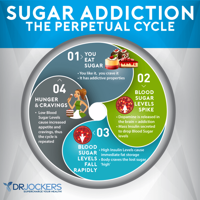
Author: Herman Swift

  • DIY Cervical Roll for Pain-Free Sleep

    DIY Cervical Roll for Pain-Free Sleep

    DIY cervical roll to manage neck pain while sleeping is a practical and affordable solution for better sleep. Neck pain can disrupt your rest, making it hard to get a good night’s sleep. Creating your own cervical roll can help support your neck in the right position, reducing pain and discomfort. This guide explores the process, covering different materials, construction techniques, and optimal use for a personalized sleep solution.

    From simple towel rolls to more complex designs using blankets and pillows, we’ll delve into various DIY cervical roll options. We’ll also discuss crucial factors like sizing, adjustments, and proper positioning to maximize comfort and effectiveness. Whether you’re looking for a quick fix or a long-term solution, understanding how to create your own cervical roll will empower you to take control of your sleep quality.

    Introduction to DIY Cervical Rolls

    Neck pain is a common ailment affecting people of all ages. It can stem from various factors, including poor posture, stress, muscle strain, or even underlying medical conditions. The pain often manifests as stiffness, soreness, or a dull ache, impacting daily activities and sleep quality. Understanding the root causes of neck pain is crucial for developing effective management strategies.Proper sleep posture plays a vital role in mitigating neck pain.

    Maintaining a neutral spine alignment during sleep is essential for preventing strain on the neck muscles. Many people find that their current sleep positions exacerbate existing neck pain or contribute to its development. DIY cervical rolls offer a practical and affordable solution to support the neck during sleep, promoting better spinal alignment and reducing discomfort. They provide a customized support system tailored to individual needs, allowing for a more comfortable and restorative sleep experience.

    Materials Needed for a DIY Cervical Roll

    Creating a DIY cervical roll is surprisingly straightforward and requires minimal materials. The most crucial aspect is selecting materials that provide adequate support without being overly rigid or uncomfortable. Here’s a list of common and effective options:

    • Soft, supportive fabric: Choose a fabric that’s comfortable against your skin, yet firm enough to provide adequate support. Examples include fleece, flannel, or a soft cotton blend. Avoid overly thin fabrics like silk or sheer cotton, as they won’t offer sufficient support.
    • Stuffing material: A variety of materials can be used for stuffing, each offering a different level of support and comfort. Consider options like buckwheat hulls, rice, or dried beans. These materials provide a gentle, supportive cushion. Avoid materials that may be allergenic or pose a choking hazard. A combination of stuffing materials can also be considered to optimize the support and comfort level.

    • A sturdy, but soft, container or form: This will hold the stuffing in place and determine the shape of the cervical roll. A pillowcase, a clean sock, or a small rectangular piece of cardboard can serve as the container. Ensure the container is soft enough to not exert undue pressure on the neck.

    Crafting Your Cervical Roll

    Crafting a cervical roll involves careful consideration of the materials and the desired firmness. The roll should be neither too firm nor too soft. An ideal firmness allows for proper support without impeding airflow or circulation.

    1. Preparing the container: If using a pillowcase or sock, ensure it’s clean and free of any imperfections that might cause discomfort. If using cardboard, ensure it’s a soft and flexible type.
    2. Filling the container: Gradually fill the container with the chosen stuffing material, leaving some space for adjustment. Overfilling can lead to discomfort, while underfilling can compromise the roll’s support.
    3. Adjusting the firmness: Once filled, gently squeeze or compress the roll to adjust its firmness. Experiment with the amount of stuffing to find the ideal balance between support and comfort.
    4. Finishing touches: Secure the opening of the container (e.g., tying the pillowcase or sock). This will help to maintain the shape of the cervical roll. Alternatively, you can use fabric glue to secure the stuffing in place.

    Benefits of DIY Cervical Rolls

    Customizable support, cost-effectiveness, and tailored comfort are among the key benefits of DIY cervical rolls. They can be adjusted to perfectly suit your individual needs, unlike store-bought rolls that might not provide the desired support. This customization ensures that the roll aligns with your neck’s unique contours, minimizing pressure points and maximizing comfort.

    Different Types of DIY Cervical Rolls

    Crafting a supportive cervical roll at home offers a cost-effective and personalized approach to managing neck pain during sleep. This flexibility allows you to tailor the support to your specific needs and preferences. Understanding the various materials and construction techniques is key to maximizing comfort and effectiveness.Various materials can be used to create a supportive cervical roll, each with its own set of advantages and disadvantages.

    Choosing the right material depends on factors like comfort, support, and budget. A crucial consideration is the ability of the material to maintain its shape and provide consistent support throughout the night.

    Materials for DIY Cervical Rolls, Diy cervical roll to manage neck pain while sleeping

    Different materials offer varying degrees of support and comfort. Towels, blankets, and pillows are common household items that can be adapted for cervical roll construction. The choice of material influences the roll’s firmness and the level of support it provides.

    • Towels: Soft and absorbent, towels are readily available and affordable. Their versatility allows for quick adjustments and modifications to the roll’s shape. However, their limited support might not be ideal for individuals needing a firmer, more substantial roll. For example, a single towel might not offer sufficient support for someone with severe neck pain.
    • Blankets: Blankets provide a more substantial and supportive structure compared to towels. Their bulkiness can offer a comforting warmth and a greater degree of pressure relief for the neck. However, blankets can be bulkier and might not be as easily shaped or adjusted as towels.
    • Pillows: Pillows, particularly those designed for specific sleeping positions or neck support, offer tailored support. Their pre-formed structure can offer a reliable level of support. However, pillows can be expensive and might not fit everyone’s needs or sleeping preferences.

    Construction Techniques

    The way a cervical roll is constructed directly impacts its effectiveness and comfort. Different techniques cater to various needs and preferences.

    • Rolling a Towel: Simply roll a towel or several towels tightly to create a cylindrical shape. This method is straightforward and adaptable to different thicknesses. Adjust the tightness of the roll to customize the support level.
    • Layering Blankets: Stack multiple blankets, carefully arranging them to create a firm and supportive structure. This technique provides more bulk and support compared to a single blanket or towel. For instance, layering three blankets might be appropriate for a heavier individual needing significant support.
    • Using Pillows: Position pillows to support the neck and head. For example, one pillow under the head and another slightly under the neck creates a customizable support system. Experiment with different pillow heights and positions to find the optimal configuration.

    Comparison of Materials

    Each material has its own advantages and disadvantages when constructing a cervical roll. The best choice depends on personal preferences and the desired level of support.

    Material Pros Cons
    Towels Affordable, readily available, easily adjustable Limited support, may not be suitable for all needs
    Blankets More substantial support, comfortable warmth Bulkier, may not be as easily adjustable
    Pillows Tailored support, pre-formed structure More expensive, might not fit everyone’s needs

    Creating a Comfortable Cervical Roll

    Diy cervical roll to manage neck pain while sleeping

    A well-made cervical roll can significantly enhance your sleep comfort and alleviate neck pain. Crucially, the roll’s effectiveness hinges on its proper size, fit, and customization to your unique needs. This section will guide you through crafting a supportive and personalized cervical roll.Creating a supportive cervical roll is not a one-size-fits-all process. Careful consideration of sizing and adjustment is vital for achieving optimal neck alignment and comfort during sleep.

    Understanding how to tailor your cervical roll to your specific needs will ensure maximum effectiveness in relieving pain and promoting restful sleep.

    Importance of Proper Sizing and Fitting

    Proper sizing and fitting of the cervical roll are essential for achieving the desired support and preventing discomfort. A roll that’s too small won’t provide adequate support, potentially exacerbating neck pain. Conversely, a roll that’s too large can lead to an unnatural curve, hindering spinal alignment and resulting in discomfort. The roll should cradle your neck naturally, allowing for a gentle curve and avoiding any pressure points.

    Adjusting the Cervical Roll for Optimal Comfort

    Several methods can be employed to adjust a cervical roll for optimal comfort. Experimentation is key. Trial and error will help you determine the ideal position and firmness for your neck. Consider using different types of padding materials or adjusting the overall length and width of the roll. A cervical roll should allow your head and neck to rest in a neutral, comfortable position without undue pressure or strain.

    Customizing the Cervical Roll for Individual Needs

    Customization is crucial for ensuring the cervical roll provides targeted support. Various methods can be used to personalize the roll to meet your unique needs. Adding padding, such as a thin layer of fleece or a small pillow, can increase the roll’s cushioning and comfort. Experiment with different materials and thicknesses to find the perfect combination. A cervical roll that’s too firm might not adequately support your neck, while a roll that’s too soft might not provide enough stability.

    Creating a Cervical Roll for Proper Spinal Alignment

    The goal is to create a roll that promotes proper spinal alignment. A properly positioned cervical roll should help maintain a natural curve in the neck, preventing strain and supporting the head. This involves ensuring the roll’s height and width align with the natural curvature of your neck. Begin by positioning the roll in the space between your shoulders and head, ensuring the roll is positioned to maintain a neutral spinal alignment.

    Ensure the roll does not force your head into an unnatural position. A good cervical roll allows the neck to rest in a comfortable and natural position.

    Using DIY Cervical Rolls for Sleep

    Snuggling into a comfy bed, ready for a restful night’s sleep, shouldn’t be marred by neck pain. A DIY cervical roll, crafted with care and tailored to your needs, can significantly enhance your sleep experience by providing optimal support and alignment. This section dives into how to strategically utilize your homemade cervical roll to maximize comfort and alleviate potential discomfort.Correct positioning of your cervical roll is crucial for maximizing its benefits.

    Proper placement ensures the roll supports your neck’s natural curvature, preventing strain and promoting a more comfortable sleep posture.

    Sleep Positions and Cervical Roll Placement

    Different sleep positions interact differently with the body’s alignment. Understanding these interactions allows you to position your DIY cervical roll for optimal support and comfort. By considering your preferred sleep position and the roll’s placement, you can achieve a better night’s sleep.

    Back Sleep

    Sleeping on your back is often recommended for maintaining spinal alignment. To maximize this, place the cervical roll directly beneath your neck, ensuring it comfortably fills the gap between your shoulders and the bed. The roll should be firm enough to maintain your neck’s natural curve without feeling overly rigid or constricting. Ensure the roll is snug enough to provide support without being too tight.

    Side Sleep

    Side sleeping can also be beneficial with a cervical roll. Position the roll along the curve of your neck, supporting your head and neck in a neutral position. If you’re a side sleeper, place the roll on the side of your head and neck to avoid putting pressure on one side of the neck. Consider the roll’s size and your body type to ensure comfort.

    The roll should not press against your jawline.

    Stomach Sleep

    Sleeping on your stomach is generally not ideal for neck health. While a cervical rollmight* offer some support, it’s unlikely to provide significant benefit compared to back or side sleeping. The unnatural curvature of the spine during stomach sleeping can put stress on the neck.

    Optimal Sleep Positions and Cervical Roll Usage

    Sleep Position Cervical Roll Placement Benefits
    Back Sleep Directly beneath the neck, filling the gap between shoulders and bed Maintains spinal alignment, promotes comfort
    Side Sleep Along the curve of the neck, supporting head and neck in a neutral position Supports head and neck, prevents strain on one side
    Stomach Sleep Potentially, but not ideal Minimal support, can exacerbate neck strain

    Common Mistakes to Avoid

    Using a cervical roll incorrectly can lead to discomfort and potentially worsen existing neck pain. Carefully consider these points to avoid potential issues.

    • Using a roll that’s too firm or too soft: A roll that’s too firm can restrict movement and cause discomfort, while a too-soft roll won’t provide adequate support.
    • Incorrect placement: The roll should be positioned directly beneath the neck, aligning with the natural curve of the spine. Incorrect positioning can result in neck pain.
    • Using the roll for extended periods: If you’re experiencing chronic neck pain, consult with a healthcare professional. Using a cervical roll for prolonged periods without proper guidance could be detrimental.
    • Ignoring personal comfort: Pay attention to your body’s feedback. If the roll isn’t providing comfort or support, adjust its placement or try a different size.

    Safety and Considerations: Diy Cervical Roll To Manage Neck Pain While Sleeping

    DIY cervical rolls can be a helpful tool for managing neck pain, but it’s crucial to approach them with caution and awareness of potential risks. Understanding the limitations of homemade solutions and prioritizing professional guidance is vital for ensuring your safety and well-being. This section explores the importance of responsible DIY practices and highlights when to seek expert medical advice.

    Potential Risks of DIY Cervical Rolls

    Improperly constructed or sized cervical rolls can lead to discomfort, further neck pain, or even complications. A poorly fitted roll may not provide adequate support, exacerbating existing neck issues or potentially causing new ones. Inaccurate measurements and unsuitable materials can lead to pressure points or uneven support, hindering the intended therapeutic effect. Furthermore, homemade rolls might not meet the same quality standards as professionally manufactured products, potentially compromising the long-term effectiveness and safety of the support.

    Importance of Professional Consultation

    Before embarking on a DIY cervical roll project, consulting a healthcare professional is paramount. A doctor or physical therapist can assess your specific needs and diagnose the underlying cause of your neck pain. They can provide tailored recommendations for appropriate support, ensuring that any cervical support you use aligns with your individual condition and needs. A professional evaluation is essential for determining the correct type, size, and firmness of the support needed to promote healing and prevent further injury.

    This personalized approach is crucial for avoiding potential harm and achieving optimal results.

    Recognizing Limitations of DIY Cervical Rolls

    DIY cervical rolls are not a substitute for professional medical care. They are designed for temporary support and comfort, not for long-term treatment of chronic neck pain or severe injuries. While they can provide temporary relief from discomfort, they may not address the root cause of the problem. For instance, a DIY roll might offer comfort during sleep, but it won’t address underlying issues like muscle imbalances or spinal misalignments that require more comprehensive treatment strategies.

    Their effectiveness is contingent upon proper use and understanding of the limitations.

    Making a DIY cervical roll for neck pain relief while sleeping is a great idea, but for optimal results, consider how long melatonin lasts in your body. Understanding how long melatonin supplements stay active in your system might influence when you use them alongside your cervical roll. Ideally, you want to align your sleep routine to maximize the effectiveness of both methods for managing your neck pain.

    A well-fitted cervical roll supports your neck while you sleep, promoting better posture and reducing stiffness. Knowing how long does melatonin last can help you strategize when to take it, and to ensure you get the best sleep possible. This can help ensure your neck pain is managed effectively and you can wake up feeling refreshed.

    Warning Signs Requiring Professional Medical Advice

    If you experience any of the following symptoms while using a DIY cervical roll or after initiating use, seek immediate medical attention:

    • Increased or persistent neck pain:
    • Persistent or worsening neck pain despite using the cervical roll may indicate a more serious underlying condition. If the pain intensifies, it is crucial to seek professional help to identify and address the root cause.

      Ever tried a DIY cervical roll for better neck support while sleeping? It’s a fantastic way to alleviate aches and pains. While you’re focusing on your neck health, it’s also important to consider other potential issues. For example, a lingering cough can be a real pain point. If you’re experiencing one, exploring effective treatments like those outlined in this article on treatments for lingering cough might be beneficial.

      Ultimately, a comfortable neck position at night is key, and a DIY cervical roll can be a game-changer in your sleep routine.

    • Numbness or tingling in the arms or hands:
    • These symptoms could signify nerve compression or damage. Prompt medical intervention is vital to prevent potential long-term complications.

    • Headaches or dizziness:
    • Headaches or dizziness associated with neck pain might be related to underlying structural issues in the neck or head. Consult a healthcare professional to rule out potential causes and receive appropriate guidance.

    • Difficulties with swallowing or breathing:
    • Any difficulty with swallowing or breathing should immediately trigger a visit to a healthcare provider. Such symptoms can signal serious medical conditions requiring immediate attention.

    • Signs of infection or inflammation around the neck area:
    • Redness, swelling, warmth, or pus around the neck area are potential indicators of infection. Seek immediate medical attention to prevent the spread of infection and complications.

    Alternatives and Comparisons

    Finding the right support for your neck pain while sleeping can feel like a treasure hunt. Sometimes, a DIY cervical roll is perfect, but other times, a different approach might be more suitable. This section explores alternatives and comparisons, helping you weigh the pros and cons of various options.While a DIY cervical roll can be an effective and budget-friendly solution, store-bought supports and other sleep strategies also offer unique advantages.

    Understanding these differences can help you make an informed decision that best addresses your individual needs.

    Comparing DIY Cervical Rolls with Store-Bought Neck Supports

    DIY cervical rolls offer the benefit of customization, allowing you to tailor the support to your exact preferences. However, store-bought options often come pre-shaped and pre-filled with materials designed for a more standardized fit. This can be beneficial for those who prefer a consistent level of support, or who find it challenging to create a roll that meets their specific needs.

    Advantages of Store-Bought Cervical Rolls

    Store-bought supports often provide a more consistent and predictable level of support. This can be particularly valuable for individuals who experience fluctuating neck pain or find it challenging to maintain a consistent DIY roll throughout the night. Pre-made rolls can also come with different fillings and cover materials, offering options for temperature regulation and comfort.

    Disadvantages of Store-Bought Cervical Rolls

    Store-bought neck supports can be more expensive than a DIY solution. Customization is often limited, and finding a support that perfectly addresses your unique needs and preferences can be challenging. Some store-bought supports may contain materials that aren’t as breathable or hypoallergenic as homemade options.

    Other Methods to Manage Neck Pain While Sleeping

    A cervical roll isn’t the only tool in your sleep toolkit. Various other methods can effectively manage neck pain. These range from adjusting your sleep position to incorporating other therapies.

    Adjusting Sleep Position

    Sleeping on your back can sometimes exacerbate neck pain. Experimenting with different positions, like sleeping on your side with a pillow between your knees or elevating your head with a few extra pillows, might provide more comfortable support.

    Alternative Therapies

    Consider incorporating alternative therapies alongside or instead of a DIY cervical roll. These can include:

    • Heat Therapy: Applying a heating pad or warm compress to the affected area can help relax muscles and alleviate pain.
    • Cold Therapy: Using an ice pack or cold compress can help reduce inflammation and pain, particularly in the acute phase of neck pain.
    • Massage Therapy: A professional massage therapist can target specific muscle groups in the neck and shoulders, improving circulation and releasing tension.
    • Acupuncture: This traditional Chinese medicine technique involves inserting thin needles into specific points on the body to stimulate energy flow and reduce pain.
    • Stretching Exercises: Gentle stretching exercises, recommended by a healthcare professional, can help improve neck flexibility and range of motion.

    Properly selecting a neck support, whether DIY or store-bought, is crucial for alleviating pain and improving sleep quality. Experimentation with different methods and consulting with healthcare professionals are key steps in managing neck pain effectively.

    Illustrative Examples

    Creating your own cervical roll can be a personalized solution for neck pain relief. Understanding different designs and construction methods allows you to tailor the support to your specific needs. Experimenting with various materials and shapes will help you find the perfect fit for your comfort.A well-made DIY cervical roll can provide the crucial support needed for a restful night’s sleep and help ease discomfort throughout the day.

    Properly crafted rolls are designed to maintain the natural curve of your neck, reducing strain and pressure points.

    Rolled-Up Towel Cervical Roll

    This is a simple and readily available method. A rolled-up towel offers a basic level of support.

    Materials: A soft, absorbent towel.

    Instructions:

    1. Place the towel flat on a surface.
    2. Roll the towel tightly from one end to the other, creating a cylindrical shape.
    3. Adjust the tightness of the roll to your desired firmness.
    4. Place the roll under your neck, supporting the natural curve of your spine.

    Blanket Cervical Roll

    A blanket offers a more substantial and potentially warmer support than a towel. This method provides an option for a firmer roll.

    Ever struggled with neck pain at night? A DIY cervical roll can be a game-changer for a more comfortable sleep. Finding the right support is key, and it can be a surprisingly effective solution. While we’re on the topic of health concerns, if you’re dealing with wet AMD, understanding who treats it is crucial. Fortunately, resources like who treats wet amd provide valuable information.

    Ultimately, a well-made cervical roll can significantly improve your sleep quality and overall neck health.

    Materials: A soft, comfortable blanket.

    Instructions:

    1. Lay the blanket flat on a surface.
    2. Roll the blanket tightly into a cylindrical shape, similar to rolling a towel.
    3. To enhance the firmness, you can use a pillow or a few books to further compact the roll.
    4. Place the roll under your neck, ensuring the curve of your neck is well-supported.

    Adjustable Cervical Roll

    This design offers customized support, allowing you to adapt the firmness and height.

    Materials: A soft pillowcase, filling material (e.g., buckwheat hulls, rice, or foam pellets), and a measuring tape.

    Instructions:

    1. Measure the length of the pillowcase needed for your desired neck support.
    2. Fill the pillowcase with the chosen filling material.
    3. Adjust the amount of filling material to achieve your desired firmness.
    4. Secure the pillowcase opening, either by tying it or using a closure.
    5. Place the roll under your neck, ensuring the support aligns with your neck’s natural curve.

    Diagram:

    Imagine a pillowcase. The pillowcase is filled with your chosen filling material. The filling material determines the firmness. You can adjust the filling to achieve the perfect firmness.

    Maintenance and Care

    Keeping your DIY cervical roll clean and well-maintained is crucial for its effectiveness and longevity. Proper care ensures a hygienic and comfortable support system for your neck. Regular cleaning and preventative measures will significantly extend the life of your roll and help you avoid potential health concerns.

    Proper Cleaning Methods

    Regular cleaning prevents the buildup of dirt, sweat, and potentially harmful microorganisms. The cleaning method depends on the materials used to construct your cervical roll. For rolls made of washable fabrics like cotton or linen, gentle machine washing is usually sufficient. For rolls incorporating stuffing like buckwheat hulls or dried beans, a gentle hand wash or spot cleaning is recommended to prevent damage to the filling.

    Always check the care instructions provided with the materials used.

    Preventing Mold and Mildew

    Mold and mildew growth can be a serious concern, particularly in humid environments. To prevent this, maintain good ventilation around your cervical roll, especially if you tend to sweat while sleeping. Allow your cervical roll to dry completely after washing or exposure to moisture. Avoid storing the roll in a damp or enclosed space. If your roll does show signs of mold or mildew, discard it immediately and replace it with a new one.

    Ensuring Longevity

    Several strategies can significantly extend the life of your DIY cervical roll. Regular inspection for wear and tear is essential. Look for any signs of fraying, damage to the filling, or structural weaknesses. Proper storage is also vital. Store your cervical roll in a clean, dry, and well-ventilated area, away from direct sunlight or extreme temperatures.

    Avoid excessive compression or bending, as this can damage the roll’s structure and reduce its effectiveness over time.

    When to Replace a Cervical Roll

    Replacing your DIY cervical roll is necessary when it no longer provides adequate support or comfort. If you notice a significant decrease in support or a change in the roll’s shape, it’s time for a replacement. Regular use and the material used for construction will affect how quickly a cervical roll degrades. Another important consideration is any visible signs of damage, such as tearing, fraying, or significant settling of the filling material.

    If you experience discomfort or pain when using the roll, it’s a clear sign to replace it.

    Concluding Remarks

    Diy cervical roll to manage neck pain while sleeping

    In conclusion, creating a DIY cervical roll can be a fantastic way to address neck pain and improve your sleep. By understanding the different materials, construction techniques, and proper use, you can tailor a support system that aligns with your specific needs. Remember to prioritize comfort, safety, and proper spinal alignment. If you experience persistent pain, always consult a healthcare professional.

    Experiment with various designs and find what works best for you. Good sleep is essential for overall well-being, and a DIY cervical roll can be a significant step in achieving that goal.

  • Preventing Degenerative Disc Disease A Guide

    Preventing Degenerative Disc Disease A Guide

    Preventing degenerative disc disease is crucial for maintaining a healthy spine and overall well-being. This comprehensive guide explores lifestyle modifications, the importance of spinal health, therapeutic interventions, prevention through early detection, supporting research, and illustrative examples of healthy spinal practices. We’ll delve into dietary recommendations, stress management, exercise plans, posture, and more, empowering you to take proactive steps towards a healthier spine.

    Understanding the anatomical structure of the spine, common risk factors, and physiological processes behind degenerative disc disease is fundamental. We’ll also examine the role of genetics, the link to overall well-being, and effective therapeutic interventions, including physical therapy, manual therapy, and exercise programs. A crucial aspect is early detection and monitoring, equipping you with the knowledge to recognize symptoms and seek timely intervention.

    Lifestyle Modifications for Preventing Degenerative Disc Disease

    Maintaining a healthy spine is crucial for overall well-being, and proactive measures can significantly reduce the risk of degenerative disc disease. Adopting a holistic approach that combines dietary choices, stress management techniques, regular exercise, and proper posture is essential for spinal health. This involves understanding the intricate relationship between lifestyle factors and the health of your spine.Dietary recommendations play a vital role in supporting spinal health.

    Taking care of your spine is crucial for preventing degenerative disc disease, and a healthy diet plays a big part. But sometimes, unexpected tummy troubles like diarrhea can throw off your routine and impact your overall well-being. Knowing how to manage those episodes quickly can actually help you maintain a healthier lifestyle, which is key for preventing disc problems.

    Check out this helpful guide on how to make diarrhea go away for some practical tips. Ultimately, consistent good habits, like proper nutrition and hydration, are essential to keep your spine strong and your body functioning at its best.

    A balanced diet rich in essential nutrients, combined with adequate hydration, is fundamental for maintaining the strength and integrity of the spine’s supporting tissues.

    Dietary Recommendations for Spinal Health

    A diet rich in fruits, vegetables, and whole grains provides essential vitamins, minerals, and antioxidants that promote overall health and support spinal tissue regeneration. Include foods high in collagen, such as bone broth and gelatin, which are vital for maintaining the structural integrity of cartilage and discs. Lean protein sources like fish, poultry, and beans are crucial for building and repairing tissues.

    Limit processed foods, sugary drinks, and excessive saturated and unhealthy fats, as these can contribute to inflammation and negatively impact spinal health. Staying hydrated is also important; water helps maintain the proper cushioning and lubrication within the spinal discs.

    Stress Management Strategies for Spinal Health

    Chronic stress can lead to muscle tension and strain in the back, potentially contributing to disc degeneration. Stress management techniques such as yoga, meditation, deep breathing exercises, and mindfulness practices can help reduce stress levels and promote relaxation, thereby mitigating the negative effects of stress on the spine. Identifying and addressing underlying stressors in daily life can also significantly contribute to long-term stress reduction.

    Sample Weekly Exercise Plan for Spinal Health

    A consistent exercise routine focusing on core strengthening and spinal flexibility is essential for preventing degenerative disc disease. This plan provides a framework, but individual needs and limitations should be considered. Adjust the intensity and duration based on your fitness level.

    • Monday: Core Strengthening (Plank variations, Russian twists, leg raises). Focus on controlled movements to avoid strain.
    • Tuesday: Flexibility and Stretching (Cat-cow stretches, spinal twists, hamstring stretches). Hold each stretch for 15-30 seconds.
    • Wednesday: Rest or Active Recovery (Light walk, gentle yoga). Allow your body to recover and rebuild.
    • Thursday: Core Strengthening (Bird-dog exercises, side planks, dead bugs). Prioritize proper form over speed.
    • Friday: Flexibility and Stretching (Child’s pose, seated forward folds, neck stretches). Maintain consistent breathing throughout the stretches.
    • Saturday: Cardiovascular Exercise (Brisk walking, swimming, cycling). Maintain a moderate intensity to improve overall fitness.
    • Sunday: Rest or Active Recovery (Gentle stretching, light yoga). Allow for complete rest and recovery.

    Importance of Posture and Ergonomics

    Maintaining good posture and ergonomic practices in daily activities is crucial for spinal health. Poor posture can lead to misalignment, placing undue stress on the spine and increasing the risk of degenerative disc disease. Ensure your workstation and seating arrangements are ergonomically sound. Use proper lifting techniques when handling objects, and avoid prolonged periods of sitting or standing in awkward positions.

    Staying active and strong is key to preventing degenerative disc disease, but sometimes a fun way to get moving is needed! Engaging in activities like sign language games and puzzles sign language games and puzzles can be a great way to boost brainpower and physical dexterity, both of which can contribute to overall well-being and help ward off issues like degenerative disc disease.

    These types of activities can make staying healthy a more enjoyable part of your routine, making it easier to maintain a healthy lifestyle and help prevent problems in the future.

    Effectiveness of Different Physical Activities

    Various forms of physical activity can contribute to spinal health. Low-impact exercises like swimming and cycling are gentle on the spine, while weight-bearing exercises like walking and strength training can build bone density and muscle strength, thereby supporting the spine. A combination of different types of physical activity, including cardiovascular exercises, core strengthening, and flexibility training, offers the most comprehensive approach to spinal health.

    Maintaining a Healthy Weight and Spinal Load

    Maintaining a healthy weight is crucial for reducing spinal load. Excess weight puts extra pressure on the spine, increasing the risk of degenerative disc disease. A healthy weight management plan that combines a balanced diet and regular exercise can help alleviate this burden on the spine and promote overall well-being. A balanced approach that prioritizes gradual weight loss and healthy habits is more sustainable and effective.

    Importance of Spinal Health

    Preventing degenerative disc disease

    The spine, our body’s central support structure, plays a crucial role in posture, movement, and overall well-being. Understanding its intricate anatomy and the factors that can compromise its health is paramount for preventing degenerative disc disease. A healthy spine allows for effortless movement and protects the delicate spinal cord and nerves. Maintaining spinal health is not just about avoiding pain; it’s about promoting a higher quality of life.The spine is a complex structure composed of interconnected vertebrae, cushioned by intervertebral discs.

    These discs act as shock absorbers, allowing for flexibility and movement while supporting the weight of the body. The spinal column’s curves provide stability and help distribute forces effectively. Maintaining these natural curves and the structural integrity of the discs is essential for spinal health.

    Anatomical Structure of the Spine, Preventing degenerative disc disease

    The spine is divided into five sections: cervical (neck), thoracic (upper back), lumbar (lower back), sacrum, and coccyx (tailbone). Each section has a specific curve and function. The cervical and lumbar regions exhibit lordotic curves, while the thoracic and sacral regions have kyphotic curves. These curves are crucial for balance, posture, and absorbing shock. The vertebrae are interconnected by facet joints and ligaments, providing stability and allowing for controlled movement.

    Risk Factors for Degenerative Disc Disease

    Several factors contribute to the development of degenerative disc disease. Age is a significant risk factor, as the natural aging process leads to changes in the discs. Repetitive strain injuries, such as those from heavy lifting or prolonged sitting, can place excessive stress on the spine. Obesity, often associated with increased body weight, exerts additional pressure on the spinal column, increasing the risk.

    Genetic predisposition also plays a role, as some individuals may inherit a susceptibility to disc degeneration.

    Physiological Processes of Degenerative Disc Disease

    Degenerative disc disease involves a gradual breakdown of the intervertebral discs. The discs lose their water content and become less elastic, leading to reduced cushioning capacity. This loss of cushioning can cause the discs to bulge or herniate, putting pressure on the spinal nerves. Furthermore, the surrounding tissues, such as ligaments and muscles, may undergo structural changes, contributing to the overall degeneration process.

    Bone spurs may also develop, causing further narrowing of the spinal canal and impingement of nerves.

    Genetics and Degenerative Disc Disease

    Genetic predisposition plays a role in individual susceptibility to degenerative disc disease. Studies have shown that certain genes may influence the rate of disc degeneration. This genetic predisposition, combined with environmental factors, can increase the risk of developing the condition. Individuals with a family history of degenerative disc disease may be at a higher risk. Genetic factors can influence the structure and composition of the discs, making them more susceptible to wear and tear.

    Spinal Health and Overall Well-being

    Maintaining spinal health has far-reaching implications for overall well-being. A healthy spine supports optimal posture and movement, allowing for a wider range of activities. It protects the spinal cord and nerves, ensuring proper transmission of signals throughout the body. Chronic back pain associated with degenerative disc disease can significantly impact daily activities, mood, and overall quality of life.

    A healthy spine allows for a more active and fulfilling life, enabling individuals to engage in physical activities, work, and social interactions without limitations.

    Therapeutic Interventions

    Addressing degenerative disc disease requires a multifaceted approach encompassing lifestyle modifications, preventative measures, and, when necessary, therapeutic interventions. Effective management often involves a combination of strategies to alleviate pain, improve function, and slow the progression of the condition. These interventions aim to reduce pain and stiffness, enhance spinal stability, and restore mobility.

    Staying active and maintaining good posture are key to preventing degenerative disc disease. While we’re on the topic of health mysteries, have you ever wondered if vivid dreams are a sign of mental illness? Exploring this question can be fascinating, and you might find some interesting insights in this article on are vivid dreams a sign of mental illness.

    Ultimately, a healthy lifestyle, including regular exercise and proper ergonomics, is crucial for keeping your spine in tip-top shape and warding off disc issues.

    Effectiveness of Physical Therapy

    Physical therapy plays a crucial role in managing degenerative disc disease. Specific exercises and techniques can strengthen supporting muscles, improve posture, and enhance spinal flexibility. Therapeutic exercises, such as core strengthening and flexibility exercises, can improve spinal stability and reduce pain. Manual therapy, including spinal manipulation, can help reduce pain and stiffness by improving joint mobility. The effectiveness of physical therapy varies depending on the individual’s specific condition, but it can significantly improve function and quality of life for many.

    Role of Manual Therapy

    Manual therapy, including spinal manipulation and mobilization techniques, can address spinal pain and stiffness associated with degenerative disc disease. These techniques aim to restore proper joint mechanics and reduce pain by improving joint mobility and reducing muscle spasms. By addressing the underlying biomechanical issues, manual therapy can enhance the effectiveness of other interventions. Properly trained therapists can perform these techniques to reduce pain and improve mobility in the affected area.

    Benefits of Exercise Programs

    Tailored exercise programs designed for spinal issues can significantly improve function and reduce pain associated with degenerative disc disease. These programs typically include exercises focusing on core strengthening, flexibility, and posture correction. Exercises targeting the core muscles enhance spinal stability, thereby reducing pressure on the affected discs. Flexibility exercises improve range of motion and reduce stiffness, while posture exercises help maintain correct spinal alignment, which can prevent further deterioration.

    Comparison of Pain Management Strategies

    Various pain management strategies are available for degenerative disc disease. Pharmacological interventions, including over-the-counter pain relievers and prescription medications, can effectively manage pain and inflammation. Non-pharmacological approaches, such as heat or cold therapy, acupuncture, and massage therapy, can also provide relief from pain and stiffness. The most effective approach often depends on the individual’s specific condition and response to different treatments.

    The choice of pain management strategy should be made in consultation with a healthcare professional.

    Summary of Interventions

    Intervention Type Description Effectiveness Potential Risks
    Physical Therapy Includes exercises, manual therapy (manipulation/mobilization), and education on posture and lifestyle modifications. Generally effective in improving function, reducing pain, and increasing spinal stability. Results vary based on individual adherence and the severity of the condition. Potential for mild discomfort during treatment, but serious complications are rare with proper technique.
    Medications Includes over-the-counter pain relievers (NSAIDs), muscle relaxants, and potentially stronger prescription medications for severe pain. Effective in managing pain and inflammation, but may have side effects. Effectiveness varies depending on the individual and the medication used. Potential for side effects such as stomach upset, kidney problems, or allergic reactions. Long-term use should be monitored by a physician.
    Injections Corticosteroid injections into the affected area can provide temporary pain relief. Can be effective for short-term pain relief, but may not be suitable for long-term management. Potential for infection, nerve damage, or other complications. May not be appropriate for all patients.
    Surgery Involves procedures like spinal fusion or disc replacement for severe cases with persistent and debilitating pain. Can be effective in relieving severe pain and restoring spinal stability, but carries risks. Potential for complications such as infection, nerve damage, implant failure, or persistent pain. Not suitable for all patients.

    Prevention through Early Detection and Monitoring: Preventing Degenerative Disc Disease

    Staying proactive in spinal health is crucial, especially for those at risk of degenerative disc disease. Regular check-ups, coupled with an understanding of potential symptoms and diagnostic tools, can significantly impact the progression of this condition. Early intervention can slow the disease’s development and potentially improve long-term outcomes.Early detection plays a vital role in managing degenerative disc disease.

    By recognizing the subtle signs and undergoing appropriate testing, individuals can work with healthcare professionals to implement strategies that may slow or halt the progression of the condition. This proactive approach can be incredibly beneficial in maintaining spinal health and quality of life.

    Importance of Regular Check-ups

    Regular check-ups, particularly for individuals with a family history of degenerative disc disease, are essential for preventative measures. These visits allow healthcare professionals to assess spinal health, identify potential risk factors, and implement preventative strategies. Proactive monitoring can help identify early signs of the condition, allowing for timely intervention. This proactive approach empowers individuals to address potential issues before they escalate.

    Symptoms to Watch Out For

    Recognizing the symptoms associated with degenerative disc disease is key to early detection. A variety of symptoms can manifest, ranging from mild discomfort to severe pain. Awareness of these signs can prompt individuals to seek professional evaluation and initiate treatment. Early intervention can significantly impact the progression of the condition.

    • Persistent lower back pain, often radiating to the buttocks, thighs, or legs (sciatica).
    • Numbness or tingling in the affected areas.
    • Muscle weakness in the legs or feet.
    • Stiffness in the lower back, especially in the morning.
    • Limited range of motion in the lower back.
    • Pain that worsens with prolonged sitting or standing.
    • Pain that is exacerbated by bending, lifting, or twisting.

    Diagnostic Tools for Early Detection

    Various diagnostic tools are available to aid in the early detection of degenerative disc disease. These methods, when combined with a thorough patient history and physical examination, provide a comprehensive assessment of spinal health.

    • Physical Examination: A physical examination allows the healthcare professional to assess posture, range of motion, and muscle strength. This provides valuable information about the current state of the spine and can highlight potential issues.
    • Imaging Techniques: Imaging techniques are crucial for visualizing the spinal structures and identifying any abnormalities. These techniques are invaluable in assessing the condition of the discs and the surrounding tissues.
    • Neurological Tests: Neurological tests evaluate the function of the nerves in the affected areas. This can help determine if the pain is stemming from nerve compression or other neurological issues.

    Role of Imaging Techniques in Assessing Spinal Health

    Imaging techniques provide crucial insights into the condition of the spine and surrounding structures. They can reveal signs of disc degeneration, herniation, or other abnormalities. These images are instrumental in the diagnostic process and help guide treatment decisions.

    • X-rays: X-rays primarily show the bony structures of the spine, allowing visualization of alignment, fractures, and other skeletal abnormalities. They are less effective at visualizing soft tissues like discs.
    • Magnetic Resonance Imaging (MRI): MRI provides detailed images of soft tissues, including the spinal cord, nerves, and discs. MRI is particularly valuable for identifying disc herniations, bulges, and other soft tissue abnormalities.
    • Computed Tomography (CT) Scans: CT scans provide detailed cross-sectional images of the spine, useful for assessing bone structures, fractures, and joint spaces. CT scans can be helpful in situations where detailed bony structures need to be evaluated.

    Importance of Early Intervention to Slow Disease Progression

    Early intervention is paramount in slowing the progression of degenerative disc disease. Prompt diagnosis and appropriate treatment strategies can effectively manage symptoms, prevent further deterioration, and preserve spinal function. By taking a proactive approach to spinal health, individuals can significantly improve their long-term well-being.

    Supporting Research and Innovations

    Preventing degenerative disc disease

    Degenerative disc disease (DDD) is a complex condition, and ongoing research plays a crucial role in understanding its mechanisms and developing effective treatments and preventative strategies. This section explores the latest advancements in research, highlighting innovative approaches and areas needing further investigation. The quest for better management of DDD involves exploring various avenues, from identifying biomarkers to employing cutting-edge therapies.Research into DDD is multifaceted, encompassing a range of methodologies and focusing on different aspects of the disease.

    Studies are designed to investigate the underlying causes, progression, and potential interventions. This exploration aims to provide a deeper understanding of the disease’s complex interplay of factors, paving the way for more effective strategies for prevention and treatment.

    Current Research Advancements

    Research in DDD is actively exploring new avenues to enhance our understanding of the disease’s mechanisms. Studies are investigating the role of genetics, inflammation, and biomechanical factors in the development and progression of DDD. This includes exploring the impact of lifestyle factors, such as posture, physical activity, and nutrition, on disc health. Furthermore, researchers are investigating the potential of novel biomarkers to aid in early detection and monitoring of DDD progression.

    Innovative Approaches

    Numerous innovative approaches are being investigated for preventing and managing DDD. These include non-invasive therapies like targeted drug delivery systems, utilizing growth factors to stimulate disc regeneration, and exploring the application of stem cell therapies. Researchers are also developing advanced imaging techniques to provide more detailed assessments of the spine and discs, enabling earlier and more accurate diagnoses.

    Furthermore, studies are exploring the use of personalized medicine approaches tailored to individual patient needs and genetic predispositions.

    Areas Requiring Further Research

    While significant progress has been made, several areas require further investigation to improve the understanding and management of DDD. These include:

    • Developing more accurate diagnostic tools for early detection of DDD, particularly in its asymptomatic stages. This includes identifying specific biomarkers that can indicate early disc degeneration and facilitate timely intervention.
    • Investigating the efficacy and safety of novel therapies, particularly non-invasive interventions, for slowing or reversing the progression of DDD. This includes further exploration of the use of stem cells and growth factors in regenerative therapies.
    • Defining the long-term effects of various treatment approaches, including the impact on patient quality of life and functional outcomes. Understanding the long-term effectiveness of different interventions is essential for guiding clinical practice and optimizing patient care.

    Research Methodologies

    Different methodologies are employed in studying DDD. Understanding the strengths and limitations of each approach is crucial for interpreting research findings.

    Methodology Description Advantages Disadvantages
    Clinical Trials Rigorous studies involving human participants to evaluate the effectiveness and safety of interventions. This includes randomized controlled trials (RCTs) where participants are randomly assigned to different treatment groups. Provides strong evidence regarding treatment efficacy and safety. Allows for objective comparison of different interventions. Can be time-consuming and expensive. May not always translate real-world applicability.
    Observational Studies Studies that observe and collect data on participants without manipulating variables. This includes cohort studies that follow a group of individuals over time to examine risk factors and outcomes. Useful for identifying potential risk factors and trends in disease progression. Can be more feasible and cost-effective than clinical trials. Cannot establish causality. Potential for confounding factors to influence results.
    In Vitro Studies Studies conducted in a laboratory setting using cells or tissues to investigate specific biological mechanisms. This includes studies on disc cell cultures to understand the effects of different factors on disc degeneration. Allows for controlled manipulation of variables and investigation of cellular processes. Can be a useful preliminary step for developing new therapies. Results may not always translate to the complex in vivo environment of the human body. Difficult to model the entire spectrum of DDD factors.

    Illustrative Examples of Healthy Spinal Practices

    Maintaining a healthy spine is crucial for overall well-being. Proper posture, lifting techniques, and daily routines directly impact spinal health, significantly reducing the risk of degenerative disc disease. Consistent healthy habits contribute to long-term spinal health, preventing pain and discomfort.Understanding the importance of spinal health goes beyond avoiding pain. A healthy spine allows for full range of motion, enabling activities of daily living and promoting an active lifestyle.

    This proactive approach to spinal care is essential for maintaining quality of life.

    Daily Routines Promoting Spinal Health

    Consistent, mindful habits are key to preserving spinal health. Regular physical activity, proper posture, and mindful movements throughout the day can significantly contribute to spinal well-being. Activities that strengthen core muscles and promote flexibility are paramount.

    • Morning Stretches: Begin your day with gentle spinal stretches to improve flexibility and circulation. Hold each stretch for 15-30 seconds, focusing on movements that engage the entire spine. This includes gentle twists, forward bends, and side stretches. Incorporating these stretches into your morning routine can significantly improve spinal health and flexibility.
    • Ergonomic Workspace Setup: A well-designed workspace is essential for spinal health during prolonged periods of sitting. Ensure your chair provides proper lumbar support, your monitor is at eye level, and your keyboard and mouse are positioned comfortably. This proactive approach minimizes stress on the spine during extended periods of work.
    • Regular Physical Activity: Engaging in regular physical activity, such as walking, swimming, or yoga, strengthens core muscles and improves spinal flexibility. Aim for at least 30 minutes of moderate-intensity exercise most days of the week. This strengthens the muscles supporting the spine, reducing the risk of injury.

    Proper Posture and Lifting Techniques

    Maintaining proper posture and using correct lifting techniques are essential to prevent spinal strain. Poor posture and improper lifting can lead to chronic pain and spinal problems. Proper Posture

    Image Description: The image depicts a person standing with good posture. The individual’s head is aligned with their spine, shoulders are relaxed, and their core is engaged. The back is straight, and the knees are slightly bent. This posture minimizes stress on the spine and promotes proper spinal alignment.

    Correct Lifting Technique

    Image Description: The image illustrates the correct technique for lifting an object. The lifter is bending at the knees, keeping their back straight, and using their leg muscles to lift the weight. The object is close to their body, minimizing stress on the lower back. This method minimizes the risk of injury during lifting activities.

    Ideal Spinal Alignment Infographics

    Visual representations of ideal spinal alignment can aid in understanding and implementing proper posture. These infographics illustrate the natural curves of the spine and the importance of maintaining a neutral spine. Ideal Spinal Alignment Infographic

    Image Description: The infographic displays a side view of the spine with its natural curves. The spine is shown in a neutral position, highlighting the importance of maintaining these curves. The infographic visually depicts the alignment of the cervical, thoracic, and lumbar spine. The image helps visualize the importance of maintaining these natural curves to promote spinal health.

    Common Mistakes Affecting Spinal Health

    Recognizing common mistakes that negatively impact spinal health is vital for preventive measures. Addressing these issues proactively can help prevent spinal problems.

    Mistake Explanation How to Correct
    Slouching Sustained poor posture, often while sitting, leading to spinal curvature and muscle strain. Use lumbar support, maintain a neutral spine, and take regular breaks.
    Lifting with a bent back Lifting heavy objects without bending the knees, putting excessive stress on the lower back. Bend at the knees, keep the back straight, and lift with leg muscles.
    Carrying heavy bags on one shoulder Uneven weight distribution causing spinal misalignment and strain. Distribute weight evenly, use a backpack or bag with adjustable straps.
    Prolonged sitting without breaks Lack of movement and engagement of spinal muscles, leading to stiffness and discomfort. Take regular breaks to stand up, stretch, and move around.

    Ending Remarks

    In conclusion, preventing degenerative disc disease is a multifaceted approach encompassing lifestyle changes, proactive healthcare, and a commitment to spinal health. This guide provides a comprehensive roadmap to understanding the condition, its prevention, and the available therapeutic interventions. By integrating the strategies discussed, you can significantly reduce your risk and maintain a healthy spine for years to come.

  • Treating Diastasis Recti with Physical Therapy A Guide

    Treating Diastasis Recti with Physical Therapy A Guide

    Treating diastasis recti with physical therapy is a comprehensive approach to managing this common postpartum condition. It involves a personalized plan encompassing assessment, targeted exercises, and patient education to effectively address the separation of abdominal muscles, often accompanied by associated symptoms like back pain and pelvic floor dysfunction. This guide delves into the various aspects of physical therapy interventions, from initial assessments to long-term management strategies, empowering individuals to understand and effectively recover from diastasis recti.

    The program begins with a thorough assessment of the condition, pinpointing the specific degree of separation and the patient’s overall physical capabilities. This personalized evaluation helps tailor a treatment plan that includes a variety of exercises focusing on core strength, breathing techniques, and pelvic floor rehabilitation. The plan also encompasses strategies for gradual progression, modifications for different fitness levels, and recognizing potential complications.

    Introduction to Diastasis Recti

    Diastasis recti is a common separation of the abdominal muscles, specifically the rectus abdominis, along the linea alba. This separation often occurs during pregnancy, but it can also develop after significant weight fluctuations or other factors. It’s a condition that affects many women, and understanding its causes, symptoms, and management is crucial for proactive health.This separation is usually a result of the stretching of the abdominal muscles during pregnancy.

    The increasing pressure on the abdomen during pregnancy, combined with hormonal changes, can weaken the connective tissue (linea alba) that holds the rectus abdominis muscles together. Post-pregnancy, factors like poor posture, improper lifting techniques, and lack of core strength can exacerbate or prolong the separation. In some cases, diastasis recti can occur in individuals who are not pregnant.

    Causes of Diastasis Recti

    Diastasis recti is often associated with pregnancy, due to the stretching of the abdominal muscles and the release of hormones. However, other factors contribute to its development, such as significant weight fluctuations, chronic coughing, or conditions that cause increased abdominal pressure. The connective tissue (linea alba) that joins the rectus abdominis muscles may weaken over time, leading to a separation.

    This can occur from improper lifting techniques or chronic conditions like asthma.

    Symptoms of Diastasis Recti

    A noticeable bulge or gap in the abdominal area, particularly when flexing the abdomen or performing certain movements, is a common symptom. Individuals may also experience discomfort or pain in the lower back, abdomen, or pelvis. This is because the weakened abdominal muscles can cause the organs to shift, leading to a potential imbalance in the body’s posture.

    Furthermore, difficulty with core exercises or everyday movements may also be experienced. In some cases, there may be no noticeable symptoms at all.

    Prevalence of Diastasis Recti, Treating diastasis recti with physical therapy

    Studies suggest that a significant portion of pregnant women experience diastasis recti, often ranging between 50-60%. While pregnancy is a common trigger, diastasis recti can affect people of all ages and genders, though the exact prevalence in the general population is less precisely measured and varies depending on the population studied. The condition can impact anyone who experiences significant abdominal strain.

    Importance of Early Intervention and Management

    Addressing diastasis recti early on is important to prevent long-term complications, such as chronic back pain, pelvic floor dysfunction, or persistent abdominal discomfort. Prompt intervention can lead to more effective treatment outcomes. Prompt diagnosis and management can improve the quality of life for individuals with this condition.

    Physical Therapy’s Role in Treating Diastasis Recti

    Physical therapy plays a crucial role in managing diastasis recti by addressing the underlying causes and symptoms. Physical therapists can design individualized exercise programs that strengthen the abdominal muscles, improve posture, and reduce pain. These programs often focus on core stabilization and strengthening exercises that do not exacerbate the separation. Physical therapists can also educate individuals on proper lifting techniques and body mechanics to prevent further separation.

    By working with the patient to identify the root cause, therapists can create a tailored plan for recovery.

    Assessment and Evaluation

    Treating diastasis recti with physical therapy

    Assessing diastasis recti involves a multifaceted approach to accurately evaluate the separation of the rectus abdominis muscles and the associated core strength. Physical therapists utilize a combination of visual observations, tactile examinations, and objective measurements to gain a comprehensive understanding of the patient’s condition and tailor an effective treatment plan.

    Visual Inspection

    A crucial initial step is visual inspection. The therapist observes the abdomen in various positions, such as supine (lying on the back), to identify any visible bulging or separation. The presence and degree of diastasis recti are noted, paying close attention to the mid-line separation. This visual assessment provides a preliminary indication of the severity of the condition.

    Palpation

    Palpation, or the feeling of the area with the hands, plays a vital role in assessing the depth and extent of the diastasis. The therapist gently feels the rectus abdominis muscles along the midline, noting the separation and the texture of the tissues. This helps determine the degree of muscle separation and the presence of any tenderness or pain.

    Measurement Techniques

    Objective measurements are essential for quantifying the diastasis recti. Various methods are employed, each offering a different perspective on the separation. These techniques help track progress and ensure the effectiveness of the treatment.

    • Tape Measures: Standard tape measures are used to measure the distance between the two rectus abdominis muscles at various points along the midline, typically at the umbilicus (belly button) and above and below it. This provides a quantifiable assessment of the separation.
    • Imaging Techniques: While not always necessary, imaging, such as ultrasound, can provide a deeper look into the tissue. It allows for a more precise assessment of the depth of separation, and the thickness of the connective tissue (linea alba) between the muscles. This can be especially helpful in complex cases or for monitoring the progression of treatment.

    Core Strength Evaluation

    Assessing core strength is integral to understanding the patient’s overall ability to stabilize the abdominal region. Specific tests evaluate the activation and function of the abdominal muscles, including the rectus abdominis, transverse abdominis, and obliques. These tests are crucial for designing targeted exercises to strengthen the core and address the diastasis recti.

    • Manual Muscle Testing: The therapist assesses the strength of each abdominal muscle group by applying resistance during various contractions. This gives insight into the strength of the core and the potential imbalances that may exist.
    • Functional Movement Assessments: These assessments observe how the patient performs everyday movements, like lifting, twisting, and bending. These tests reveal how the core functions during functional activities, helping identify weaknesses and potential compensations.

    Assessment Tools and Applications

    Assessment Tool Application
    Tape Measure Measuring the distance between the rectus abdominis muscles at different points.
    Ultrasound Visualizing the depth of separation and thickness of the linea alba, providing detailed anatomical information.
    Manual Muscle Testing Assessing the strength of individual abdominal muscles.
    Functional Movement Assessments Evaluating how the core functions during everyday movements.

    Comparison of Diastasis Recti Measurement Methods

    Method Description Advantages Disadvantages
    Visual Inspection Observing the abdomen for visible separation. Quick, initial assessment. Subjective, can be influenced by body fat.
    Palpation Feeling the muscles for separation. Provides tactile feedback about the separation’s depth and texture. Subjective, may not provide precise measurements.
    Tape Measure Measuring the distance between the muscles. Objective measurement, quantifiable data. May not capture the full extent of the separation, dependent on positioning.
    Ultrasound Using sound waves to visualize the separation. Detailed visualization, can show the connective tissue. Requires specialized equipment, may not be accessible in all settings.

    Physical Therapy Interventions

    Physical therapy plays a crucial role in managing diastasis recti, focusing on restoring abdominal strength and function while promoting healing and preventing future issues. A tailored physical therapy program addresses the specific needs of each individual, considering factors like the severity of the diastasis, pre-existing conditions, and overall health. This approach emphasizes gradual progression, ensuring the body can adapt and recover without causing further strain.Effective physical therapy for diastasis recti involves a multifaceted approach encompassing various techniques, including specific exercises, proper breathing, and core strengthening.

    Pelvic floor rehabilitation is also an essential component, as it helps maintain stability and support in the entire core region. Manual therapy techniques can be used to address any restrictions or imbalances in the surrounding muscles and connective tissues.

    Physical therapy is a great way to treat diastasis recti, focusing on strengthening core muscles. While we’re on the subject of healthy habits, did you know that a handful of walnuts every day for health can contribute to overall well-being? handful of walnuts every day for health are packed with nutrients. Ultimately, incorporating these healthy habits, along with physical therapy, can significantly improve your recovery from diastasis recti.

    Common Exercises and Techniques

    A variety of exercises are employed in physical therapy to address diastasis recti. These include gentle abdominal contractions, targeted core strengthening exercises, and pelvic floor muscle exercises. The goal is to gradually increase strength and improve the body’s ability to support the abdominal wall. Breathing techniques are integral to these exercises, optimizing oxygen intake and promoting proper core engagement.

    Importance of Proper Breathing Techniques

    Proper breathing techniques are fundamental to diastasis recti treatment. Diaphragmatic breathing, or deep belly breathing, helps engage the core muscles and reduce strain on the abdominal wall. It allows for controlled movement and prevents excessive pressure on the separated rectus abdominis muscles. Incorporating controlled breathing patterns during exercise promotes efficient core engagement and prevents exacerbation of the condition.

    Core Strengthening Exercises

    Core strengthening exercises are vital for restoring stability and support to the abdominal wall. Effective exercises focus on engaging the deep core muscles, such as the transverse abdominis, which acts as a natural support system for the spine and abdominal contents. Examples include pelvic tilts, bird-dog exercises, and variations of planks. These exercises are tailored to progressively challenge the core muscles without putting excessive strain on the diastasis.

    The goal is to strengthen the deep core muscles first, before introducing more complex movements.

    Physical therapy can be a game-changer for treating diastasis recti, helping to strengthen abdominal muscles and improve core stability. While working on those core muscles, it’s also important to consider how to tighten skin after weight loss, which can often be a concern for those recovering from a significant weight change. A holistic approach, including targeted exercises and understanding techniques like those outlined in how to tighten skin after weight loss , is crucial for overall recovery and achieving a healthy physique.

    This comprehensive approach helps you understand and address all aspects of your recovery journey, including the impact of weight loss on your skin, and will ultimately lead to a stronger, more confident you.

    Significance of Pelvic Floor Rehabilitation

    Pelvic floor rehabilitation is essential in treating diastasis recti, as the pelvic floor muscles play a crucial role in maintaining core stability. Weakness or dysfunction in these muscles can contribute to the separation of the rectus abdominis, and vice versa. Exercises targeting the pelvic floor muscles can improve support and stability, indirectly benefiting the diastasis recti. A comprehensive approach often combines pelvic floor exercises with abdominal strengthening to promote holistic recovery.

    Role of Manual Therapy Techniques

    Manual therapy techniques, such as soft tissue mobilization and joint mobilization, can aid in treating diastasis recti. These techniques address any restrictions or imbalances in the surrounding muscles and connective tissues, which can contribute to the separation. This can include myofascial release techniques or gentle mobilization of the lumbar spine. A physical therapist can assess the specific needs and apply appropriate manual therapy techniques to promote optimal recovery.

    Summary Table of Exercises

    Exercise Type Targeting Muscles Benefits for Diastasis Recti
    Pelvic Tilts Transverse Abdominis, Pelvic Floor Muscles Strengthens deep core, improves pelvic stability, reduces strain on diastasis
    Bird-Dog Core Muscles (including back muscles), Transverse Abdominis Improves spinal stability, strengthens core, reduces pressure on diastasis
    Plank Variations Core Muscles, Transverse Abdominis, Rectus Abdominis (as tolerated) Gradually builds core strength, improves stability, enhances postural alignment
    Diaphragmatic Breathing Diaphragm, Transverse Abdominis Optimizes oxygen intake, reduces abdominal pressure, promotes core engagement
    Pelvic Floor Exercises Pelvic Floor Muscles Improves pelvic floor strength, enhances core stability, supports diastasis

    Exercise Progressions and Modifications

    A crucial aspect of diastasis recti recovery is understanding and implementing progressive exercise progressions. This approach allows the abdominal muscles to gradually adapt and strengthen, minimizing the risk of re-injury and maximizing the effectiveness of treatment. It’s vital to tailor exercises to individual needs and abilities, progressing from simpler movements to more complex ones as strength and stability improve.

    This approach ensures a safe and effective recovery journey.

    Importance of Progressive Exercise Progressions

    Progressive exercise progressions are essential for diastasis recti recovery because they allow the muscles to adapt gradually. Starting with simple exercises and gradually increasing the intensity, duration, and complexity of movements helps prevent injury and promotes optimal healing. This approach also fosters a greater sense of accomplishment and motivation as patients witness their progress, which is critical for adherence to the exercise program.

    Modifying Exercises for Different Levels

    Adapting exercises to accommodate varying levels of ability and experience is crucial for successful diastasis recti recovery. Modifications ensure that individuals of all fitness levels can participate in the exercises without compromising their safety or risking further injury. This approach promotes inclusivity and helps patients maintain a consistent exercise routine.

    Examples of Exercises for Different Stages

    Choosing exercises appropriate for different stages of recovery and healing is essential. Early stages should focus on core activation and gentle movements. As the condition improves, exercises should progress to include more challenging postures and movements. This progression allows the muscles to strengthen gradually while protecting the integrity of the abdominal wall.

    Physical therapy is a great way to treat diastasis recti, focusing on strengthening core muscles. While the ideal weight for women varies greatly based on factors like height and bone structure, understanding the average weight for women can sometimes help determine if a patient is carrying extra weight that could contribute to diastasis recti. Ultimately, a physical therapist can assess the individual needs of the patient and create a tailored treatment plan that addresses any underlying issues and strengthens the core to effectively treat diastasis recti.

    Principles of Exercise Progression

    The principles of exercise progression in diastasis recti recovery are built on gradual increases in intensity, duration, and complexity of exercises. A key element is listening to the body and modifying or stopping exercises if pain or discomfort arises. Progressive overload, where the body is challenged but not overwhelmed, is essential. It allows the muscles to adapt and grow stronger without causing damage.

    Listening to the Body and Recognizing Signs of Discomfort

    Recognizing signs of pain or discomfort during exercise is crucial. This involves paying attention to subtle sensations in the abdominal area. Pain is a signal that the body is being pushed beyond its current capacity. If pain occurs, the exercise should be modified or stopped to prevent injury. Rest and recovery are equally important components of the progression.

    Progressive Exercise Progressions Table

    Stage Description Example Exercises Modifications
    Basic Gentle core engagement and activation. Focus on proper breathing techniques and spinal alignment. Pelvic tilts, abdominal contractions, bird-dog pose, knee-to-chest. Reduce repetitions, slow down movements, use support like pillows or bolsters.
    Intermediate Gradually increasing intensity and complexity. Introduce exercises that engage multiple muscle groups. Side planks, bird-dog with arm and leg raise, controlled leg lifts. Reduce the duration of the exercise, maintain neutral spine, avoid rounding the back.
    Advanced Challenging exercises focusing on core strength, stability, and endurance. Side plank with hip dips, Russian twists, dead bugs, hollow body holds. Use light weights or resistance bands, maintain proper form throughout the exercise.

    Addressing Associated Conditions

    Diastasis recti is often more than just a separation of abdominal muscles. It frequently presents alongside other musculoskeletal issues, impacting posture, gait, and overall well-being. Addressing these associated conditions is crucial for a comprehensive physical therapy approach. A holistic treatment plan, incorporating targeted exercises and interventions, can significantly improve patient outcomes and reduce long-term complications.

    Potential Associated Conditions

    Diastasis recti can be a symptom of underlying issues or a contributing factor to the development of other conditions. Recognizing and addressing these co-existing conditions is essential for effective management. This includes understanding how diastasis recti can influence various aspects of musculoskeletal health.

    Relationship Between Diastasis Recti and Low Back Pain

    Diastasis recti often contributes to low back pain. The weakened abdominal muscles, characteristic of diastasis recti, can lead to an imbalance in core stability. This imbalance can strain the lumbar spine, increasing the risk of low back pain and potentially exacerbating existing conditions. Physical therapy interventions can address low back pain by strengthening the abdominal muscles, improving posture, and restoring core stability, thereby reducing pain and discomfort.

    Impact of Diastasis Recti on Posture and Gait

    Diastasis recti can negatively affect posture and gait. The weakened abdominal muscles make it challenging to maintain proper spinal alignment, potentially leading to a swayback posture or other postural deviations. Impaired core stability can also impact gait, potentially causing compensatory movements and increasing the risk of falls. Physical therapy can help improve posture through targeted exercises, addressing muscle imbalances and restoring core stability, which ultimately improves gait mechanics.

    Connection Between Diastasis Recti and Pelvic Floor Dysfunction

    Diastasis recti can influence pelvic floor health. The weakened core muscles can indirectly impact the pelvic floor, potentially contributing to pelvic floor dysfunction. This dysfunction can manifest as urinary incontinence, pelvic pain, or other related symptoms. Physical therapy plays a vital role in addressing pelvic floor dysfunction by strengthening core muscles, including the pelvic floor muscles, thereby restoring proper function and alleviating associated symptoms.

    Potential Associated Conditions and Physical Therapy Interventions

    Associated Condition Symptoms Physical Therapy Interventions
    Low Back Pain Pain in the lower back, often radiating to the legs, stiffness, limited range of motion Core strengthening exercises, posture correction, manual therapy, pain management techniques, education on proper lifting mechanics
    Pelvic Floor Dysfunction Urinary incontinence (stress, urge, or mixed), pelvic pain, constipation, sexual dysfunction Pelvic floor muscle training, core strengthening exercises, biofeedback, manual therapy, education on lifestyle modifications
    Postural Deviations Swayback posture, rounded shoulders, head forward posture, asymmetry in the body Postural correction exercises, core strengthening, stretching, manual therapy, education on proper posture and ergonomics
    Gait Abnormalities Uneven gait, limping, difficulty with balance Core strengthening, proprioceptive exercises, gait retraining, balance training, education on safe movement patterns

    Patient Education and Compliance

    Understanding diastasis recti and actively participating in the physical therapy process is crucial for successful recovery. Patient education plays a vital role in empowering individuals to take ownership of their healing journey, fostering compliance with the treatment plan, and achieving long-term results. A strong therapeutic alliance, built on clear communication and shared understanding, is key to navigating the complexities of this condition.Effective patient education equips individuals with the knowledge and skills necessary to manage their diastasis recti at home, maximizing the impact of physical therapy sessions and accelerating the healing process.

    It also helps to address any anxieties or concerns they might have, fostering a sense of control and optimism throughout their recovery.

    Strategies for Educating Patients

    Patient education goes beyond simply explaining the condition. It involves creating a supportive environment where patients feel empowered to ask questions and actively participate in their treatment. This involves using clear, concise language, avoiding medical jargon, and tailoring the information to the individual’s comprehension level. Visual aids, such as diagrams and videos, can significantly enhance understanding.

    Importance of Compliance

    Adherence to the prescribed exercise program is paramount to achieving positive outcomes. A well-structured program, combined with consistent effort, is vital for strengthening the abdominal muscles, reducing separation, and restoring core stability. Encouraging patients to view their recovery as a gradual process, rather than an immediate fix, is essential. This fosters realistic expectations and reduces the likelihood of frustration or discouragement.

    Practical Tips and Resources for Home Recovery

    Providing patients with practical tools and resources is critical to support their recovery at home. This includes a detailed home exercise program, clear instructions, and visual aids. These resources can be readily available online, through downloadable PDFs, or even as physical handouts. Sharing success stories from other patients can also provide motivation and reassurance.

    Importance of Regular Follow-Up Appointments and Communication

    Regular follow-up appointments are crucial for monitoring progress, adjusting the treatment plan as needed, and addressing any concerns or challenges that arise. Open communication between the patient and physical therapist is essential for this process. Regular feedback from the therapist provides valuable insights and encourages the patient to stay on track.

    Key Elements of Patient Education Materials

    Element Description Example
    Clear Explanation of Diastasis Recti Detailed explanation of the condition, its causes, and potential long-term effects. “Diastasis recti is a separation of the abdominal muscles…It can be caused by pregnancy, weight gain, or other factors.”
    Importance of Compliance Emphasize the critical role of adherence to the prescribed exercises and lifestyle recommendations. “Consistent exercise is vital for strengthening the abdominal muscles and reducing separation.”
    Home Exercise Program Provide a detailed and visual home exercise program with clear instructions and modifications. “Include detailed instructions, photos, and videos demonstrating each exercise.”
    Expected Outcomes and Timeline Provide realistic expectations about the healing process, avoiding unrealistic promises. “Healing takes time. Expect gradual improvement over several weeks or months.”
    Addressing Concerns and Questions Create a safe space for patients to ask questions and express concerns. “Offer a dedicated Q&A session or encourage patients to schedule a phone call for clarifications.”

    Long-Term Management and Prevention: Treating Diastasis Recti With Physical Therapy

    Diastasis recti, while often resolving after pregnancy, requires proactive long-term management to prevent recurrence and maintain core strength. A comprehensive approach involves consistent exercise, lifestyle modifications, and understanding the role of nutrition. This section Artikels strategies for sustained recovery and well-being.

    Ongoing Exercise Routines

    Maintaining a strong core is crucial for long-term diastasis recti management. A tailored exercise program that prioritizes proper form and gradual progression is essential. This should include exercises targeting the deep core muscles, such as the transverse abdominis, along with exercises that engage the entire core, improving stability and posture. Progressive overload, where you gradually increase the intensity or duration of exercises over time, is important to continue challenging your muscles and prevent plateaus.

    A physical therapist can guide you in designing an effective program specific to your needs and recovery stage.

    Lifestyle Modifications for Prevention

    Beyond exercise, lifestyle modifications play a significant role in preventing diastasis recti recurrence. Maintaining a healthy weight is crucial, as excess weight can put extra stress on the abdominal muscles. Good posture habits, avoiding prolonged periods of sitting or standing, and proper lifting techniques are also essential to prevent further strain. Addressing underlying stress and practicing relaxation techniques, such as yoga or meditation, can contribute to overall well-being and reduce muscle tension.

    Chronic stress can also negatively impact core stability.

    Pregnancy and Postpartum Advice for Reduced Risk

    During pregnancy and postpartum, prioritizing core strengthening and proper body mechanics is crucial for reducing the risk of diastasis recti. Consult with a healthcare professional or physical therapist about safe and effective exercises tailored to your specific needs during each trimester. Postpartum, focus on gradual return to exercise, and avoid excessive strain on the abdominal muscles during the initial weeks.

    Addressing any underlying issues such as pelvic floor dysfunction can also be beneficial. Understanding the importance of gradual return to exercise, rather than jumping back into strenuous activities, can significantly reduce the risk of diastasis recti.

    Incorporating Core Strengthening into Daily Activities

    Integrating core strengthening into daily activities can enhance long-term recovery. Engage your core muscles during everyday tasks such as lifting, carrying, or bending. Consciously drawing your belly button towards your spine during these actions helps to activate the deep core muscles. Practicing mindful movement and proper body mechanics throughout the day can prevent strain and improve overall core strength.

    Incorporate these core strengthening habits into everyday activities like carrying groceries or doing household chores.

    Nutrition for Recovery and Prevention

    Proper nutrition plays a crucial role in supporting recovery and preventing diastasis recti. A balanced diet rich in protein, vitamins, and minerals supports muscle repair and overall health. Staying hydrated is essential for optimal muscle function. Adequate protein intake is vital for muscle repair and growth. Hydration supports muscle performance and reduces inflammation.

    Avoiding processed foods and excessive sugar intake is beneficial for overall health and can help manage weight.

    Key Long-Term Management Strategies

    Strategy Description
    Consistent Exercise Program Regularly perform core-strengthening exercises tailored to your needs and recovery stage.
    Healthy Lifestyle Maintain a healthy weight, practice good posture, and avoid prolonged periods of sitting or standing.
    Pregnancy and Postpartum Care Consult with healthcare professionals about safe exercises during pregnancy and postpartum, focusing on gradual return to exercise.
    Daily Core Engagement Actively engage your core muscles during everyday activities to maintain strength and stability.
    Balanced Nutrition Consume a balanced diet rich in protein, vitamins, and minerals, and stay hydrated.

    Last Point

    Treating diastasis recti with physical therapy

    In conclusion, treating diastasis recti with physical therapy is a journey requiring a multi-faceted approach that considers the unique needs of each individual. By combining targeted exercises, personalized assessments, and comprehensive patient education, physical therapists empower patients to reclaim their core strength and well-being. This approach ensures long-term management and prevention strategies, equipping individuals with the tools to maintain a healthy lifestyle post-recovery.

    Remember, consistent effort and open communication with your physical therapist are crucial for optimal results.

  • Pros and Cons of IUD A Comprehensive Guide

    Pros and Cons of IUD A Comprehensive Guide

    Pros and cons of IUD: This comprehensive guide dives deep into the world of intrauterine devices (IUDs), exploring their various types, advantages, potential drawbacks, and crucial factors to consider before choosing one. We’ll examine the effectiveness, convenience, and potential side effects, equipping you with the knowledge to make an informed decision about your reproductive health.

    From understanding the different types of IUDs – hormonal and copper – to exploring their mechanisms of action, we’ll unravel the complexities behind this popular birth control method. We’ll also discuss the potential complications, risks, and essential steps involved in insertion and removal, ensuring you’re well-versed in all aspects of IUD use.

    Introduction to Intrauterine Devices (IUDs)

    Intrauterine devices (IUDs) are small, T-shaped devices inserted into the uterus to prevent pregnancy. They are a highly effective and long-lasting form of birth control, offering significant advantages over other methods. The mechanism of action varies depending on the specific type of IUD, but they all work by creating a hostile environment for sperm or preventing the implantation of a fertilized egg.Understanding the different types of IUDs and their mechanisms is crucial for making informed decisions about contraception.

    Choosing the right IUD can depend on individual needs and preferences, and consulting with a healthcare provider is essential for determining the most suitable option.

    Types of Intrauterine Devices

    Various IUDs are available, each with unique characteristics and mechanisms of action. The most common types are hormonal and copper IUDs.

    Hormonal IUDs

    Hormonal IUDs, such as the Mirena and Liletta, release a small amount of progestin hormone into the uterus. This hormone thickens the cervical mucus, making it difficult for sperm to reach the egg. It also thins the uterine lining, reducing the likelihood of implantation. This continuous release of hormones is a key factor in their long-term effectiveness.

    Copper IUDs

    Copper IUDs, like the ParaGard, contain copper within their structure. The copper ions released by the device create a hostile environment for sperm, preventing fertilization. They do not contain hormones, making them a suitable option for individuals who prefer non-hormonal methods.

    Comparison of IUD Types

    Characteristic Hormonal IUD (e.g., Mirena) Copper IUD (e.g., ParaGard)
    Mechanism of Action Releases progestin hormone, thickens cervical mucus, thins uterine lining. Releases copper ions, creating a hostile environment for sperm.
    Effectiveness >99% effective at preventing pregnancy. >99% effective at preventing pregnancy.
    Side Effects Possible side effects include irregular periods, spotting, or cramping in the initial weeks. Some users may experience mood changes or breast tenderness. Possible side effects include heavier or more frequent periods, cramping, and spotting.
    Duration Effective for 5-7 years. Effective for 10 years.

    Advantages of Using IUDs

    Intrauterine devices (IUDs) offer a highly effective and convenient method of contraception for many women. Their long-term effectiveness, coupled with their low maintenance, makes them a popular choice for those seeking reliable birth control. This section delves into the specific advantages of IUDs, exploring their long-term efficacy, ease of use, and overall impact on reproductive health.IUDs are designed to prevent pregnancy by creating a hostile environment for sperm and/or by preventing implantation.

    Their sustained action offers a significant advantage over other methods that require daily or monthly attention. This prolonged protection makes them a highly practical choice for women who value ease and reliability in their contraceptive regimen.

    Long-Term Effectiveness as a Contraceptive

    IUDs are remarkably effective at preventing pregnancy over extended periods. They provide continuous protection, often for several years, without the need for daily reminders or the risk of user error associated with other methods. This sustained effectiveness is a key advantage for women who desire a reliable contraceptive option with minimal intervention. A properly inserted and functioning IUD is highly effective, with failure rates significantly lower than other methods.

    For example, the copper IUD has a failure rate of less than 1% over a 10-year period. This consistent efficacy is a significant draw for women who prioritize long-term reliability.

    Convenience and Low Maintenance

    One of the most significant advantages of IUDs is their convenience. Once inserted by a healthcare professional, the IUD requires minimal ongoing effort from the user. This contrasts sharply with other methods that necessitate daily or weekly actions, such as taking pills or using condoms. Users do not need to remember to take a pill every day or worry about remembering to use protection each time they engage in sexual activity.

    Thinking about an IUD? Weighing the pros and cons is key. While some women experience fewer periods and reduced cramping, others might face complications. It’s important to consider your overall health, including potential links to other conditions. For instance, understanding how ear infections and COVID-19 can be connected, as discussed in this article ear infection and covid 19 , can help you make informed decisions.

    Ultimately, the best choice depends on your individual circumstances and what feels right for you.

    This low maintenance factor significantly enhances the user experience, making it a time-saving and convenient option. This aspect of ease is a substantial advantage for busy individuals who value convenience.

    Reduced Risk of Pregnancy Compared to Other Methods

    The effectiveness of IUDs results in a dramatically reduced risk of pregnancy compared to many other birth control methods. Factors such as user error and inconsistent use can significantly impact the effectiveness of methods like birth control pills or condoms. IUDs, on the other hand, provide a consistent barrier to pregnancy without the need for daily or regular actions.

    This predictable efficacy is critical for women who want to be assured of their contraceptive protection. For instance, IUDs have a significantly lower failure rate than birth control pills or barrier methods like condoms, particularly when considering long-term use.

    Potential Benefits for Women Using IUDs

    IUDs offer a range of potential benefits beyond their contraceptive function. Some women report a reduction in menstrual cramps and a decrease in the amount of menstrual bleeding. This can improve overall comfort and well-being. Additionally, some IUDs can be left in place for several years, significantly reducing the need for repeated visits to a healthcare provider for new prescriptions or methods.

    Thinking about an IUD? Weighing the pros and cons is key. While they’re generally a very effective and convenient birth control option, like any medical device, there are potential side effects. Interestingly, a recent study on intermittent fasting, specifically the 4:3 method ( 4 3 intermittent fasting outperforms daily calorie restriction in weight loss study ), highlights how different approaches to weight management can impact health.

    Ultimately, the best choice for birth control always depends on your individual needs and health status, making careful consideration of the pros and cons of IUDs even more crucial.

    This consistency can also offer a level of financial savings for women.

    • Reduced menstrual cramps: Some women experience a reduction in the intensity and frequency of menstrual cramps with IUD use.
    • Decreased menstrual bleeding: Some women report a decrease in the amount of menstrual blood flow with IUD use.
    • Long-term convenience: IUDs can be left in place for several years, minimizing the need for frequent follow-up appointments and reducing the need for ongoing purchases of birth control.
    • Reduced risk of pregnancy: The high effectiveness of IUDs significantly lowers the chances of unintended pregnancy compared to other methods.

    Comparison of Effectiveness Over Time

    Birth Control Method Typical Use Failure Rate (Annual Percentage) Perfect Use Failure Rate (Annual Percentage)
    IUD (Hormonal) 0.2 – 0.8 0.0
    IUD (Copper) 0.8 – 1.0 0.0
    Birth Control Pills 9.0 0.3
    Condoms 15.0 2.0

    Note: Failure rates represent the probability of pregnancy per 100 women using the method over one year. The failure rates for IUDs are exceptionally low, demonstrating their reliability and long-term effectiveness.

    Disadvantages of Using IUDs

    Pros and cons of iud

    While intrauterine devices (IUDs) are a highly effective form of contraception, they are not without potential drawbacks. Understanding the potential side effects and complications is crucial for making an informed decision about whether an IUD is the right choice for you. Careful consideration of these aspects can help ensure a positive experience and minimize potential issues.

    Potential Side Effects

    IUDs can sometimes cause discomfort or changes in normal bodily functions. These side effects are generally temporary and often manageable with proper care and communication with your healthcare provider. It’s important to remember that individual experiences can vary greatly.

    • Pain and Cramping: Some women experience pain or cramping, especially during the first few weeks after insertion. This is often related to the IUD’s placement and your body’s adjustment to the foreign object. Over-the-counter pain relievers may help manage this discomfort.
    • Bleeding Irregularities: Changes in menstrual bleeding patterns, such as heavier or more frequent periods, are possible. This can sometimes be a temporary adjustment, but in some cases, it might persist. Consult your healthcare provider if you have concerns.
    • Pelvic Pain: Persistent or severe pelvic pain unrelated to menstruation could signal a potential complication. It’s essential to report any such pain to your doctor immediately.

    Expulsion and Perforation

    IUDs, though generally safe, have a small risk of expulsion or perforation. Expulsion means the IUD comes out of the uterus, rendering it ineffective as contraception. Perforation is a puncture of the uterus wall, which is a rare but serious complication.

    • Expulsion: Risk factors for expulsion include improper insertion technique, certain physical characteristics, or excessive physical activity shortly after insertion. Symptoms of potential expulsion may include unusual cramping, bleeding, or feeling the IUD. Frequent check-ups are crucial for early detection and appropriate management.
    • Perforation: This is a rare but serious complication that requires immediate medical attention. Perforation can occur during IUD insertion. Factors like anatomical variations or improper technique can contribute to the risk. Early diagnosis and prompt treatment are essential to prevent severe complications.

    Potential Complications During Insertion and Removal

    While IUD insertion and removal are typically safe procedures, potential complications can arise. These complications are usually mild and resolved with prompt medical attention.

    • Insertion Complications: During insertion, there’s a slight risk of infection or injury to surrounding tissues. These complications are often short-lived and easily managed.
    • Removal Complications: Difficulties in removing the IUD can arise. This may be due to improper positioning or the IUD becoming embedded. Proper technique during removal can minimize these risks.

    Infections and Other Health Issues

    Although rare, infections or other health issues can potentially be associated with IUD use. These complications are usually mild and treatable. However, prompt medical attention is important for early intervention.

    • Infections: The risk of infection, like pelvic inflammatory disease (PID), is slightly increased in the initial period after IUD insertion. However, PID is not common, and early treatment can prevent serious complications. Following your healthcare provider’s instructions regarding hygiene is vital.
    • Other Health Issues: Some women might experience unusual symptoms or changes in health after IUD insertion. It is crucial to communicate any concerns or unusual symptoms with your doctor promptly.

    Frequency of Side Effects

    Side Effect Frequency
    Pain and Cramping (first few weeks) Common
    Bleeding Irregularities Possible
    Pelvic Pain (unrelated to menstruation) Uncommon
    Expulsion Rare
    Perforation Extremely Rare
    Insertion Complications Rare
    Removal Complications Rare
    Infections (e.g., PID) Rare
    Other Health Issues Rare

    Factors to Consider Before Choosing an IUD

    Choosing the right intrauterine device (IUD) is a significant decision, requiring careful consideration of various factors. It’s not a one-size-fits-all solution, and understanding your individual circumstances is crucial for a successful and comfortable experience. Ultimately, a personalized approach, guided by your healthcare provider, is key to ensuring the IUD is the best contraceptive option for you.Before committing to an IUD, a thorough evaluation of your medical history, lifestyle, and preferences is essential.

    This proactive approach helps in determining the suitability of the IUD for your specific needs. It’s also important to understand the potential impact of various medical conditions on the IUD’s effectiveness and safety.

    Medical History

    Understanding your medical history is fundamental to assessing the appropriateness of an IUD. Certain conditions may influence the suitability of an IUD. For example, a history of pelvic inflammatory disease (PID) or certain sexually transmitted infections (STIs) may require careful consideration. A history of uterine abnormalities, such as fibroids or polyps, might also necessitate a more in-depth discussion with your healthcare provider.

    Other conditions, like bleeding disorders, may also affect the choice of IUD and the monitoring process.

    Lifestyle Factors

    Your lifestyle plays a significant role in determining the best IUD for you. Factors such as frequency of sexual activity, desire for future pregnancies, and general health concerns all influence the decision-making process. For example, if you have a demanding career or travel frequently, the convenience and reliability of the IUD are important considerations. Conversely, if you are considering future pregnancies, the reversibility of different IUD types becomes crucial.

    Individual Preferences

    Individual preferences are vital to consider when choosing an IUD. Factors such as the type of IUD (hormonal or copper), the level of comfort with potential side effects, and the desired length of contraception influence the decision. For example, some women may prefer the convenience of a long-term method, while others might be more comfortable with a shorter-term solution.

    Different women have different levels of comfort with potential side effects, such as cramping or bleeding irregularities.

    Consulting a Healthcare Provider

    A crucial aspect of choosing an IUD is consulting with a healthcare provider. A healthcare provider can evaluate your medical history, lifestyle, and preferences to provide personalized advice. They can discuss the potential benefits and risks of different IUD types and address any concerns you may have. A thorough discussion is essential to ensure the IUD is the most suitable choice for your health and well-being.

    Table: Factors to Consider Before Choosing an IUD

    Factor Description How it Affects IUD Suitability
    Medical History Past conditions like PID, STIs, or uterine abnormalities. Certain conditions may require a more cautious approach to IUD selection and monitoring.
    Lifestyle Frequency of sexual activity, travel, and career demands. Lifestyle factors impact the convenience and reliability of the IUD.
    Preferences Comfort level with potential side effects, desired duration of contraception, and IUD type. Preferences directly influence the selection of the most comfortable and suitable IUD.
    Healthcare Provider Consultation Personalized assessment of medical history, lifestyle, and preferences. Provides crucial insights and recommendations for the best IUD choice.

    Comparison of IUD Types

    Intrauterine devices (IUDs) come in various forms, each with its unique hormonal or copper composition. Understanding these differences is crucial for making an informed decision about which IUD type might best suit your individual needs and preferences. Different IUDs can have varying effects on menstrual cycles, overall health, and duration of effectiveness. This section will delve into the specifics of each type, highlighting key considerations.

    Hormonal IUDs

    Hormonal IUDs release a small amount of progestin hormone into the uterus. This hormone works to prevent pregnancy by thickening the cervical mucus, making it difficult for sperm to reach the egg. It also thins the uterine lining, further reducing the likelihood of implantation. This consistent release of hormones leads to significant changes in menstrual cycles and can impact overall health in different ways.

    • Reduced bleeding: Hormonal IUDs are often associated with significantly lighter or even absent periods. This can be a significant benefit for some women, reducing discomfort and potential iron deficiency.
    • Potential side effects: While reduced bleeding is a common benefit, some women experience spotting or other changes in their menstrual flow. Other potential side effects may include headaches, mood changes, or breast tenderness. These effects are usually mild and temporary.
    • Duration of effectiveness: Hormonal IUDs typically offer protection for three to seven years, depending on the specific type. This extended protection eliminates the need for frequent replacements.
    • Potential long-term implications: Long-term use of hormonal IUDs has not been linked to major health risks. However, some women may experience changes in their menstrual cycle patterns, or experience other side effects.

    Copper IUDs

    Copper IUDs, unlike hormonal IUDs, do not contain hormones. Instead, they rely on the copper component to prevent pregnancy. The copper creates an inhospitable environment for sperm, inhibiting their ability to fertilize an egg.

    • No hormonal effects: This is a significant advantage for women who prefer not to use hormonal methods or have experienced negative reactions to hormones.
    • Menstrual cycle changes: Copper IUDs can sometimes lead to heavier or more prolonged periods, along with more cramping in the first few months. However, the experience can vary significantly between individuals.
    • Duration of effectiveness: Copper IUDs provide protection for up to 10 years. This longer duration is a considerable advantage in terms of convenience and cost.
    • Potential long-term implications: Studies have not revealed major health risks associated with long-term use of copper IUDs. However, some women may experience prolonged or intense menstrual cramping, requiring a doctor’s consultation if symptoms become severe.

    Comparison Table

    Feature Hormonal IUD Copper IUD
    Hormonal Content Yes (Progestin) No
    Mechanism of Action Thickens cervical mucus, thins uterine lining Creates inhospitable environment for sperm
    Menstrual Cycle Effects Reduced or absent bleeding; potential spotting Heavier or prolonged periods; potential cramping
    Duration of Effectiveness 3-7 years 10 years
    Potential Side Effects Spotting, headaches, mood changes Heavier bleeding, cramping

    Insertion and Removal Procedures

    Pros and cons of iud

    Intrauterine devices (IUDs) offer a long-term, effective method of contraception. Understanding the insertion and removal procedures is crucial for both patients and healthcare providers. Proper technique ensures the device’s effectiveness and minimizes potential discomfort.The procedures for inserting and removing an IUD are typically performed by a healthcare professional, such as a gynecologist or nurse practitioner. The specific steps and sensations may vary slightly depending on the type of IUD and the individual’s anatomy.

    A thorough understanding of these procedures can help alleviate anxiety and promote a positive experience.

    Weighing the pros and cons of an IUD can be a big decision, and it’s definitely something to think about carefully. While some women find it incredibly convenient and effective, others might experience side effects. For example, if you’re exploring ways to manage your health, consider how changing your diet might help treat psoriasis. Changing your diet to cure psoriasis could be a holistic approach, but it’s important to remember that IUDs offer a different kind of birth control solution and should be discussed with a healthcare provider.

    IUD Insertion Procedure

    The insertion process typically involves several steps designed to ensure the IUD’s proper placement and minimize discomfort. Careful preparation and communication with the healthcare provider are essential for a smooth experience.

    The insertion process is generally quick, taking between 5-10 minutes.

    • Preparation: The healthcare provider will typically conduct a pelvic exam to assess the cervix and uterus. They may use a speculum to visualize the cervix and apply a local anesthetic to reduce discomfort.
    • Measuring and Positioning: The provider will measure the length and position of the uterine cavity to ensure proper IUD placement. A sterile lubricant may be used to help with insertion.
    • Insertion: The provider will carefully introduce the IUD through the cervix and into the uterus. They will then use specialized tools to expand and position the device correctly. There may be cramping or mild discomfort during this stage.
    • Post-Insertion Check: Once the IUD is in place, the provider will verify its correct position using ultrasound or other imaging techniques. This is crucial to ensure the device is functioning correctly.

    IUD Removal Procedure

    Removing an IUD is a relatively straightforward procedure, usually taking only a few minutes. The process is often similar to insertion, but focuses on dislodging the device.

    • Preparation: The healthcare provider will conduct a pelvic exam to locate the IUD. A speculum will be used to visualize the cervix.
    • Grasping and Removal: Using specialized instruments, the provider will carefully grasp the IUD’s strings and gently remove it from the uterus. There may be some mild cramping or discomfort as the device is withdrawn.
    • Post-Removal Check: The provider will confirm the IUD has been completely removed. This step ensures the device is not lodged in the uterus.

    Potential Discomfort and Pain

    Both IUD insertion and removal can cause some degree of discomfort or cramping. The intensity of the pain varies from person to person.

    Some women experience mild cramping, while others may experience more significant pain. Factors such as the individual’s pain tolerance and the type of IUD can influence the level of discomfort.

    Many healthcare providers use local anesthesia to minimize pain during insertion and removal. Over-the-counter pain relievers, like ibuprofen, can also help manage any discomfort.

    Recovery Time

    The recovery time after IUD insertion or removal is typically short. Most women can resume normal activities within a few hours.

    Slight cramping or spotting may occur for a few days following the procedure. Resting and taking over-the-counter pain medication can help manage any discomfort.

    The typical recovery time is usually a few hours to a day or two, but some women may experience longer recovery periods.

    Potential Complications and Risks

    Intrauterine devices (IUDs) are generally safe and effective methods of contraception. However, like any medical procedure, they carry potential complications and risks. Understanding these potential issues, their associated risk factors, and the importance of regular check-ups can help women make informed decisions about using IUDs.While rare, complications can occur. Factors such as individual health history, the specific type of IUD used, and adherence to follow-up appointments all play a role in the likelihood of experiencing a complication.

    This section delves into the potential complications, outlining their causes, frequencies, and how proactive monitoring can minimize risks.

    Infection

    IUD insertion can introduce a small risk of infection. This risk is generally low, but factors like existing pelvic inflammatory disease (PID) or poor hygiene practices can increase the likelihood of infection. The frequency of IUD-related infections is relatively low, with studies showing it occurring in a small percentage of cases.

    Expulsion

    IUD expulsion, where the IUD shifts or completely leaves the uterus, is a possible complication. This can occur more frequently in women who have had multiple pregnancies or in those with a history of pelvic inflammatory disease or abnormalities in the uterine cavity. While expulsion is not a significant health risk, it renders the IUD ineffective as contraception.

    The frequency of expulsion varies depending on the type of IUD and the individual woman’s anatomy and medical history.

    Perforation

    Perforation, where the IUD punctures the uterine wall, is a rare but serious complication. Factors that can increase the risk include a smaller uterine cavity, anatomical variations, or improper insertion technique. The incidence of perforation is quite low, and proper training and technique for insertion play a significant role in reducing the risk. It’s crucial for both the healthcare provider and the patient to understand the procedure’s nuances to minimize this risk.

    Other Potential Complications

    Other less frequent complications include heavier or more painful periods, cramping, and bleeding between periods.

    These issues are usually temporary and can be managed with over-the-counter pain relievers or other methods as prescribed by a healthcare provider.

    Regular Check-ups

    Regular check-ups are crucial for women using IUDs. These appointments allow healthcare providers to monitor the IUD’s position, assess for any signs of infection or other complications, and address any concerns the woman may have. Early detection and intervention can often prevent potential problems from escalating.

    Table of Potential Complications and Risks

    Potential Complication Associated Risk Factors Frequency (Approximate)
    Infection Existing PID, poor hygiene, other infections Low
    Expulsion Multiple pregnancies, PID, uterine abnormalities Variable, depends on IUD type
    Perforation Small uterine cavity, anatomical variations, improper insertion Very low
    Other (e.g., heavier bleeding) Individual variations, IUD type Variable, often temporary

    Long-Term Effects and Maintenance: Pros And Cons Of Iud

    The long-term use of an intrauterine device (IUD) can significantly impact a woman’s menstrual cycle and overall well-being. Understanding these effects and the importance of regular check-ups are crucial for ensuring the IUD’s continued effectiveness and safety. Proper maintenance and prompt attention to any unusual symptoms are vital for a positive experience.

    Long-Term Effects on Menstrual Cycles

    IUDs can alter menstrual patterns in various ways. Some women experience lighter bleeding or spotting between periods, while others may experience heavier bleeding during their periods. These changes are often temporary and typically normalize within a few months of insertion. However, persistent or significant changes warrant a discussion with your healthcare provider.

    Importance of Regular Check-ups

    Regular check-ups are essential for monitoring the IUD’s position and ensuring its continued effectiveness. These appointments allow your healthcare provider to assess the IUD’s placement, detect any signs of complications, and address any concerns you may have.

    Frequency of Check-ups

    The recommended frequency of check-ups varies depending on the specific IUD and individual circumstances. For instance, some IUDs might require yearly check-ups, while others might have a different schedule. Your healthcare provider will discuss the appropriate frequency based on your individual needs.

    Situations Requiring Immediate Medical Attention

    Certain situations require immediate medical attention. These include severe abdominal pain, fever, unusual vaginal discharge, heavy bleeding, and persistent pain or discomfort. Any of these symptoms should prompt an immediate call to your healthcare provider.

    Recommended Follow-up Schedules, Pros and cons of iud

    IUD Type Initial Check-up Follow-up Appointments (Frequency)
    Hormonal IUD (e.g., Mirena) Within 4-6 weeks post-insertion Annually, or sooner if needed
    Copper IUD (e.g., ParaGard) Within 4-6 weeks post-insertion Annually, or sooner if needed
    Other IUD Types Within 4-6 weeks post-insertion Follow your healthcare provider’s specific recommendations

    Note: This table provides general guidelines. Your specific follow-up schedule will depend on your individual circumstances and the type of IUD used. Consult your healthcare provider for personalized recommendations.

    Final Thoughts

    In conclusion, while IUDs offer a highly effective and convenient long-term birth control option, it’s crucial to weigh the advantages against potential disadvantages and consider individual factors before making a decision. Thorough research, consultations with healthcare providers, and careful consideration of personal circumstances are paramount. This guide aims to provide a balanced perspective, enabling you to navigate the decision-making process with confidence and make an informed choice that aligns with your needs and well-being.

  • Tuberculous Meningitis Overview and More

    Tuberculous Meningitis Overview and More

    Tuberculous meningitis overview and more delves into the complexities of this often-overlooked neurological disease. From its insidious origins to the intricate diagnostic processes, we’ll explore the multifaceted nature of this infection, highlighting its unique challenges and the critical need for early intervention.

    This comprehensive overview covers everything from the defining symptoms and diagnostic methods to the intricate pathophysiology, treatment approaches, and ultimately, the potential long-term outcomes. We’ll examine the crucial role of a multidisciplinary approach in patient care and offer a glimpse into the challenges of managing such a complex case.

    Introduction to Tuberculous Meningitis

    Tuberculous meningitis (TBM) is a serious and potentially life-threatening infection of the membranes surrounding the brain and spinal cord. It’s a form of meningitis, caused by the bacteriumMycobacterium tuberculosis*, the same organism responsible for tuberculosis. Understanding its causes, symptoms, and diagnosis is crucial for timely intervention and improved patient outcomes.Tuberculous meningitis arises when theMycobacterium tuberculosis* bacteria, typically present in the lungs, spreads to the central nervous system (CNS).

    The exact pathogenesis, or how the disease develops, is complex, involving the body’s immune response to the infection. In essence, the immune system’s attempts to fight off the bacteria can cause inflammation and damage to the meninges, leading to a range of neurological symptoms.

    Definition of Tuberculous Meningitis

    Tuberculous meningitis (TBM) is an inflammation of the meninges (the protective membranes surrounding the brain and spinal cord) caused by the bacteriumMycobacterium tuberculosis*. This inflammatory response leads to a range of neurological symptoms and complications.

    Etiology and Pathogenesis

    Tuberculosis, caused byMycobacterium tuberculosis*, is the underlying cause of TBM. The bacteria often initially infect the lungs, but they can spread to other parts of the body, including the central nervous system. This dissemination happens through the bloodstream, leading to the inflammation of the meninges. The immune system’s response to the infection plays a crucial role in the pathogenesis.

    While the immune system attempts to control the infection, it can also contribute to the damage of the meninges, resulting in the characteristic symptoms of TBM.

    Typical Presentation of Symptoms

    Patients with TBM typically present with a gradual onset of symptoms. Early symptoms may mimic other illnesses, making diagnosis challenging. Common symptoms include:

    • Headache: Often persistent and worsening over time, sometimes described as a throbbing or band-like sensation around the head.
    • Fever: Usually high, and often accompanied by chills and sweating.
    • Nausea and vomiting: Can be severe and occur frequently.
    • Stiff neck (meningismus): A common symptom indicative of meningeal irritation.
    • Neurological dysfunction: Varying symptoms, including confusion, lethargy, seizures, focal neurological deficits (e.g., weakness on one side of the body), and cranial nerve palsies (e.g., impaired vision or hearing).

    These symptoms often develop subtly, with a progression that can be insidious.

    Common Diagnostic Methods

    Diagnosis of TBM relies on a combination of clinical evaluation, laboratory tests, and imaging studies. Key methods include:

    • Lumbar puncture (spinal tap): A crucial diagnostic procedure that involves obtaining cerebrospinal fluid (CSF) for analysis. Examination of the CSF for presence of bacteria, inflammatory cells, and other markers can aid in diagnosis.
    • Chest X-ray: Used to look for evidence of active pulmonary tuberculosis, a common source of the infection.
    • Blood tests: May reveal evidence of an infection, but aren’t specific to TBM.
    • Polymerase Chain Reaction (PCR) testing: A molecular test that can detect the presence of
      -Mycobacterium tuberculosis* DNA in CSF, providing a rapid and highly sensitive method for diagnosis.

    Comparison of Tuberculous Meningitis Symptoms with Other Meningitis Types, Tuberculous meningitis overview and more

    Symptom Tuberculous Meningitis Viral Meningitis Bacterial Meningitis
    Headache Often persistent, worsening May occur, often less severe Sudden, severe
    Fever Often high, gradual onset May occur, often mild Often high, rapid onset
    Neck Stiffness Present May be present Present
    Neurological Dysfunction Common, progressive Less common, generally mild Common, potentially severe and rapidly evolving
    CSF Findings Lymphocytic pleocytosis, elevated protein, low glucose Lymphocytic pleocytosis, normal or slightly elevated protein, normal glucose Neutrophilic pleocytosis, elevated protein, low glucose

    Note that the CSF findings (cerebrospinal fluid) are particularly important in differentiating between different types of meningitis. Variations in cell counts, protein levels, and glucose levels provide clues about the underlying cause of the meningitis.

    Pathophysiology

    Tuberculous meningitis (TBM) arises from the complex interplay between Mycobacterium tuberculosis (Mtb) infection and the host’s immune response within the central nervous system (CNS). The infection’s progression isn’t a straightforward process but involves a cascade of events, ultimately leading to inflammation and neurological damage. Understanding the pathophysiology is crucial for developing effective diagnostic and therapeutic strategies.The immune system’s initial response to Mtb in the CNS is characterized by the activation of macrophages and the release of inflammatory cytokines.

    These initial inflammatory responses, while crucial in containing the infection, can also contribute to the development of the disease. The subsequent immune response within the CNS leads to the formation of granulomas, which can cause localized damage. The presence of Mtb within the CNS triggers a series of events that ultimately lead to the clinical manifestations of TBM.

    Immune Response to Mycobacterium tuberculosis in the CNS

    The immune response to Mtb in the CNS is multifaceted and involves various cell types and mediators. Macrophages, a crucial part of the innate immune system, engulf Mtb and initiate an inflammatory response. This response, while intended to eliminate the pathogen, can lead to significant inflammation and tissue damage within the CNS. The subsequent activation of T cells and the release of cytokines further contribute to the inflammatory cascade.

    Inflammatory Processes in the Development of TBM

    The inflammatory processes in TBM are complex and involve multiple signaling pathways. The release of cytokines like TNF-α, IL-1β, and IFN-γ, crucial components of the immune response, promotes inflammation. These cytokines can cause increased vascular permeability, leading to edema and further tissue damage within the CNS. The presence of inflammatory cells, such as neutrophils and lymphocytes, contributes to the inflammatory infiltrate within the CNS, further exacerbating the damage.

    Mechanisms of Neurological Damage

    Neurological damage in TBM stems from several factors. The inflammatory process itself causes direct damage to neuronal cells and their supporting structures. The accumulation of inflammatory cells and the resulting edema lead to compression of brain structures, causing neurological dysfunction. The presence of granulomas and the formation of fibrous tissue can obstruct cerebrospinal fluid flow and contribute to hydrocephalus.

    In severe cases, this damage can lead to permanent neurological sequelae.

    Role of Granuloma Formation

    Granuloma formation is a key characteristic of TBM. These structures, formed by immune cells and containing Mtb, attempt to contain the infection. However, the presence of granulomas within the CNS can cause mechanical damage, disrupting brain tissue and leading to neurological deficits. The size and location of granulomas significantly influence the severity of the neurological manifestations. Granulomas can also cause compression of vital structures within the brain, potentially resulting in focal neurological deficits.

    Stages of Tuberculous Meningitis Progression

    Stage Characteristics Clinical Manifestations
    Early Stage Initial infection and mild inflammation; often asymptomatic or with nonspecific symptoms Headache, fever, malaise, anorexia, lethargy
    Intermediate Stage Progression of inflammation; increased intracranial pressure Severe headache, stiff neck (meningismus), photophobia, nausea, vomiting, seizures, cranial nerve palsies
    Late Stage Extensive inflammation and neurological damage; potential for complications Focal neurological deficits (e.g., hemiparesis, aphasia), hydrocephalus, coma, death

    The progression of TBM is highly variable, influenced by the host’s immune response, the virulence of the infecting Mtb strain, and the timely initiation of treatment. Monitoring and tracking the disease’s progression are crucial to ensure appropriate therapeutic intervention and minimize long-term neurological sequelae.

    Clinical Manifestations

    Tuberculous meningitis (TBM) presents a complex tapestry of neurological and systemic symptoms, often varying significantly between individuals and even within the same patient over time. Recognizing these manifestations, both subtle and dramatic, is crucial for timely diagnosis and appropriate management. The spectrum of presentations underscores the importance of a high index of suspicion, particularly in individuals with risk factors for TB exposure.

    Early diagnosis and intervention are vital to mitigate long-term neurological sequelae.The clinical picture of TBM is often characterized by insidious onset, with initial symptoms frequently subtle and easily overlooked. This insidious nature often leads to delayed diagnosis. Recognizing the potential for these subtle manifestations and the possibility of TBM in vulnerable populations is critical. Prompt evaluation is crucial to avoid potential neurological complications.

    Neurological Symptoms

    A wide array of neurological symptoms can emerge in TBM. These range from mild headache and fever to more severe manifestations like seizures, cranial nerve palsies, and altered mental status. The progression of neurological dysfunction is often gradual, but can also exhibit sudden exacerbations.

    • Headache: A persistent headache, often described as throbbing or severe, is a frequent initial symptom. It may be accompanied by fever and stiffness of the neck. Its intensity may fluctuate, and its localization may be diffuse or focal, mimicking other neurological conditions.
    • Fever: High fever is a common presenting symptom. Its presence, coupled with other symptoms, may heighten suspicion for TBM.
    • Neck Stiffness (Nuchal Rigidity): This symptom often accompanies meningitis, reflecting inflammation of the meninges. It is usually a late symptom in the progression of TBM.
    • Cranial Nerve Palsies: Inflammation around the cranial nerves can lead to a range of impairments. These can include blurred vision, difficulty with eye movements, facial weakness, and difficulty swallowing. The specific cranial nerve affected can offer clues about the location of the inflammatory process.
    • Seizures: These can be focal or generalized, reflecting the underlying inflammatory process and potential involvement of brain tissue.
    • Altered Mental Status: This can manifest as confusion, drowsiness, lethargy, or even coma. The degree of mental status change can fluctuate, sometimes correlating with the severity of the infection.
    • Focal neurological deficits: These can include weakness, numbness, or sensory loss in specific body parts, indicating localized brain involvement.

    Associated Systemic Symptoms

    Beyond neurological manifestations, TBM can also present with systemic symptoms, which can often mimic other infectious illnesses. These may be present in varying degrees of severity.

    • Weight loss: This can be significant, reflecting the body’s response to the chronic inflammatory process.
    • Fatigue: Persistent fatigue and weakness are common, contributing to the overall decline in function.
    • Anorexia: Loss of appetite is often present, compounding the patient’s overall decline.
    • Night sweats: These can be a prominent symptom, adding to the diagnostic considerations.
    • Cough: In some cases, a cough may be present, possibly reflecting pulmonary involvement in the disease.
    • General malaise: This is a non-specific symptom encompassing a feeling of discomfort and unease, often accompanying various illnesses.

    Variability in Clinical Presentation

    The clinical presentation of TBM can vary considerably, depending on factors such as age, immune status, and the extent of the disease. This variability can complicate early diagnosis.

    • Age: Infants and young children may present with nonspecific symptoms such as irritability, feeding difficulties, and lethargy, making diagnosis even more challenging.
    • Immunocompromised individuals: Patients with weakened immune systems, such as those with HIV/AIDS, may have atypical or muted presentations, making it harder to differentiate TBM from other infections.
    • Co-morbidities: Pre-existing conditions can influence the presentation and severity of TBM symptoms.

    Importance of Early Diagnosis

    Early diagnosis of TBM is paramount. Prompt treatment significantly reduces the risk of long-term neurological complications. Delay in diagnosis can lead to irreversible brain damage and even death.

    • Reduced neurological sequelae: Early treatment can help minimize permanent neurological damage.
    • Improved patient outcomes: Treatment initiated early can significantly enhance patient survival and functional recovery.

    Differentiating from Other Neurological Conditions

    Differentiating TBM from other neurological conditions requires a comprehensive evaluation, including detailed history, physical examination, and appropriate laboratory investigations.

    • Other types of meningitis: Bacterial meningitis, viral meningitis, and fungal meningitis may present with overlapping symptoms, necessitating careful laboratory investigation.
    • Brain tumors: Some brain tumors can mimic the symptoms of meningitis, but usually present with more persistent and slowly evolving symptoms.
    • Neurodegenerative diseases: Certain neurodegenerative conditions may exhibit symptoms that overlap with those of TBM. However, the progression and presentation are usually different.

    Diagnosis

    Diagnosing tuberculous meningitis (TBM) can be challenging, often requiring a multifaceted approach. The presentation can mimic other types of meningitis, making a definitive diagnosis crucial for timely and appropriate treatment. Accurate diagnosis hinges on a thorough history, physical examination, and a battery of laboratory investigations, particularly cerebrospinal fluid (CSF) analysis.A crucial aspect of the diagnostic process is recognizing the subtle but important clues in the patient’s history and physical findings.

    Early recognition and prompt investigation are essential to reduce morbidity and mortality associated with this condition.

    Diagnostic Criteria

    Tuberculous meningitis diagnosis relies on a combination of clinical and laboratory findings. No single test definitively confirms TBM. The diagnosis often involves ruling out other potential causes of meningitis while accumulating suggestive evidence. The presence of granulomas and acid-fast bacilli (AFB) in the cerebrospinal fluid (CSF) is highly suggestive, though not always present.

    Importance of History and Physical Examination

    A detailed history, focusing on recent or prolonged exposure to individuals with tuberculosis (TB), is crucial. This includes evaluating any potential contact with individuals exhibiting symptoms of pulmonary TB, or even asymptomatic individuals. Physical examination should meticulously assess neurological status, looking for signs of meningeal irritation, such as nuchal rigidity, or focal neurological deficits. These subtle clinical findings, combined with the patient’s history, can significantly aid in the diagnostic process.

    Laboratory Investigations

    Several laboratory investigations are employed to aid in the diagnosis of TBM. A comprehensive blood work-up, including a complete blood count (CBC), erythrocyte sedimentation rate (ESR), and C-reactive protein (CRP), can offer insights into the patient’s overall health and inflammatory state. Importantly, the cerebrospinal fluid (CSF) analysis is paramount.

    CSF Analysis

    Cerebrospinal fluid (CSF) analysis is a cornerstone of TBM diagnosis. It’s crucial to analyze the CSF for various parameters. This includes an examination for cells, protein, glucose, and other biomarkers. The typical CSF findings in TBM often include an elevated white blood cell count (WBC), with a predominance of lymphocytes, an elevated protein level, and a decreased glucose level.

    These findings, when considered alongside the clinical picture, provide valuable clues.

    CSF Findings Comparison Table

    | Feature | Tuberculous Meningitis | Other Meningitis Types ||—|—|—|| Cell Count | Lymphocytic pleocytosis (predominantly lymphocytes) | Variable, can include neutrophils or lymphocytes || Protein | Elevated | Variable, can be elevated || Glucose | Decreased | Normal or slightly decreased (in some cases) || Gram Stain | Negative | Variable, may be positive || Culture | Positive for

    Mycobacterium tuberculosis* | Variable |

    | Acid-Fast Bacilli (AFB) | Positive | Negative |

    Radiological Findings

    Neuroimaging plays a crucial role in evaluating the extent of the disease. Computed tomography (CT) scans and magnetic resonance imaging (MRI) scans are valuable tools in assessing the brain and meninges. CT scans may reveal hydrocephalus, which is often observed in severe cases. MRI scans can depict the presence of inflammatory lesions, which are characteristic of TBM.

    In addition, the location of the lesions within the brain can offer clues to the underlying pathology.

    Treatment and Management

    Tuberculous meningitis, a severe form of infection, necessitates a multifaceted approach to treatment and management. Early diagnosis and aggressive intervention are crucial to minimizing neurological damage and improving patient outcomes. This includes a combination of anti-tubercular drugs, supportive care, and careful monitoring. Successful management relies heavily on adherence to the prescribed treatment regimen.Standard treatment for tuberculous meningitis typically involves a combination of multiple drugs targeting the causative bacteria, Mycobacterium tuberculosis.

    This multi-drug therapy (MDT) approach is vital in combating the development of drug resistance. Supportive care plays a significant role in addressing the symptoms and complications that often accompany this disease.

    Standard Treatment Regimens

    The standard treatment for tuberculous meningitis encompasses a multi-drug regimen, typically lasting for 12 to 24 months. This extended duration reflects the need for thorough eradication of the bacteria from the central nervous system. The specific medications and dosages are carefully tailored to individual patient needs and response. A crucial aspect of treatment is close monitoring of the patient’s condition throughout the duration of therapy.

    Importance of Multi-Drug Therapy

    Multi-drug therapy (MDT) is crucial in the treatment of tuberculous meningitis. It significantly reduces the risk of developing drug resistance in Mycobacterium tuberculosis, a critical factor in the long-term effectiveness of the treatment. This approach utilizes a combination of medications with different mechanisms of action, which hinders the emergence of resistant strains and enhances the overall success rate of the treatment.

    Failure to adhere to MDT can lead to treatment failure and potentially life-threatening complications.

    Role of Supportive Care

    Supportive care is integral to the management of tuberculous meningitis. It addresses the various symptoms and complications that may arise, including fever, headache, seizures, and altered mental status. Close monitoring of vital signs, fluid balance, and nutritional status is essential. Appropriate management of seizures, if they occur, is crucial to prevent further neurological damage. Adequate hydration and nutrition are paramount to supporting the body’s response to treatment.

    Tuberculous meningitis, a serious infection, requires careful medical attention. Understanding the causes and symptoms is crucial for early diagnosis and treatment. While researching this, I stumbled upon an interesting question: can blood donation lower cholesterol levels? The answer, as explored in this resource, can blood donation lower cholesterol levels , is complex and multifaceted. Ultimately, the best approach to managing tuberculous meningitis, and overall health, is a holistic one involving medical guidance and proactive lifestyle choices.

    Potential Complications and Management

    Tuberculous meningitis can lead to a range of complications, including hydrocephalus, cranial nerve palsies, and cerebral infarction. Hydrocephalus, characterized by the accumulation of cerebrospinal fluid in the brain, may necessitate surgical intervention to relieve pressure. Cranial nerve palsies can result in impaired motor or sensory function, requiring close monitoring and supportive care. Cerebral infarction, a blockage of blood flow to the brain, can lead to permanent neurological deficits.

    Treatment focuses on addressing the underlying cause, managing symptoms, and preventing further complications. Early detection and prompt intervention are vital in mitigating the severity of these complications.

    Specific Medications in the Treatment Regimen

    Medication Mechanism of Action Potential Side Effects
    Isoniazid (INH) Inhibits the synthesis of mycolic acids, essential components of the mycobacterial cell wall. Peripheral neuropathy, liver damage
    Rifampicin (RIF) Inhibits RNA synthesis in mycobacteria. Hepatotoxicity, discoloration of body fluids
    Pyrazinamide (PZA) Inhibits bacterial growth at an acidic pH. Hepatotoxicity, hyperuricemia
    Ethambutol (EMB) Disrupts the synthesis of the mycobacterial cell wall. Optic neuritis
    Streptomycin (STR) Inhibits protein synthesis in mycobacteria. Ototoxicity, nephrotoxicity

    Note: This table provides a general overview. Specific dosages and durations of treatment should be determined by a qualified healthcare professional based on individual patient factors and response to therapy.

    Prognosis and Outcomes

    Tuberculous meningitis (TBM) is a serious and potentially devastating infection of the brain and spinal cord. The prognosis for TBM varies significantly depending on several factors, including the speed of diagnosis and treatment initiation, the severity of the initial infection, and the individual’s overall health. Understanding these factors is crucial for developing effective management strategies and improving patient outcomes.

    Factors Influencing Prognosis

    The prognosis of TBM is significantly influenced by several key factors. These include the time taken to diagnose and initiate appropriate treatment, the extent of brain involvement, the presence of complications like hydrocephalus, and the patient’s pre-existing health conditions. Early detection and prompt initiation of therapy are paramount to minimize long-term neurological damage. Patients with more extensive involvement of the meninges or brain parenchyma tend to have a poorer prognosis.

    Furthermore, the presence of hydrocephalus, a buildup of cerebrospinal fluid, often complicates the clinical course and necessitates surgical intervention to reduce pressure on the brain. Pre-existing medical conditions, such as HIV infection or malnutrition, can exacerbate the severity of TBM and negatively impact the prognosis.

    Potential Long-Term Neurological Sequelae

    TBM can lead to a range of long-term neurological sequelae, affecting various cognitive, motor, and sensory functions. These sequelae can include cognitive impairment, such as memory loss, difficulty concentrating, or learning disabilities. Motor deficits, like paralysis or weakness in limbs, are also common. Sensory disturbances, including vision or hearing problems, can also occur. The severity of these sequelae is highly dependent on the extent of brain damage caused by the infection and the effectiveness of treatment.

    For instance, a patient with minimal involvement and prompt treatment might experience only mild cognitive difficulties, whereas someone with widespread infection and delayed treatment could suffer significant, long-lasting neurological impairments.

    Importance of Early Intervention and Appropriate Treatment

    Early diagnosis and treatment are crucial for minimizing the risk of long-term neurological damage. Prompt initiation of anti-tuberculosis therapy, often administered intravenously, is essential to control the infection and prevent further damage to the brain. Early recognition of symptoms, such as headache, fever, stiff neck, and altered mental status, is vital to initiate timely intervention. Proper adherence to the prescribed treatment regimen is equally important to ensure the effectiveness of therapy and prevent relapse.

    A well-structured treatment plan, carefully monitored by medical professionals, is crucial for optimizing patient outcomes.

    Monitoring Procedures for Assessing Treatment Effectiveness and Prognosis

    Monitoring the effectiveness of treatment and prognosis involves a combination of clinical assessments and laboratory tests. Neurological examinations, including assessments of motor function, sensory perception, and cognitive abilities, are routinely performed to evaluate the progression or improvement in the patient’s condition. Serial cerebrospinal fluid (CSF) analyses are essential to monitor the decline in the number of inflammatory cells and the reduction in the presence of bacteria.

    Tuberculous meningitis, a serious infection, often starts subtly like a typical cold. But unlike a common cold, it requires immediate medical attention. Learning the subtle signs your cold is getting better, like a reduced cough or a clearer nose, can be a helpful comparison, but it’s crucial to remember that tuberculous meningitis needs a doctor’s diagnosis. Understanding the overview and more about this condition is vital for early detection and treatment.

    For a detailed guide on recognizing cold improvement, check out this helpful resource: signs your cold is getting better. Ultimately, any unusual symptoms should prompt a visit to your doctor.

    Regular monitoring of vital signs, such as temperature, blood pressure, and heart rate, is also crucial for identifying any complications or deterioration. Radiological imaging, such as CT scans or MRI, may be used to evaluate brain structure and identify any signs of complications.

    Correlation Between Treatment Duration and Recovery Outcomes

    The duration of treatment for TBM significantly correlates with the likelihood of full recovery. Patients who adhere to the complete treatment regimen, typically lasting for several months, have a higher chance of achieving a full recovery. A shorter duration of treatment might be associated with a higher risk of relapse or incomplete recovery. Early intervention, prompt initiation of therapy, and consistent monitoring are critical to maximizing the chances of a successful outcome.

    Treatment Duration (Months) Recovery Outcome
    6-9 Good recovery, minimal sequelae
    9-12 Moderate recovery, some sequelae
    >12 Poor recovery, significant sequelae

    The table above provides a general guideline. Individual responses to treatment vary, and the actual outcomes may differ based on the specific patient’s condition and adherence to treatment.

    Prevention and Public Health

    Tuberculous meningitis, a devastating complication of tuberculosis, underscores the critical need for proactive prevention and robust public health strategies. Effective interventions at individual and societal levels are essential to minimize the risk of transmission and the devastating consequences of this disease. This section will Artikel key preventive measures, emphasizing the importance of early detection and treatment, and highlighting public health strategies for controlling outbreaks.Effective prevention hinges on understanding the transmission pathways and implementing appropriate measures.

    The transmission ofMycobacterium tuberculosis* occurs primarily through respiratory droplets. This means individuals who are infected and coughing or sneezing can spread the bacteria to those nearby. Preventing transmission involves mitigating exposure to these droplets.

    Measures to Prevent Transmission

    Preventing the transmission ofMycobacterium tuberculosis* relies on multiple strategies. These strategies aim to reduce exposure to the bacteria and to ensure prompt treatment of active cases. Individuals with active tuberculosis must take precautions to prevent the spread of the disease. This often involves the use of respiratory protection, such as masks, and isolation to minimize contact with others.

    Role of Vaccination

    Vaccination plays a crucial role in preventing tuberculosis, particularly in high-risk populations. The BCG vaccine, while not completely protective against all forms of tuberculosis, has proven effective in reducing the incidence of severe forms of the disease, including meningitis. This vaccine is frequently used in many parts of the world, especially in children.

    Importance of Early Detection and Treatment

    Early detection and treatment of tuberculosis are critical in preventing the development of tuberculous meningitis. Early diagnosis allows for prompt initiation of treatment, reducing the risk of transmission and the development of severe complications. Regular screening and monitoring of high-risk populations are essential components of this strategy.

    Public Health Strategies for Controlling Tuberculosis Outbreaks

    Public health strategies for controlling tuberculosis outbreaks encompass a wide range of measures. These strategies are multifaceted and involve community engagement, contact tracing, and the provision of appropriate healthcare services. Comprehensive tuberculosis control programs are essential for effective disease management and prevention of outbreaks. Such programs often include:

    • Contact Tracing: Identifying and monitoring individuals who have had close contact with individuals with active tuberculosis is crucial for preventing further transmission. This involves interviewing individuals who were exposed to determine their risk and providing them with necessary preventative measures or testing.
    • Screening and Testing: Regular screening and testing of high-risk populations, such as healthcare workers and individuals with HIV, are important for early detection of tuberculosis and preventing the development of severe complications like meningitis.
    • Treatment Adherence Support: Ensuring patients with tuberculosis complete their treatment regimen is essential to prevent the emergence of drug-resistant strains and the spread of the disease. This includes addressing social and economic barriers that may hinder treatment adherence.
    • Improved Infrastructure and Resources: Strengthening healthcare infrastructure and ensuring adequate resources for tuberculosis diagnosis and treatment are vital for effective disease control. This includes ensuring access to diagnostic tools and trained healthcare personnel.

    Preventive Measures for Individuals at Risk

    Individuals at increased risk of developing tuberculosis, such as those with weakened immune systems or those with close contact to individuals with active tuberculosis, should take proactive measures to minimize their risk of infection. These measures include:

    • Hygiene Practices: Maintaining good hygiene practices, such as frequent handwashing, covering coughs and sneezes, and avoiding close contact with individuals with active tuberculosis, is crucial for preventing the spread of the disease.
    • Healthy Lifestyle Choices: Maintaining a healthy lifestyle, including adequate nutrition and regular exercise, can strengthen the immune system and make individuals less susceptible to tuberculosis infection.
    • Regular Check-ups: Regular check-ups with healthcare providers, particularly for those with underlying health conditions or known exposures, can facilitate early detection of any potential tuberculosis infection.
    • Vaccination (if eligible): Individuals eligible for vaccination, particularly those in high-risk populations, should consider vaccination with BCG.

    Illustrative Case Studies

    Tuberculous meningitis overview and more

    Tuberculous meningitis (TBM) presents a complex diagnostic and therapeutic challenge, often requiring a multidisciplinary approach. Early recognition and prompt treatment are crucial to minimize neurological sequelae. The following case studies illustrate the diverse presentations, diagnostic dilemmas, and management strategies involved in TBM.

    Tuberculous meningitis, a serious infection of the brain’s membranes, requires careful medical attention. Understanding the various symptoms is crucial, but sometimes similar symptoms can arise from other conditions. For instance, back pain and nerve-related issues, like those stemming from herniated discs, what are herniated disc symptoms , can mimic certain aspects of meningitis. This is why a proper diagnosis is essential to ensure the right treatment.

    Further research into the different causes and management strategies for tuberculous meningitis is important to promote awareness and effective care.

    Case Study 1: A Young Adult with Gradual Neurological Deterioration

    This case involves a 25-year-old male presenting with a progressive headache, fever, and gradually worsening confusion over a 3-week period. He denied any recent travel or contact with individuals with known infections. Physical examination revealed meningeal signs, including stiff neck and Brudzinski’s sign. A lumbar puncture yielded cerebrospinal fluid (CSF) analysis showing elevated protein, lymphocytic pleocytosis, and a positive acid-fast bacilli smear.

    PCR for Mycobacterium tuberculosis was also positive. The patient was diagnosed with tuberculous meningitis and started on a four-drug regimen (isoniazid, rifampicin, pyrazinamide, and ethambutol). He was also administered corticosteroids to reduce inflammation.Challenges encountered included the insidious onset of symptoms, which delayed the diagnosis. The absence of travel history or obvious risk factors further complicated the initial assessment.

    The patient’s progressive neurological deterioration required intensive monitoring and supportive care, including respiratory support.A multidisciplinary approach, involving neurologists, infectious disease specialists, and pulmonologists, was crucial for optimal patient management. The team coordinated treatment, monitored for side effects, and addressed the patient’s psychosocial needs.The patient responded well to treatment, showing gradual improvement in neurological function and resolution of symptoms.

    Follow-up care and monitoring were essential to ensure long-term recovery.

    Case Study 2: A Child with Acute Meningitis

    A 6-year-old child presented with high fever, severe headache, and vomiting. The child exhibited stiff neck, altered consciousness, and a positive Kernig’s sign. The CSF analysis revealed elevated protein, lymphocytic pleocytosis, and a positive culture for Mycobacterium tuberculosis. Diagnosis was further confirmed with PCR. The patient received the same four-drug regimen as in Case Study 1, supplemented with close monitoring for complications.

    The child’s condition was initially critical, requiring intensive care and close observation for signs of increased intracranial pressure.The challenges in this case included the difficulty in distinguishing TBM from other forms of acute meningitis, especially in a pediatric population. Early diagnosis was crucial to prevent severe neurological damage. The multidisciplinary approach ensured close collaboration between pediatric specialists, neurologists, and infectious disease specialists.The child responded to treatment with a positive trend in the CSF analysis.

    However, significant neurological sequelae, including cognitive impairment, could not be entirely ruled out, highlighting the importance of long-term rehabilitation.

    Tuberculous meningitis requires a comprehensive approach encompassing early diagnosis, aggressive treatment, and a strong multidisciplinary team. Factors like the patient’s age, underlying health conditions, and adherence to treatment play crucial roles in the outcome. Proper follow-up care is essential to assess and address potential complications.

    Differential Diagnosis: Tuberculous Meningitis Overview And More

    Tuberculous meningitis overview and more

    Tuberculous meningitis (TBM) can mimic other neurological conditions, making accurate diagnosis crucial. The overlapping symptoms and subtle differences in presentation can lead to diagnostic challenges. Early and precise identification of TBM is vital for timely treatment and favorable outcomes. This necessitates a thorough understanding of the differential diagnoses and the specific diagnostic tests used to distinguish TBM from other causes of meningitis.Differentiating TBM from other forms of meningitis and neurological illnesses is essential to provide appropriate and effective treatment.

    Failure to accurately identify TBM can lead to delays in initiating crucial anti-tuberculosis therapy, potentially resulting in poor neurological outcomes. A systematic approach, considering the clinical presentation, epidemiological factors, and specific diagnostic tests, is paramount in arriving at a definitive diagnosis.

    Comparison with Other Meningitis Causes

    Identifying the specific cause of meningitis is a critical aspect of patient management. Different types of meningitis have varying clinical presentations, diagnostic findings, and treatment strategies. Accurate differentiation is crucial for initiating the appropriate management plan.

    • Viral Meningitis: Viral meningitis is frequently characterized by a more benign course compared to TBM. Symptoms are often less severe and typically resolve within a few weeks. Laboratory findings, such as cerebrospinal fluid (CSF) analysis, often show a lymphocytic pleocytosis, with a higher proportion of lymphocytes compared to TBM. The presence of characteristic viral pathogens in the CSF can further aid in diagnosis.

      Viral meningitis is typically self-limiting, and treatment focuses on symptom management.

    • Bacterial Meningitis: Bacterial meningitis, often caused by pathogens like
      -Streptococcus pneumoniae* or
      -Neisseria meningitidis*, presents with a more rapid and severe onset of symptoms compared to TBM. CSF analysis typically reveals a neutrophilic pleocytosis, high protein levels, and low glucose levels. Rapid initiation of broad-spectrum antibiotics is crucial for bacterial meningitis. The clinical presentation, particularly the rapid progression and severe symptoms, helps distinguish it from TBM.

    • Fungal Meningitis: Fungal meningitis, often caused by
      -Cryptococcus neoformans*, may present with a subacute or chronic course. CSF analysis may reveal lymphocytic pleocytosis, but the presence of specific fungal elements can aid in diagnosis. Treatment strategies differ significantly from TBM and involve antifungal agents.
    • Other Neurological Conditions: Conditions such as brain abscesses, stroke, and encephalitis can present with symptoms overlapping with TBM, especially concerning neurological deficits. Imaging studies, like CT scans and MRIs, can aid in distinguishing these conditions from TBM. The presence of focal neurological deficits or specific imaging findings is key to differentiating them from the more diffuse involvement seen in TBM.

    Diagnostic Tests for Differential Diagnosis

    A comprehensive diagnostic approach is crucial in distinguishing TBM from other neurological conditions. This involves a combination of clinical evaluation, laboratory tests, and imaging studies.

    • Cerebrospinal Fluid (CSF) Analysis: CSF analysis is a fundamental diagnostic tool. The presence of lymphocytes, low glucose levels, and elevated protein levels, along with the presence of acid-fast bacilli (AFB) in the CSF, is indicative of TBM. Comparing these findings with other causes of meningitis helps establish a differential diagnosis. Furthermore, the cellular composition and other markers in the CSF help differentiate between the various causes of meningitis.

    • Imaging Studies: Imaging studies, such as CT scans and MRIs, are essential to rule out other neurological conditions. These scans can reveal abnormalities indicative of brain abscesses, stroke, or other focal lesions. CT scans can help identify signs of intracranial pressure elevation, which is common in TBM, and MRIs can further delineate the extent of the pathology and identify specific regions of involvement.

    • Sputum and Tissue Culture: Culture of sputum or tissue samples for
      -Mycobacterium tuberculosis* can confirm the diagnosis of TBM. This is important for accurate identification of the causative agent, which is critical for effective treatment.

    Overlap of Symptoms and Challenges

    The overlapping symptoms between TBM and other neurological conditions can make differentiation challenging. For example, both TBM and viral meningitis can manifest with fever, headache, and altered mental status. A meticulous clinical history, a thorough neurological examination, and the combination of laboratory findings are crucial for distinguishing TBM from other causes.

    Table of Key Differences

    Characteristic Tuberculous Meningitis Viral Meningitis Bacterial Meningitis
    CSF Findings Lymphocytic pleocytosis, low glucose, elevated protein, AFB Lymphocytic pleocytosis, normal glucose, normal or slightly elevated protein Neutrophilic pleocytosis, low glucose, elevated protein
    Course Insidious, subacute Acute, self-limiting Rapid, severe
    Symptoms Headache, fever, altered mental status, neurological deficits Headache, fever, altered mental status, malaise Severe headache, high fever, stiff neck, altered mental status

    Conclusion

    In conclusion, tuberculous meningitis, though a formidable adversary, is not insurmountable. By understanding its multifaceted nature, from its insidious onset to the potential for long-term sequelae, we can better equip ourselves to combat this disease. Early diagnosis and appropriate treatment remain paramount, underscoring the vital role of public health initiatives and research in prevention and improved outcomes.

  • Parkinsons Dystonia vs Dyskinesia A Deep Dive

    Parkinsons Dystonia vs Dyskinesia A Deep Dive

    Dystonia vs dyskinesia in parkinsons is a crucial distinction for understanding and managing this complex neurological condition. These movement disorders, often presenting with involuntary muscle contractions and abnormal movements, can significantly impact a Parkinson’s patient’s quality of life. This exploration delves into the specifics of each, their differences, and how they manifest in Parkinson’s disease. We’ll examine their underlying causes, diagnostic methods, and treatment approaches, ultimately offering a clearer understanding of these challenging symptoms.

    This comprehensive guide aims to equip readers with a deeper understanding of the nuances between dystonia and dyskinesia in Parkinson’s. We will cover the various types of each, their prevalence, diagnostic criteria, and typical presentations. Moreover, we’ll delve into potential risk factors, treatment strategies, and long-term management, ultimately providing a well-rounded perspective on these often-overlooked aspects of Parkinson’s disease.

    Defining Dystonia and Dyskinesia

    Dystonia vs dyskinesia in parkinsons

    Understanding the differences between dystonia and dyskinesia is crucial for accurate diagnosis and effective management in neurological conditions like Parkinson’s disease. Both conditions involve involuntary movements, but their underlying mechanisms and clinical presentations differ significantly. This section delves into the specifics of each, highlighting their characteristics, symptoms, and the mechanisms driving them.

    Defining Dystonia

    Dystonia is a neurological movement disorder characterized by sustained or intermittent muscle contractions. These contractions cause twisting, repetitive movements or abnormal postures. Symptoms can vary significantly in severity and location, affecting different parts of the body. For example, someone might experience twisting of the neck, abnormal postures of the hands or feet, or even widespread muscle contractions across multiple areas.

    The spasms often cause pain and discomfort, and can significantly impact daily activities. The movements can be slow, sustained, or rapid, depending on the individual and the specific type of dystonia.

    Defining Dyskinesia

    Dyskinesia encompasses a group of neurological movement disorders characterized by involuntary, irregular, and often repetitive movements. These movements can range from subtle fidgeting to severe, uncontrollable jerking. The movements are typically non-rhythmic and can affect various parts of the body, including the face, arms, legs, and trunk. Dyskinesia is often associated with certain medical conditions, like Parkinson’s disease, and can be triggered or worsened by medications or other factors.

    For instance, individuals with Parkinson’s disease may experience dyskinesia as a side effect of levodopa treatment.

    Comparing Clinical Presentations

    Dystonia and dyskinesia, while both involving involuntary movements, differ in their presentation. Dystonia often manifests as sustained, patterned movements that produce abnormal postures. Dyskinesia, on the other hand, typically presents as irregular, unpredictable movements, sometimes described as writhing or jerking. The timing and location of these movements in dyskinesia are often variable and can fluctuate over time.

    The speed and pattern of dyskinetic movements can change rapidly. Distinguishing between the two conditions often relies on careful observation of the patient’s movements, noting their consistency and pattern.

    Underlying Mechanisms, Dystonia vs dyskinesia in parkinsons

    The precise mechanisms underlying dystonia and dyskinesia are still being investigated. However, current research suggests that dystonia may arise from abnormal neuronal activity in the basal ganglia, leading to sustained muscle contractions. This imbalance can be triggered by various factors, including genetic predisposition, environmental toxins, or brain injury. Dyskinesia, in contrast, may be linked to abnormal dopamine signaling in the brain, particularly in the basal ganglia.

    Understanding the difference between dystonia and dyskinesia in Parkinson’s can be tricky, but it’s crucial for effective management. While exploring ways to manage these symptoms, it’s important to remember that sometimes lifestyle changes, like focusing on losing weight with IBS , can significantly impact overall well-being and potentially ease certain symptoms. This can, in turn, influence the progression and severity of dystonia and dyskinesia in Parkinson’s.

    This can be a consequence of prolonged or excessive dopamine receptor stimulation, as seen with certain medications used to treat Parkinson’s disease.

    Differences Summarized

    Feature Dystonia Dyskinesia
    Movement Pattern Sustained, twisting, repetitive, often producing abnormal postures Irregular, unpredictable, often repetitive, jerking or writhing
    Muscle Involvement Localized or widespread muscle contractions Variable, can affect various body parts
    Symptoms Sustained muscle contractions, abnormal postures, pain Involuntary, irregular movements, varying in severity
    Underlying Mechanism Abnormal neuronal activity in the basal ganglia Abnormal dopamine signaling in the basal ganglia

    Dystonia in Parkinson’s Disease

    Dystonia, a movement disorder characterized by sustained muscle contractions, can significantly impact the quality of life for individuals with Parkinson’s disease (PD). While tremor and rigidity are more commonly associated with PD, dystonia can co-occur, often adding a complex layer to the clinical presentation and management. Understanding the nuances of dystonia in PD is crucial for accurate diagnosis and effective treatment strategies.Parkinson’s disease itself is a progressive neurodegenerative disorder, primarily affecting dopamine-producing neurons in the brain.

    This dopamine deficiency leads to motor symptoms such as tremors, slowness of movement (bradykinesia), and rigidity. Dystonia, in this context, represents an additional motor complication, often manifesting as twisting or repetitive movements.

    Types of Dystonia in Parkinson’s

    Dystonia in Parkinson’s disease can manifest in various forms. Common types include focal dystonia, affecting a specific muscle group, and segmental dystonia, involving adjacent muscle groups. Generalized dystonia, which affects multiple muscle groups, and cranial dystonia, impacting the muscles of the head and neck, can also occur. The specific type of dystonia in a given patient can vary and may evolve over time.

    Prevalence and Incidence of Dystonia in Parkinson’s

    The prevalence of dystonia in Parkinson’s disease is estimated to range from 10% to 40% across different studies. The incidence, meaning the rate at which new cases occur, varies depending on the specific subtype and the patient population studied. Factors like disease duration and age of onset can influence the likelihood of developing dystonia. It is important to note that these figures represent estimates and can differ based on study methodology and inclusion criteria.

    Diagnostic Criteria for Dystonia in Parkinson’s

    Diagnosing dystonia in Parkinson’s involves a multi-faceted approach. Clinicians consider the patient’s medical history, physical examination findings, and potentially supporting diagnostic tests. The diagnostic criteria emphasize the presence of sustained muscle contractions, twisting movements, or postures, and their impact on daily functioning. Differentiating dystonia from other movement disorders in PD is crucial for appropriate management.

    Typical Presentation of Dystonia in Parkinson’s Patients

    The presentation of dystonia in Parkinson’s patients can vary. It often presents as twisting or repetitive movements, such as twisting of the neck, or abnormal postures, like a fixed position of the arm or leg. These symptoms can fluctuate in severity and may be exacerbated by factors like stress, fatigue, or changes in medication dosage. The symptoms may also be more prominent during specific times of the day.

    Methods Used to Diagnose Dystonia in Parkinson’s

    Various methods are used to diagnose dystonia in Parkinson’s disease, including a comprehensive neurological examination, which assesses muscle tone, reflexes, and range of motion. Electrodiagnostic studies, like electromyography (EMG), can help differentiate dystonia from other movement disorders by evaluating the electrical activity in muscles. Brain imaging techniques, such as MRI or CT scans, can assist in ruling out other potential causes of movement disorders.

    Ever wondered about the difference between dystonia and dyskinesia in Parkinson’s? It’s a tricky one, but essentially, dystonia involves sustained muscle contractions, while dyskinesia is more about involuntary movements. Trying to figure out the best ways to manage these symptoms can be tough, and it’s important to talk to your doctor. This can often involve careful monitoring and adjustments to treatment plans, just like understanding how long you can safely stay in a sauna can involve knowing your body’s limits.

    How long can you stay in a sauna depends on various factors, and similarly, managing Parkinson’s symptoms requires a personalized approach. Ultimately, the goal is to find what works best for you and your individual needs, and the key to this is thorough consultation with your healthcare professional.

    Risk Factors for Dystonia in Parkinson’s

    Risk Factor Description
    Age of Onset Earlier age of Parkinson’s onset may increase the risk of developing dystonia.
    Disease Duration Longer duration of Parkinson’s disease is often associated with a higher chance of developing dystonia.
    Medication Dosage Specific medications for Parkinson’s disease, especially levodopa, can potentially trigger or worsen dystonia in some individuals.
    Genetic Predisposition A family history of dystonia or other neurological disorders might increase the risk of developing dystonia in Parkinson’s patients.
    Underlying Medical Conditions Presence of other medical conditions can interact with Parkinson’s and potentially increase the likelihood of dystonia.

    Dyskinesia in Parkinson’s Disease

    Parkinson’s disease, a progressive neurodegenerative disorder, often presents with motor symptoms like tremors and rigidity. However, a significant proportion of individuals with Parkinson’s also experience dyskinesias, involuntary movements that can significantly impact their quality of life. Understanding the different types, prevalence, and diagnostic criteria of dyskinesia is crucial for effective management and support.

    Types of Dyskinesia in Parkinson’s

    Dyskinesias in Parkinson’s can manifest in various forms, impacting different parts of the body and varying in severity. Common types include:

    • Chorea: Characterized by rapid, jerky, and unpredictable movements, often involving multiple parts of the body simultaneously. These movements can be subtle or quite prominent.
    • Athetosis: Involves slow, writhing, and involuntary movements, primarily affecting the extremities and face. The movements tend to be more continuous than chorea.
    • Dystonia: Involves sustained muscle contractions, resulting in twisting or repetitive movements. These movements can lead to abnormal postures and impair function.
    • Myoclonus: Brief, shock-like muscle contractions, often affecting the limbs. These movements are typically less severe than chorea or athetosis.

    Prevalence and Incidence of Dyskinesia

    The prevalence of dyskinesia in Parkinson’s disease is significant, impacting a substantial portion of patients over the long term. Studies indicate that the risk of developing dyskinesia increases with the duration of levodopa therapy, a common medication used to manage Parkinson’s symptoms. The precise incidence varies depending on factors such as age at diagnosis, the specific type of dyskinesia, and the individual’s response to treatment.

    It’s important to note that not all Parkinson’s patients develop dyskinesia, and the severity can differ widely.

    Diagnostic Criteria for Dyskinesia

    A diagnosis of dyskinesia in Parkinson’s relies on a combination of clinical evaluation and observation. Clinicians assess the nature, frequency, and distribution of involuntary movements. A detailed medical history, including the duration and type of Parkinson’s symptoms and medication history, is crucial. No single test definitively diagnoses dyskinesia; instead, the diagnosis is based on a thorough evaluation of symptoms.

    The observation of abnormal involuntary movements is central to diagnosis.

    Typical Presentation of Dyskinesia in Parkinson’s

    The presentation of dyskinesia in Parkinson’s patients varies greatly. Symptoms often emerge after long-term levodopa therapy, becoming more pronounced as the disease progresses. Patients may experience dyskinesia during periods of activity or rest. Some individuals experience dyskinesia that is intermittent, while others have continuous, fluctuating movements. Symptoms are typically assessed using standardized scales, which evaluate the severity and impact of the involuntary movements.

    Methods Used to Diagnose Dyskinesia

    Diagnosis involves a comprehensive clinical assessment. A neurological examination, focused on observing involuntary movements, is essential. Neuroimaging techniques, such as magnetic resonance imaging (MRI) or computed tomography (CT), can sometimes help rule out other neurological conditions. Moreover, detailed documentation of the movements, including frequency, location, and intensity, is crucial. Specific scales are used to quantitatively assess the severity and impact of dyskinesia on daily activities.

    Potential Risk Factors for Dyskinesia

    Factors influencing the development of dyskinesia are complex and not fully understood. Here’s a table summarizing potential risk factors:

    Risk Factor Description
    Levodopa Dose and Duration Higher doses and longer durations of levodopa therapy are strongly associated with a greater risk of dyskinesia.
    Age at Parkinson’s Onset Younger age at diagnosis might be a factor, although more research is needed to confirm this.
    Disease Duration The progression of the disease itself, measured in time, may play a role in the development of dyskinesia.
    Genetic Predisposition Some genetic variations may increase the susceptibility to dyskinesia.
    Other Medications Concurrent use of other medications, particularly those that interact with levodopa, can influence dyskinesia risk.

    Distinguishing Dystonia from Dyskinesia in Parkinson’s

    Differentiating between dystonia and dyskinesia in Parkinson’s disease can be challenging, as both can manifest with involuntary movements. Accurate diagnosis is crucial for appropriate management and treatment. These conditions, though often co-occurring, have distinct characteristics that can be identified with careful neurological examination and, sometimes, imaging studies. Understanding these differences is vital for optimizing patient care.While both dystonia and dyskinesia involve involuntary movements, their underlying mechanisms and clinical presentation differ significantly.

    Dystonia is characterized by sustained muscle contractions, often leading to twisting or repetitive movements, while dyskinesia involves involuntary, irregular movements. These differences are key in establishing a precise diagnosis.

    Key Differences in Clinical Presentation

    Dystonia and dyskinesia differ in the nature of the involuntary movements they produce. Dystonia typically involves sustained muscle contractions, resulting in postures or twisting movements that are often asymmetrical and can be painful. Dyskinesia, on the other hand, presents as irregular, unpredictable, and often more rapid movements. These movements can be choreiform (dance-like), athetoid (slow, writhing), or ballism (violent, jerky).

    Recognizing these patterns helps distinguish between the two conditions.

    Neurological Examination

    A comprehensive neurological examination plays a crucial role in differentiating dystonia from dyskinesia. A thorough assessment involves observing the patient’s posture, range of motion, and involuntary movements. The examiner should carefully evaluate the distribution of the involuntary movements, their speed, and the presence of any associated features such as pain or discomfort. For example, a patient with dystonia may exhibit a characteristic posture, while a patient with dyskinesia may have more generalized, irregular movements.

    Imaging Studies

    Imaging studies, such as Magnetic Resonance Imaging (MRI), may provide additional insights. While MRI typically doesn’t offer definitive diagnoses, it can help rule out other neurological conditions that might mimic dystonia or dyskinesia. MRI may be helpful in cases where the clinical presentation is ambiguous, particularly to identify structural brain abnormalities that could contribute to the symptoms.

    Treatment Strategies

    Treatment approaches for dystonia and dyskinesia in Parkinson’s disease often differ. For dystonia, strategies might include medication adjustments, botulinum toxin injections, or surgical interventions like deep brain stimulation (DBS). Dyskinesia management often involves medication adjustments, such as lowering the dose of levodopa, or exploring other medication strategies. Individualized treatment plans are essential, as the effectiveness of various therapies can vary greatly among patients.

    Figuring out dystonia versus dyskinesia in Parkinson’s can be tricky, but it’s important for proper treatment. While those are neurological issues, sometimes foot pain can be a red herring. For instance, Morton’s neuroma, a common cause of foot pain, mortons neuroma common cause of foot pain , can mimic the symptoms of movement disorders. Ultimately, accurate diagnosis of dystonia and dyskinesia is crucial for effective management of Parkinson’s symptoms.

    Progression Over Time

    The progression of dystonia and dyskinesia in Parkinson’s disease can vary considerably. Dystonia may initially manifest as intermittent spasms that gradually become more frequent and sustained. Dyskinesia, often associated with long-term levodopa therapy, typically appears later in the disease course, potentially increasing in severity over time. Monitoring the progression of these conditions is crucial for tailoring treatment strategies and managing potential complications.

    Treatment Approaches

    Dystonia vs dyskinesia in parkinsons

    Managing dystonia and dyskinesia in Parkinson’s disease requires a multifaceted approach tailored to the individual patient. The effectiveness of interventions varies greatly, highlighting the need for personalized strategies. Treatment aims to alleviate symptoms, improve quality of life, and minimize the impact of these debilitating motor fluctuations.Addressing these motor complications often involves a combination of pharmacological and non-pharmacological strategies.

    Finding the optimal balance and adjusting treatments over time is crucial for managing these conditions effectively.

    Treatment Strategies for Dystonia in Parkinson’s Disease

    Various treatment strategies are employed to manage dystonia in Parkinson’s disease. These approaches aim to reduce the severity and frequency of dystonic spasms.

    • Pharmacological Interventions: Adjustments to Parkinson’s medications, such as increasing levodopa dosage or adding dopamine agonists, are frequently attempted. Sometimes, increasing the dose of medications that are known to help with dystonia can provide relief. However, these adjustments can also increase the risk of dyskinesia.
    • Botulinum Toxin (Botox): Injections of botulinum toxin are a common treatment option. These injections can temporarily relax muscles affected by dystonia, reducing spasms and improving movement. However, the effects are temporary, requiring repeated injections. Side effects from Botox include pain at the injection site and bruising.
    • Surgical Interventions: Deep brain stimulation (DBS) is a surgical option that may be considered in cases of severe, medication-resistant dystonia. DBS involves implanting electrodes in specific brain regions to deliver electrical impulses, modulating abnormal neuronal activity. The effectiveness of DBS in managing dystonia in Parkinson’s disease can vary considerably.
    • Physical Therapy and Occupational Therapy: These non-pharmacological therapies can significantly improve functional ability and reduce the impact of dystonia. Strategies like stretching, range-of-motion exercises, and assistive devices can enhance daily activities and reduce discomfort. Occupational therapy can adapt daily routines to accommodate dystonic movements and improve independence.

    Treatment Strategies for Dyskinesia in Parkinson’s Disease

    Managing dyskinesia in Parkinson’s disease focuses on reducing the severity and frequency of involuntary movements.

    • Pharmacological Adjustments: Reducing levodopa dosage or adjusting the timing of medication administration can sometimes reduce dyskinesia. Adding medications such as amantadine or adjusting the dose of other medications may also be considered. This approach aims to strike a balance between managing Parkinson’s symptoms and minimizing dyskinesia. However, reducing levodopa can worsen Parkinson’s symptoms.
    • Lowering Levodopa Dose: Reducing the dosage of levodopa, the primary medication for Parkinson’s disease, is a common strategy to manage dyskinesia. This reduction aims to decrease the intensity of dopamine fluctuations, thereby reducing the frequency and severity of dyskinesia. However, this reduction often leads to worsening of Parkinson’s motor symptoms.
    • Combination Therapy: Combining different medications, including those that affect dopamine receptors or other neurotransmitters, may provide better symptom control and minimize dyskinesia. This approach often involves careful monitoring and adjustments to dosage and timing.
    • Surgical Interventions: Deep brain stimulation (DBS) can also be considered in cases of severe, medication-resistant dyskinesia. As with dystonia, the effectiveness of DBS in managing dyskinesia can vary significantly.

    Comparison of Treatment Effectiveness

    The effectiveness of treatments for dystonia and dyskinesia in Parkinson’s varies depending on individual factors and the specific characteristics of each condition.

    Treatment Side Effects
    Levodopa Dose Adjustments Parkinson’s symptom worsening, dyskinesia fluctuations
    Botulinum Toxin (Botox) Pain at injection site, bruising
    Deep Brain Stimulation (DBS) Surgical risks, infection, device malfunction
    Amantadine Confusion, dizziness, hallucinations, nausea, sleep disturbances

    Examples of Medication

    Examples of medications used to manage dystonia include botulinum toxin (Botox), and adjustments in the dosage of Parkinson’s medications.Examples of medications used to manage dyskinesia include amantadine, and adjustments in the dosage of Parkinson’s medications.

    Illustrative Case Studies

    Understanding dystonia and dyskinesia in Parkinson’s disease requires looking at real-life examples. These case studies, while hypothetical, illustrate the diverse ways these movement disorders can manifest and affect individuals. They highlight the challenges in diagnosis and the importance of personalized treatment approaches.The following case studies detail the symptoms, diagnosis, and treatment for patients with dystonia and dyskinesia, respectively, in the context of Parkinson’s disease.

    They emphasize the physical and emotional toll these conditions can take on patients and their families.

    Case Study 1: Dystonia in Parkinson’s Disease

    This case involves a 62-year-old male patient, Mr. A, diagnosed with Parkinson’s disease five years prior. He initially presented with the typical Parkinsonian symptoms, including tremors, rigidity, and bradykinesia. However, over time, he started experiencing progressively worsening dystonic spasms in his left foot. These spasms were particularly pronounced during periods of stress or fatigue.

    The spasms made walking difficult and painful, and his gait became unsteady.Mr. A’s symptoms were initially attributed to worsening Parkinson’s disease. However, a detailed neurological examination, including specific tests for dystonia, revealed the separate nature of the spasms. A diagnosis of dystonia co-occurring with Parkinson’s disease was made.Treatment involved a combination of strategies. Medication adjustments, including levodopa optimization, were attempted.

    Physical therapy focused on improving flexibility and range of motion, along with exercises to address the specific muscle groups affected by dystonia. Botulinum toxin injections into the affected muscles provided temporary relief from the spasms. Support groups and counseling helped Mr. A cope with the emotional and psychological impact of the disease.Long-term outcomes for patients with Parkinson’s-related dystonia can vary significantly.

    While some individuals experience periods of remission or significant improvement, others may experience persistent symptoms that impact their quality of life. Mr. A, in this example, continued to experience fluctuating dystonic episodes despite treatment.

    Case Study 2: Dyskinesia in Parkinson’s Disease

    A 58-year-old female patient, Ms. B, experienced dyskinesia as a complication of long-term levodopa therapy for her Parkinson’s disease. Her initial symptoms, including tremors and rigidity, were effectively managed with medication. However, after several years of stable control, Ms. B developed increasingly frequent and severe dyskinetic movements, particularly in her arms and legs.

    These movements were involuntary, jerky, and unpredictable, making everyday tasks challenging.Ms. B’s symptoms were further assessed by a specialist to differentiate the dyskinesia from other movement disorders. A detailed medical history, including medication records and a neurological examination, confirmed the diagnosis of levodopa-induced dyskinesia.Treatment strategies focused on managing the dyskinesia while maintaining adequate Parkinson’s disease symptom control. This included adjusting the timing and dosage of levodopa, as well as exploring alternative medications that might minimize dyskinetic movements.

    Deep brain stimulation (DBS) was also considered, but its application depended on the severity and responsiveness to other treatments. The emotional impact of the dyskinesia on Ms. B’s daily life was addressed through counseling and support groups.Long-term outcomes for patients with levodopa-induced dyskinesia are variable. While some patients may experience partial or complete remission with medication adjustments, others may continue to experience significant challenges with their dyskinetic movements.

    Ms. B, in this example, saw some improvement in the frequency and severity of her dyskinesia with medication adjustments, but the symptoms persisted.

    Prognosis and Long-Term Management: Dystonia Vs Dyskinesia In Parkinsons

    Living with Parkinson’s disease (PD) can be challenging, and the presence of dystonia or dyskinesia adds another layer of complexity. Understanding the long-term outlook and developing effective management strategies is crucial for maintaining a good quality of life for individuals affected by these conditions. This section delves into the prognosis and management approaches for both dystonia and dyskinesia in the context of Parkinson’s.The long-term management of dystonia and dyskinesia in Parkinson’s involves a multifaceted approach encompassing medication adjustments, lifestyle modifications, and supportive therapies.

    The goal is to mitigate symptoms, improve functional abilities, and enhance the overall well-being of the patient.

    Long-Term Prognosis for Dystonia in Parkinson’s

    Dystonia in Parkinson’s is often a progressive condition. While the progression varies greatly from person to person, it typically involves a gradual worsening of symptoms over time. The severity of the dystonia, its location, and the overall progression of Parkinson’s disease are all contributing factors. Some individuals may experience periods of stability, while others may see a more rapid decline.

    The long-term prognosis is highly dependent on the individual’s specific response to treatment and the underlying progression of Parkinson’s disease.

    Long-Term Prognosis for Dyskinesia in Parkinson’s

    Dyskinesia, a common side effect of levodopa therapy in Parkinson’s, is often characterized by involuntary movements. The severity and type of dyskinesia can fluctuate significantly. Some individuals may experience mild, intermittent dyskinesia, while others may face more severe and debilitating movements. The long-term prognosis is influenced by the duration and dose of levodopa therapy, as well as the individual’s response to treatment adjustments.

    The progression of Parkinson’s itself also plays a role in the evolution of dyskinesia.

    Strategies for Long-Term Management of Dystonia and Dyskinesia

    Effective long-term management of dystonia and dyskinesia requires a personalized approach tailored to the individual’s needs. Medication adjustments are often a cornerstone of treatment. This might involve altering the dosage, timing, or type of medication to minimize symptoms while maintaining adequate Parkinson’s control. Furthermore, non-pharmacological strategies are also important, including physical therapy, occupational therapy, and speech therapy.

    These therapies can help improve motor skills, functional abilities, and communication, mitigating the impact of dystonia and dyskinesia. In addition, lifestyle modifications such as exercise, a balanced diet, and stress management techniques are important components of a comprehensive approach.

    Importance of Patient Education and Support

    Patient education plays a critical role in managing dystonia and dyskinesia. Educating patients about their condition, potential triggers, and treatment options empowers them to actively participate in their care. This knowledge empowers patients to make informed decisions and adapt their lifestyle to manage symptoms more effectively. Support groups and counseling services can provide valuable emotional and practical support, helping patients cope with the emotional toll of these conditions.

    Understanding the emotional impact and providing support mechanisms can significantly improve the patient’s overall quality of life.

    Potential Impact on Quality of Life

    Dystonia and dyskinesia can significantly impact a patient’s quality of life. The involuntary movements, pain, and limitations in daily activities can lead to feelings of frustration, isolation, and reduced independence. The impact varies greatly from individual to individual, depending on the severity and type of symptoms. Strategies to address these impacts, including therapy, social support, and adjustments in daily routines, can enhance the quality of life.

    Summary

    In conclusion, differentiating dystonia from dyskinesia in Parkinson’s requires a nuanced understanding of their distinct characteristics and presentations. While both can significantly impact a patient’s life, their underlying mechanisms, symptoms, and treatment strategies differ. This in-depth look at dystonia vs dyskinesia in parkinsons aims to provide a clearer picture for patients, caregivers, and healthcare professionals. By understanding these complexities, we can better support those living with Parkinson’s disease.

  • Yeast Infection vs UTI Understanding the Differences

    Yeast Infection vs UTI Understanding the Differences

    Yeast infection vs UTI: This guide delves into the often-confused conditions of yeast infections and urinary tract infections (UTIs). We’ll explore their distinct symptoms, causes, and treatment approaches, helping you understand the key differences and how to approach diagnosis and care. Knowing the specifics is crucial for getting the right treatment.

    From common symptoms like burning and itching to the potential causes, like bacteria or fungi, this detailed comparison will help you navigate the complexities of these infections. We’ll also examine risk factors, diagnostic methods, and various treatment options available. Finally, we’ll cover preventive measures and when to seek medical attention.

    Understanding Yeast Infections and UTIs

    Yeast infections and urinary tract infections (UTIs) are common conditions that can cause discomfort and disrupt daily life. While both can manifest with similar symptoms, they differ significantly in their underlying causes and affected areas. Knowing the distinctions between these conditions is crucial for seeking appropriate medical care and initiating effective treatment.

    Key Differences in Symptoms, Causes, and Affected Areas

    Yeast infections and UTIs are distinct conditions, although both can present with uncomfortable symptoms. A key difference lies in their location and the underlying causes. Yeast infections primarily affect the vagina and surrounding areas, whereas UTIs affect the urinary tract, including the bladder, ureters, and kidneys.

    Symptoms of Yeast Infections and UTIs

    Recognizing the symptoms of both conditions is vital for prompt diagnosis and treatment. Yeast infections often present with itching, burning, and a thick, white discharge. UTIs, on the other hand, frequently involve pain or burning during urination, frequent urination, and sometimes cloudy or bloody urine.

    Trying to figure out if it’s a yeast infection or a UTI can be tricky, right? Symptoms can be similar, but understanding the underlying causes is key. While exploring natural remedies, I’ve been researching how gelatin can support overall health, and its potential role in different aspects of wellness. For example, a recent article on gelatin uses nutrition and more highlighted its potential benefits.

    Ultimately, consulting a doctor is crucial for accurate diagnosis and personalized treatment plans for yeast infections or UTIs.

    Causes of Yeast Infections and UTIs

    Understanding the causes of these conditions is essential for prevention and management. Yeast infections are typically caused by an overgrowth of a type of fungus, called Candida, which is naturally present in the body. UTIs, conversely, are usually caused by bacteria entering the urinary tract, often from the outside, through the urethra.

    Treatment for Yeast Infections and UTIs

    Proper treatment for yeast infections and UTIs is essential for resolving symptoms and preventing recurrence. Effective treatments for yeast infections often include antifungal medications, either creams or oral pills. UTIs, conversely, typically require antibiotics to eliminate the bacterial infection.

    Comparison Table

    Condition Symptoms Causes Treatment
    Yeast Infection Itching, burning, thick white discharge, redness, swelling Overgrowth of Candida yeast, often due to changes in vaginal pH or immune system factors Antifungal medications (creams, oral pills)
    UTI Pain or burning during urination, frequent urination, cloudy or bloody urine, lower abdominal pain Bacteria entering the urinary tract (often E. coli), poor hygiene, sexual activity Antibiotics

    Symptoms

    Recognizing the symptoms of yeast infections and UTIs is crucial for prompt and effective treatment. Understanding the differences and similarities between these conditions can help you pinpoint the problem and seek appropriate medical advice. Accurate identification of symptoms allows for the right diagnosis and treatment, potentially preventing complications.Symptoms of yeast infections and UTIs can overlap, leading to confusion in self-diagnosis.

    However, key differences exist in the location and character of the discomfort, allowing for a more nuanced understanding of the condition. Differentiating between these infections is important to ensure appropriate treatment and prevent potential complications.

    Common Symptoms

    Many individuals experience similar initial symptoms for both yeast infections and UTIs. These shared symptoms can make diagnosis challenging, highlighting the importance of a thorough evaluation by a healthcare professional. Careful attention to accompanying symptoms and the location of discomfort can offer clues for accurate identification.

    • Burning sensation during urination: This is a common symptom in both conditions. In yeast infections, the burning sensation is typically localized to the vaginal area, whereas in UTIs, it’s more prominent during urination and may extend to the urethra.
    • Pain or discomfort in the pelvic area: This can be present in both conditions, though the degree and location of discomfort can vary. In yeast infections, pain may be concentrated in the vulva and vagina, whereas in UTIs, the pain might radiate to the lower abdomen or back.
    • Increased frequency of urination: Both yeast infections and UTIs can lead to a heightened need to urinate, particularly at night. The frequency of urination might be more pronounced with a UTI.

    Less Common Symptoms

    While the common symptoms can overlap, certain less common symptoms can offer further clues in differentiating between yeast infections and UTIs. Observing these less common symptoms can help provide a more complete picture for medical professionals.

    • Vaginal discharge: In yeast infections, the discharge is typically thick, white, and clumpy, resembling cottage cheese. In UTIs, there may be no noticeable discharge or the discharge may be clear or slightly cloudy.
    • Itching and irritation: Itching and irritation in the vaginal area are more prominent in yeast infections. These symptoms can be mild to severe and often worsen during the day or when exposed to heat.
    • Pain during sexual intercourse: Pain during sexual intercourse (dyspareunia) can be a symptom in both conditions. However, in yeast infections, the pain is usually localized to the vulva and vagina, whereas in UTIs, the pain might be more generalized or associated with urination.

    Severity and Duration

    The severity and duration of symptoms can vary greatly between individuals and even within the same individual. Factors such as the individual’s immune system, the underlying cause, and the severity of the infection all play a role in the presentation and duration of symptoms. Accurate assessment of symptoms is essential for appropriate treatment.

    Symptom Comparison

    Symptom Description (Yeast Infection vs UTI)
    Burning sensation during urination Common in both, but more localized to vaginal area in yeast infections; more prominent during urination and may extend to urethra in UTIs.
    Vaginal discharge Thick, white, clumpy (cottage cheese-like) in yeast infections; absent or clear/slightly cloudy in UTIs.
    Itching and irritation More prominent and often worsening in yeast infections; may or may not be present in UTIs.
    Pain/discomfort in pelvic area Concentrated in vulva and vagina in yeast infections; may radiate to lower abdomen or back in UTIs.
    Increased frequency of urination Can occur in both, but more pronounced in UTIs.
    Pain during sexual intercourse Usually localized to vulva and vagina in yeast infections; may be more generalized in UTIs.

    Causes

    Understanding the causes of yeast infections and UTIs is crucial for effective prevention and treatment. Different factors contribute to these conditions, and knowing these factors can help you take proactive steps to maintain your health. Knowing the root causes helps you identify potential triggers and adjust your lifestyle accordingly.Yeast infections and urinary tract infections (UTIs) are common, yet distinct conditions.

    Both can cause discomfort and require attention, but their underlying causes differ significantly. While both can be frustrating, understanding their origins can lead to better management and prevention.

    Primary Factors of Yeast Infections

    Yeast infections, primarily caused by the overgrowth of

    • Candida albicans*, are often linked to several factors. A disruption in the delicate balance of the vaginal microbiome is a key contributor. This imbalance can result from various influences. For example, antibiotics, which can kill beneficial bacteria, can create an environment where
    • Candida* flourishes. Furthermore, hormonal changes, such as those experienced during pregnancy or menopause, can also affect the vaginal pH, making it easier for
    • Candida* to proliferate. A diet high in sugar, or a weakened immune system can also play a role.

    Various Causes of UTIs

    Urinary tract infections (UTIs) are predominantly caused by bacteria, most commonlyEscherichia coli* (E. coli). The bacteria typically enter the urinary tract through the urethra and multiply in the bladder. Factors like sexual activity, poor hygiene, and holding urine for extended periods can increase the risk of bacterial colonization. Also, pre-existing conditions such as kidney stones or anatomical abnormalities can also increase the susceptibility to UTIs.

    Figuring out if it’s a yeast infection or a UTI can be tricky, especially with overlapping symptoms. Sometimes, a positive herpes IGG test result can be confusing, leading to worry and questions about other potential conditions. Understanding what a positive herpes IGG test means is crucial to accurately diagnosing the underlying issue, and ultimately helping to determine if you have a yeast infection or a UTI.

    Thankfully, there are resources to help you understand the nuances of these conditions. For a detailed explanation of what a positive herpes IGG test result signifies, check out this helpful resource: what does a positive herpes igg test mean. Ultimately, accurate diagnosis and treatment are key, and knowing the difference between a yeast infection and a UTI remains important.

    Certain medications and medical devices can also disrupt the normal urinary tract environment.

    Bacteria vs. Fungi

    The roles of bacteria and fungi in these conditions differ significantly. Yeast infections are primarily caused by an overgrowth of a fungal species,

    • Candida*, while UTIs are predominantly caused by bacteria.
    • Candida* normally exists in small numbers in the body, and its overgrowth occurs due to an imbalance in the vaginal microbiome. On the other hand, bacteria like E. coli are typically not part of the normal vaginal flora and enter the urinary tract, causing infection when the conditions allow. The differing mechanisms of infection and the types of organisms involved distinguish the two conditions.

    Comparison of Common Causes

    Characteristic Yeast Infections UTIs
    Primary Pathogen Candida albicans (fungus) Escherichia coli (bacteria)
    Contributing Factors Antibiotics, hormonal changes, high sugar diet, weakened immune system, tight clothing Sexual activity, poor hygiene, holding urine, pre-existing conditions, certain medications, medical devices
    Entry Point Vagina Urethra

    Risk Factors

    Understanding the risk factors for yeast infections and UTIs is crucial for prevention and early intervention. Knowing what increases your susceptibility allows you to make informed lifestyle choices to minimize your chances of developing these common conditions. Different factors play a role in each condition, and often, these factors overlap.

    Factors Increasing Yeast Infection Risk

    Certain factors significantly increase the risk of yeast infections. A weakened immune system, often due to illness or stress, allows the overgrowth of yeast, a naturally occurring fungus in the body. Antibiotics, by disrupting the natural balance of bacteria in the vagina, can also create an environment favorable to yeast proliferation. Wearing tight, non-breathable clothing, particularly in warm, humid conditions, can trap moisture, creating an ideal breeding ground for yeast.

    Furthermore, hormonal changes, such as those associated with pregnancy or menopause, can alter the vaginal environment, increasing the risk. Poor hygiene practices, while not a primary cause, can contribute to the growth of yeast if not coupled with other preventative measures.

    Factors Increasing UTI Risk

    Several factors can elevate the risk of urinary tract infections (UTIs). Sexual activity, particularly unprotected sex, can introduce bacteria into the urinary tract. Holding urine for extended periods can also increase the risk, as bacteria can multiply in the stagnant urine. Certain medical conditions, such as diabetes, can impair the body’s ability to fight off infection. Furthermore, anatomical factors, such as a shorter urethra in women, can make it easier for bacteria to reach the bladder.

    The use of spermicides or diaphragms can alter the vaginal environment and may slightly increase the risk of UTI.

    Comparison of Risk Factors

    Risk Factor Explanation (Yeast Infection vs. UTI)
    Weakened Immune System Yeast Infection: Allows overgrowth of existing yeast.
    UTI: Reduces the body’s ability to fight off bacteria.
    Antibiotic Use Yeast Infection: Disrupts the natural vaginal bacterial balance, promoting yeast overgrowth.
    UTI: Can kill beneficial bacteria, making the body more susceptible to infection.
    Tight Clothing Yeast Infection: Creates a warm, moist environment for yeast to thrive.
    UTI: Not a direct cause, but may contribute to discomfort and improper hygiene practices.
    Hormonal Changes Yeast Infection: Alters the vaginal environment, making it more conducive to yeast growth.
    UTI: Not a direct cause, but can contribute to changes in vaginal pH.
    Sexual Activity Yeast Infection: Not a direct cause, but sexual activity may increase risk if other factors are present.
    UTI: Can introduce bacteria into the urinary tract.
    Holding Urine Yeast Infection: Not a direct cause.
    UTI: Stagnant urine provides an environment for bacterial growth.
    Medical Conditions (e.g., Diabetes) Yeast Infection: May impact immune response, increasing risk.
    UTI: Compromised immune function makes the body more susceptible to infection.
    Anatomical Factors (e.g., Short Urethra) Yeast Infection: Not a direct cause.
    UTI: Makes it easier for bacteria to reach the bladder.
    Use of Spermicides or Diaphragms Yeast Infection: May indirectly affect vaginal pH, but not a primary cause.
    UTI: May slightly alter the vaginal environment, increasing risk.

    Diagnosis

    Figuring out if you have a yeast infection or a UTI can be tricky, as the symptoms can overlap. Accurate diagnosis is crucial for getting the right treatment and preventing potential complications. A healthcare professional can distinguish between these conditions and guide you towards the best course of action.Proper diagnosis involves a careful evaluation of symptoms, medical history, and sometimes, additional tests.

    Understanding the specific diagnostic methods for each condition helps in ensuring the right treatment plan.

    Diagnostic Methods for Yeast Infections, Yeast infection vs uti

    A healthcare provider typically begins by asking about your symptoms, including their duration, severity, and any other associated issues. They will also want to know your medical history and any medications you’re currently taking. A physical examination may be conducted to look for any visible signs of the infection, such as redness, swelling, or discharge. In some cases, a sample of the discharge might be collected for laboratory analysis to confirm the presence of yeast.

    Diagnostic Methods for UTIs

    Similar to yeast infections, a healthcare provider will gather information about your symptoms, medical history, and any medications. A physical examination, often including checking for tenderness in the lower abdomen or pelvis, may be performed. For a definitive diagnosis, a urine sample is crucial. The sample is sent to a lab to look for signs of infection, such as bacteria, white blood cells, or unusual substances.

    Some cases might require additional tests, such as a urine culture, to identify the specific type of bacteria causing the infection and its susceptibility to antibiotics.

    Importance of a Medical Professional

    A healthcare provider is essential in diagnosing yeast infections and UTIs. Their expertise allows them to accurately assess symptoms, rule out other potential conditions, and determine the appropriate diagnostic tests. They can interpret the results of those tests and make informed decisions about treatment options. Without professional guidance, you might misdiagnose yourself, leading to delayed or inappropriate treatment, which could worsen the condition or cause complications.

    Comparison of Diagnostic Procedures

    Characteristic Yeast Infection UTI
    Initial Assessment Symptom evaluation, medical history, physical examination (visual inspection for signs of infection) Symptom evaluation, medical history, physical examination (checking for tenderness in the lower abdomen or pelvis)
    Key Diagnostic Test Microscopic examination of discharge sample, possibly culture Urine sample analysis (looking for bacteria, white blood cells, or unusual substances), urine culture (if needed)
    Additional Considerations History of recent antibiotic use, sexual activity, or other factors that might influence the infection History of recent sexual activity, recent urinary tract procedures, or other factors that might influence the infection
    Importance of Professional Diagnosis Accurate identification of the cause, preventing complications from improper treatment, and ruling out other potential conditions. Accurate identification of the causative bacteria, selection of the appropriate antibiotic, preventing complications from improper treatment, and ruling out other potential conditions.

    Treatment Options

    Treating yeast infections and UTIs effectively depends on accurate diagnosis. Different treatment approaches are tailored to the specific type of infection and its severity. This section Artikels common treatment options for both conditions, highlighting the distinctions in approach.

    Yeast Infection Treatments

    Yeast infections, typically caused byCandida albicans*, are often manageable with over-the-counter (OTC) antifungal medications. These medications work by inhibiting the growth of the yeast.

    Trying to figure out if it’s a yeast infection or a UTI can be tricky, especially when you’re also dealing with other symptoms. Sometimes, those symptoms can mimic other illnesses, like the flu. For example, children experiencing flu symptoms, like fever and body aches, might also have similar symptoms to a yeast infection or UTI. But, importantly, if you’re concerned about a possible yeast infection or UTI, it’s always best to consult a healthcare professional for accurate diagnosis and treatment.

    Check out this resource for more info on flu symptoms in children to get a better idea of the differences in symptoms.

    • Topical Antifungal Creams/Ointments: These creams are applied directly to the affected area. Common examples include clotrimazole, miconazole, and butoconazole. They are generally effective for mild to moderate infections. Patients should follow the instructions on the package carefully, ensuring consistent application for the recommended duration to achieve full eradication of the infection.
    • Oral Antifungal Medications: For more severe or recurring infections, oral antifungal medications, such as fluconazole, are prescribed by a healthcare provider. These medications are systemic, targeting the infection throughout the body. The dosage and duration of treatment vary depending on the individual case.

    UTI Treatments

    UTIs, often caused by bacteria like

    Escherichia coli*, require different approaches depending on the severity.

    • Antibiotics: The cornerstone of UTI treatment is antibiotic therapy. Various antibiotics are used, with the selection depending on the specific bacteria causing the infection and the patient’s overall health. Examples include trimethoprim/sulfamethoxazole (TMP/SMZ), nitrofurantoin, and cephalexin. Strict adherence to the prescribed dosage and duration is crucial to eliminate the bacteria and prevent recurrence.
    • Over-the-Counter Pain Relievers: Pain and discomfort associated with UTIs can be alleviated with over-the-counter pain relievers like ibuprofen or acetaminophen. These medications can help manage symptoms, but they do not treat the underlying infection.
    • Cranberry Products: Some individuals use cranberry products to prevent UTIs. While some studies suggest that cranberry products may help prevent recurrent UTIs, more research is needed to definitively establish their efficacy. These should not be considered a replacement for professional medical advice.

    Treatment Differences Based on Severity

    Mild infections often respond well to OTC treatments, while severe or recurrent infections may necessitate stronger, prescribed medications. This is where a healthcare professional’s guidance is crucial. Factors such as the patient’s medical history and the severity of symptoms guide the appropriate treatment.

    Treatment Option Description (Yeast Infection vs UTI)
    Topical Antifungal Creams/Ointments Effective for mild yeast infections. Applied directly to the affected area.
    Oral Antifungal Medications Prescribed for more severe or recurring yeast infections. Systemic treatment.
    Antibiotics Essential for UTI treatment, targeting the causative bacteria.
    Over-the-Counter Pain Relievers Alleviate pain and discomfort associated with both yeast infections and UTIs, but do not treat the infection itself.
    Cranberry Products May help prevent recurrent UTIs, but more research is needed. Not a substitute for professional medical advice.

    Prevention: Yeast Infection Vs Uti

    Staying healthy and preventing yeast infections and UTIs is achievable with the right knowledge and lifestyle adjustments. Simple changes in your daily routine can significantly reduce your risk of these common issues. By understanding the triggers and adopting preventative measures, you empower yourself to maintain optimal vaginal and urinary health.Maintaining a healthy balance of good bacteria in the vagina and urinary tract is key to preventing both yeast infections and UTIs.

    These conditions often arise when the natural environment is disrupted, allowing harmful microorganisms to overgrow.

    Strategies for Preventing Yeast Infections

    Understanding the factors that contribute to yeast overgrowth is crucial for prevention. Maintaining a healthy vaginal pH, avoiding excessive use of harsh soaps or douches, and wearing breathable clothing are important steps.

    • Maintaining a Healthy Vaginal pH: A balanced vaginal pH, typically around 3.8-4.5, helps prevent the overgrowth of yeast. Factors like stress, antibiotics, and hormonal changes can disrupt this balance. Avoiding overly alkaline products like scented soaps and douches, and wearing cotton underwear, which allows for better airflow, can help maintain a healthy pH.
    • Avoiding Tight Clothing: Wearing tight-fitting clothing, especially synthetic fabrics, can trap moisture and heat in the genital area, creating a breeding ground for yeast. Opting for loose-fitting, breathable cotton clothing is beneficial.
    • Managing Stress: Stress can impact the immune system, making you more susceptible to yeast infections. Incorporating stress-reducing activities like exercise, yoga, or meditation can be helpful.
    • Probiotics: Taking probiotics, which contain beneficial bacteria, can help restore the balance of vaginal flora and prevent yeast overgrowth. Consult with a healthcare professional before adding probiotics to your routine.

    Methods for Preventing UTIs

    Preventing UTIs often involves maintaining good hygiene practices and making smart dietary choices.

    • Proper Hygiene: Maintaining good hygiene practices is essential in preventing UTIs. Wiping from front to back after urination is crucial to avoid transferring bacteria from the anal area to the urethra.
    • Staying Hydrated: Drinking plenty of water helps flush out bacteria from the urinary tract, reducing the risk of infection. Aim for 8 glasses of water per day, or more depending on activity level.
    • Avoiding Tight Clothing: Similar to yeast infections, tight clothing can restrict blood flow and increase the risk of UTIs. Wearing loose-fitting clothing, especially in the groin area, can help.
    • Cranberry Juice: Consuming cranberry juice or products containing cranberries may help prevent UTIs by preventing bacteria from sticking to the walls of the urinary tract. However, some people may experience digestive discomfort from consuming high amounts of cranberry products.

    Lifestyle Changes for Minimizing Risk

    Implementing certain lifestyle changes can significantly reduce the risk of both yeast infections and UTIs.

    • Diet: A balanced diet rich in fruits, vegetables, and whole grains can support overall health, potentially reducing the risk of infections. Avoid excessive sugar intake, as it can promote yeast overgrowth.
    • Exercise: Regular exercise can improve blood circulation and overall health, potentially decreasing the risk of infections. Aim for at least 30 minutes of moderate-intensity exercise most days of the week.
    • Hygiene: Practicing good hygiene, such as wiping from front to back after urination, and avoiding harsh soaps or douches, can help maintain a healthy environment and reduce the risk of infection.

    Preventive Measures

    Preventive Measure Explanation (Yeast Infection vs UTI)
    Maintaining a healthy vaginal pH For yeast infections, a balanced pH helps prevent yeast overgrowth. For UTIs, a healthy pH in the urinary tract is important to avoid bacterial growth.
    Avoiding tight clothing Tight clothing traps moisture and heat, increasing the risk of both yeast infections and UTIs.
    Drinking plenty of water Drinking water helps flush bacteria from the urinary tract, preventing UTIs. While not directly impacting yeast, it’s a general health benefit.
    Practicing good hygiene Good hygiene practices, like wiping from front to back, are crucial to prevent the spread of bacteria.
    Avoiding excessive sugar intake High sugar intake can promote yeast overgrowth, increasing the risk of yeast infections.

    When to Seek Medical Attention

    Yeast infection vs uti

    Navigating the complexities of yeast infections and urinary tract infections (UTIs) can be daunting. Knowing when to seek medical attention is crucial for prompt and effective treatment, preventing complications, and ensuring your overall well-being. Proper medical guidance can differentiate between manageable conditions and those requiring immediate intervention.

    Urgent Situations for Yeast Infections

    Yeast infections, while often manageable at home, can sometimes signal underlying issues or become more severe. Seeking medical attention is important if symptoms worsen rapidly or if you experience complications. Persistent, severe pain that significantly impacts daily life warrants a visit to a healthcare provider. This is particularly true if the infection spreads to other areas of the body, such as the vagina or surrounding skin.

    Similarly, if you experience fever, chills, or other systemic symptoms alongside the infection, prompt medical evaluation is recommended.

    Urgent Situations for UTIs

    Urinary tract infections (UTIs) can progress quickly and cause serious complications if left untreated. Immediate medical attention is crucial in certain scenarios. If you experience high fever (above 101°F), severe flank pain (pain in the side of the back), or painful urination that is accompanied by blood in the urine, seek immediate medical care. These symptoms may indicate a kidney infection, a more serious condition that needs prompt treatment to prevent further complications.

    Additionally, if you have a compromised immune system or have a history of recurrent or complicated UTIs, consulting a healthcare professional is vital.

    Importance of Consulting a Healthcare Professional

    Regardless of the perceived severity of symptoms, consulting a healthcare professional for any concerns related to yeast infections or UTIs is highly recommended. A healthcare provider can accurately diagnose the condition, determine the appropriate treatment, and prevent potential complications. Self-treating can sometimes mask underlying issues or delay necessary care. A healthcare professional can tailor a treatment plan to your specific needs and circumstances.

    Comparison Table: Yeast Infection vs. UTI

    Symptoms/Situations Action (Yeast Infection vs UTI)
    Severe, persistent pain; rapid symptom worsening; fever; chills; spread to other areas Seek medical attention for yeast infection
    High fever (above 101°F); severe flank pain; painful urination with blood; compromised immune system; recurrent UTIs Seek immediate medical attention for UTI
    Unusually strong odor; vaginal discharge; persistent itching; burning sensation during urination; frequent urination Consult a healthcare professional for both conditions
    Symptoms not improving or worsening after home remedies; pain interfering with daily activities Consult a healthcare professional for both conditions

    Visualizing the Affected Areas

    Yeast infection vs uti

    Understanding the precise location of a yeast infection or a urinary tract infection (UTI) is crucial for accurate diagnosis and effective treatment. Different anatomical sites affect symptom presentation and dictate the best course of action. This section provides a visual representation of the areas commonly affected by these conditions.

    Yeast Infections: A Closer Look

    Yeast infections, also known as candidiasis, primarily affect the moist, warm areas of the body where fungi thrive. These infections are typically found in the genital area, mouth, and skin folds. The most common location is the vagina in women, causing vaginal irritation and discharge. In men, yeast infections can affect the penis, often appearing as redness and swelling.

    In other parts of the body, yeast infections can manifest as skin rashes, redness, and itching in the folds of skin, such as under the breasts, in the groin, or in between the toes. The skin’s moisture content plays a significant role in the infection’s development.

    Urinary Tract Infections: Anatomical Considerations

    Urinary tract infections (UTIs) are infections that affect any part of the urinary system, including the kidneys, ureters, bladder, and urethra. The most common site of infection is the bladder (cystitis), followed by the urethra (urethritis). Kidney infections (pyelonephritis) are less common but can be severe if left untreated. The urethra, the tube that carries urine out of the body, is a common entry point for bacteria, leading to infections in the lower urinary tract.

    How Location Affects Symptoms and Treatment

    The location of the infection significantly impacts the symptoms experienced. Vaginal yeast infections often present with itching, burning, and a thick, white discharge. A UTI in the bladder can cause frequent urination, painful urination, and cloudy or strong-smelling urine. The symptoms and severity of the infection can vary depending on the location and the individual. Treatment strategies also differ based on the affected area.

    A vaginal yeast infection might be treated with topical antifungal medications, while a UTI might require oral antibiotics.

    Importance of Recognizing the Affected Area for Accurate Diagnosis

    Precise identification of the affected body part is essential for accurate diagnosis. For example, symptoms like burning and itching in the vaginal area strongly suggest a yeast infection, whereas frequent urination and painful urination are more indicative of a UTI. Misdiagnosis can lead to inappropriate treatment and potentially delay proper management of the underlying condition. A thorough understanding of the anatomical locations allows healthcare professionals to tailor the diagnostic process and treatment plan.

    Illustrative Table: Infection, Location, Symptoms, and Treatment

    Condition Body Part Affected Symptoms Treatment Approach
    Vaginal Yeast Infection Vagina Itching, burning, thick white discharge Topical antifungal creams or oral medications
    Urinary Tract Infection (UTI) Bladder, Urethra, Kidneys Frequent urination, painful urination, cloudy/strong-smelling urine Oral antibiotics
    Skin Yeast Infection Skin folds (e.g., under breasts, groin) Redness, itching, rash Topical antifungal creams or lotions
    Penile Yeast Infection Penis Redness, swelling, itching Topical antifungal creams or oral medications

    Epilogue

    In conclusion, recognizing the differences between yeast infections and UTIs is essential for prompt and effective treatment. This comprehensive overview highlights the unique characteristics of each condition, empowering you with knowledge to seek the right medical care. Remember, accurate diagnosis and appropriate treatment are key to resolving these infections and maintaining your overall well-being.

  • Right Sided Heart Failure Causes and Treatment Explained

    Right Sided Heart Failure Causes and Treatment Explained

    Right sided heart failure causes and treatment is a critical area of medical study. Understanding the complexities of this condition is essential for effective diagnosis and management. This comprehensive guide delves into the intricacies of right-sided heart failure, exploring its causes, symptoms, diagnosis, treatment strategies, and the crucial role of ongoing management. We’ll uncover the factors influencing the prognosis and provide illustrative case studies to further solidify the understanding of this challenging medical issue.

    The right ventricle, a vital component of the circulatory system, plays a crucial role in pumping deoxygenated blood to the lungs. When this vital function falters, it leads to a cascade of events, often resulting in right-sided heart failure. This failure can stem from various underlying conditions, each with its own pathophysiological mechanisms. Symptoms range from mild discomfort to severe complications, impacting the patient’s overall well-being and quality of life.

    Introduction to Right-Sided Heart Failure

    Right-sided heart failure, a less common but equally significant form of heart failure, occurs when the right ventricle of the heart struggles to pump blood effectively. This leads to a buildup of blood in the veins, causing fluid retention and impacting the body’s overall circulatory function. Understanding this condition is crucial for recognizing and treating it effectively.The right ventricle, the heart’s right pumping chamber, is responsible for receiving deoxygenated blood from the body and sending it to the lungs for oxygenation.

    In a healthy heart, this process happens smoothly and efficiently. However, when the right ventricle weakens, it can’t adequately perform this task, resulting in a backup of blood throughout the venous system. This dysfunction often stems from conditions affecting the heart’s ability to contract or relax properly.Right-sided heart failure frequently arises as a consequence of conditions affecting the left side of the heart.

    For example, if the left ventricle isn’t pumping blood effectively, the pressure in the blood vessels leading to the right side increases. This increased pressure eventually leads to the right side failing to keep up. Other conditions such as lung diseases (pulmonary hypertension) can also place excessive strain on the right side of the heart, ultimately contributing to right-sided heart failure.

    Common Symptoms of Right-Sided Heart Failure

    Recognizing the symptoms of right-sided heart failure is essential for timely diagnosis and treatment. Symptoms often develop gradually, making them sometimes overlooked. Below is a table outlining common symptoms and their descriptions.

    Symptom Description Severity
    Edema (swelling) Swelling, typically in the lower extremities (ankles, feet, legs), but can also occur in the abdomen (ascites) or other areas. The swelling is often worse at the end of the day due to fluid accumulation. Mild to severe. Significant swelling may indicate a more advanced stage of heart failure.
    Fatigue and weakness Persistent tiredness, lack of energy, and reduced ability to perform daily activities. This symptom is often overlooked, and patients may attribute it to other factors. Mild to severe. Extreme fatigue can significantly impact a patient’s quality of life.
    Shortness of breath (dyspnea) Difficulty breathing, particularly when lying down or during exertion. This symptom can also be associated with fluid buildup in the lungs. Mild to severe. Severe shortness of breath can lead to the need for supplemental oxygen.
    Abdominal bloating and pain Swelling and discomfort in the abdomen due to fluid buildup (ascites). This can lead to feelings of fullness and pressure. Mild to severe. Significant ascites may require medical intervention.
    Reduced appetite and nausea Loss of interest in food, feelings of fullness, and potential nausea. These symptoms may be related to the fluid buildup in the abdomen and overall body discomfort. Mild to moderate. Severe cases can lead to malnutrition.
    Weight gain Unintentional weight gain due to fluid retention. This can be a subtle but important indicator of right-sided heart failure. Mild to severe. Significant weight gain should be reported to a doctor.

    Right-sided heart failure can present with a range of symptoms, making diagnosis challenging. It is crucial for individuals experiencing persistent or worsening symptoms to seek medical attention.

    Causes of Right-Sided Heart Failure: Right Sided Heart Failure Causes And Treatment

    Right-sided heart failure, a condition where the right ventricle of the heart struggles to pump blood effectively, often stems from underlying issues in the circulatory system. Understanding these causes is crucial for timely diagnosis and appropriate treatment strategies. This condition frequently arises as a consequence of problems elsewhere in the body, highlighting the interconnectedness of the cardiovascular system.The pathophysiology of right-sided heart failure often involves a cascade of events.

    Blood flow becomes impeded, leading to increased pressure in the venous system. This elevated pressure forces the right ventricle to work harder, eventually leading to its weakening and reduced pumping capacity. The resulting backup of blood in the veins causes fluid accumulation in the body.

    Prevalent Causes

    Several conditions can trigger right-sided heart failure. The most common causes often stem from issues in the pulmonary circulation or conditions affecting the entire circulatory system. Left-sided heart failure is a frequent culprit, as blood backs up into the right side of the heart.

    Pulmonary Hypertension

    Pulmonary hypertension, characterized by abnormally high blood pressure in the pulmonary arteries, is a significant contributor to right-sided heart failure. The increased resistance to blood flow forces the right ventricle to work harder to pump blood into the lungs. Over time, this continuous strain leads to right ventricular hypertrophy and ultimately, right-sided heart failure. This condition can arise from various causes, including congenital heart defects, blood clots, and certain lung diseases.

    Examples include idiopathic pulmonary arterial hypertension, a form that lacks a known cause, and pulmonary hypertension secondary to chronic obstructive pulmonary disease (COPD).

    Left Ventricular Dysfunction

    Left ventricular dysfunction, a condition where the left ventricle doesn’t pump blood effectively, frequently leads to right-sided heart failure. The inability of the left ventricle to adequately pump blood into the aorta results in a back-up of blood into the left atrium and subsequently, into the right side of the heart. This increased pressure overload on the right ventricle compromises its ability to pump blood to the lungs, eventually causing right-sided heart failure.

    This is a common scenario, as the left ventricle and right ventricle are interconnected in the circulatory system.

    Chronic Obstructive Pulmonary Disease (COPD)

    Chronic obstructive pulmonary disease (COPD) is another major cause of right-sided heart failure. The chronic inflammation and obstruction of the airways in COPD lead to hypoxemia (low oxygen levels in the blood). The body compensates by constricting the pulmonary vessels, which elevates the pulmonary vascular resistance. This elevated resistance causes an increase in pressure in the pulmonary circulation, eventually overloading the right ventricle.

    This often leads to right ventricular hypertrophy and, ultimately, right-sided heart failure.

    Congenital Heart Defects

    Congenital heart defects, present from birth, can also contribute to right-sided heart failure. Certain defects, like atrial septal defects, allow blood to flow from the left atrium to the right atrium, increasing the workload on the right side of the heart. This additional pressure and volume overload gradually weaken the right ventricle, leading to right-sided heart failure. This can occur in patients with complex congenital heart defects.

    Right-sided heart failure, often stemming from conditions like lung disease or high blood pressure, can lead to fluid buildup in the body. Understanding the causes and treatments is crucial, and seeking expert advice is always a good idea. Fortunately, resources like ask an expert diagnose wet amd offer valuable insights into similar complex health issues. Ultimately, effective management of right-sided heart failure hinges on addressing the underlying cause and managing symptoms.

    Risk Factors and Their Impact

    Understanding the risk factors for right-sided heart failure is vital for preventive measures. These factors often interact to increase the risk of developing the condition.

    Risk Factor Potential Impact on Right-Sided Heart Failure
    Pulmonary Hypertension Increased pressure in the pulmonary arteries, forcing the right ventricle to work harder and leading to right ventricular hypertrophy.
    Left Ventricular Dysfunction Blood backs up into the right side of the heart, increasing pressure and workload on the right ventricle.
    Chronic Obstructive Pulmonary Disease (COPD) Increased pulmonary vascular resistance, leading to right ventricular overload and hypertrophy.
    Congenital Heart Defects Abnormal blood flow patterns increase the workload on the right ventricle.
    Cor Pulmonale Long-term right ventricular enlargement and dysfunction, often as a result of pulmonary hypertension.
    Obesity Increased workload on the heart due to increased blood volume and systemic vascular resistance.
    Smoking Increased risk of pulmonary hypertension and COPD, both major contributors to right-sided heart failure.

    Diagnosis of Right-Sided Heart Failure

    Pinpointing right-sided heart failure often requires a multifaceted approach, combining various diagnostic tools and techniques. The diagnostic journey typically starts with a thorough patient history and physical examination, followed by specialized tests to confirm the suspected diagnosis and identify the underlying cause. A precise diagnosis is crucial for developing an effective treatment plan.

    Diagnostic Procedures

    A comprehensive evaluation of right-sided heart failure involves a series of procedures designed to identify the underlying cause and assess the extent of the condition. These procedures often include a combination of physical examinations, imaging techniques, and blood tests.

    Imaging Techniques

    Imaging plays a pivotal role in diagnosing right-sided heart failure, providing crucial insights into the heart’s structure and function. Echocardiography is a common imaging technique used to visualize the heart’s chambers and valves. It can identify signs of right ventricular enlargement, a key indicator of right-sided heart failure. Additionally, chest X-rays can reveal fluid buildup in the lungs and other structures, often associated with right heart failure.

    Computed tomography (CT) scans and magnetic resonance imaging (MRI) can provide detailed anatomical images of the heart and surrounding structures. These advanced imaging modalities are often used to investigate complex cases or when other tests are inconclusive.

    Cardiac Biomarkers

    Cardiac biomarkers, specific proteins released into the bloodstream in response to heart damage, can be helpful in the diagnosis and monitoring of right-sided heart failure. Elevated levels of these biomarkers, such as natriuretic peptides (NPs), can indicate the presence and severity of the condition. NPs, including BNP and NT-proBNP, are particularly useful in evaluating heart function and identifying patients at risk for developing right-sided heart failure.

    Physical Examinations, Right sided heart failure causes and treatment

    A thorough physical examination is essential in evaluating patients suspected of having right-sided heart failure. During the physical examination, the healthcare provider assesses vital signs, including blood pressure, heart rate, and respiratory rate. Physical findings, such as jugular venous distention (JVD), edema (swelling), and abnormal lung sounds, can provide valuable clues about the presence and severity of right-sided heart failure.

    A focused examination of the abdomen, liver, and extremities is also important for assessing systemic effects.

    Summary of Diagnostic Procedures

    Diagnostic Procedure Technique Role in Diagnosis
    Echocardiography Ultrasound imaging of the heart Visualizes heart chambers, valves, and overall function, identifying right ventricular enlargement.
    Chest X-ray X-ray imaging of the chest Reveals fluid buildup in the lungs and other structures, often indicative of right heart failure.
    Computed Tomography (CT) Scan Detailed X-ray imaging using computer processing Provides detailed anatomical images of the heart and surrounding structures, particularly useful in complex cases.
    Magnetic Resonance Imaging (MRI) Magnetic field and radio waves to produce detailed images Offers high-resolution images of the heart and surrounding tissues, useful for assessing complex cases.
    Cardiac Biomarkers (e.g., BNP, NT-proBNP) Blood tests measuring specific proteins Assist in diagnosing and monitoring right-sided heart failure by detecting elevated levels indicating heart damage.
    Physical Examination Assessment of vital signs, physical findings (JVD, edema) Provides crucial initial clues about the presence and severity of right-sided heart failure.

    Treatment Strategies for Right-Sided Heart Failure

    Right-sided heart failure, a condition where the right side of the heart struggles to pump blood effectively, necessitates a multifaceted approach to treatment. The primary goal is to alleviate symptoms, improve the heart’s function, and prevent further complications. This involves a combination of pharmacological interventions, meticulous fluid management, and supportive care tailored to the individual patient’s needs.Effective treatment for right-sided heart failure hinges on understanding the underlying cause and its impact on the patient’s overall health.

    Addressing the root cause, whenever possible, alongside symptomatic relief, plays a critical role in managing the progression of the condition.

    Pharmacological Therapies

    Pharmacological therapies are crucial in treating right-sided heart failure. These medications aim to reduce fluid buildup, improve heart function, and control associated symptoms. A variety of drugs, often used in combination, are employed to achieve optimal results.

    • Diuretics: Diuretics are frequently prescribed to remove excess fluid from the body. This reduction in fluid volume helps alleviate swelling (edema) in the legs, ankles, abdomen, and lungs. Examples include furosemide (Lasix), bumetanide (Bumex), and spironolactone (Aldactone). Careful monitoring of electrolyte levels is essential when using diuretics, as they can lead to imbalances.
    • Vasodilators: Vasodilators relax blood vessels, reducing the workload on the heart. This can improve blood flow and reduce pressure in the blood vessels. Examples include hydralazine and isosorbide dinitrate. These medications can be particularly beneficial in managing hypertension, a common factor contributing to right-sided heart failure.
    • ACE Inhibitors and ARBs: Angiotensin-converting enzyme (ACE) inhibitors and angiotensin receptor blockers (ARBs) are often used to manage hypertension and reduce the strain on the heart. They also help slow the progression of the disease. These medications work by blocking the production of angiotensin II, a hormone that narrows blood vessels.
    • Beta-blockers: Beta-blockers, while not the first-line treatment for right-sided heart failure, can be beneficial in some cases. They help regulate heart rate and rhythm, reducing the heart’s workload. However, they are usually used in combination with other treatments, and careful monitoring is essential.

    Fluid Management

    Proper fluid management is paramount in treating right-sided heart failure. Excessive fluid accumulation, or fluid overload, is a major symptom and contributor to the disease’s progression. Strategies for fluid management include:

    • Dietary restrictions: Limiting sodium intake is crucial in managing fluid overload. Sodium attracts water, so reducing sodium intake helps reduce the amount of fluid retained in the body.
    • Daily weight monitoring: Patients are often advised to weigh themselves daily to track fluid fluctuations. Changes in weight can indicate fluid retention or loss, allowing for timely adjustments in treatment.
    • Fluid intake restrictions: In severe cases, limiting fluid intake may be necessary. However, this is usually done under the guidance of a healthcare professional.

    Supportive Care Options

    Supportive care plays a vital role in managing the symptoms and improving the quality of life for patients with right-sided heart failure.

    Right-sided heart failure, often stemming from conditions like pulmonary hypertension, can be tricky to manage. Understanding the underlying causes, like those related to lung disease, is key. Sometimes, lifestyle adjustments, like focusing on a healthy diet and regular exercise, can make a difference. However, for some, medication, including certain birth control options like amethyst the continuous birth control pill , might be necessary.

    Ultimately, the best approach depends on the individual’s specific situation and requires careful consultation with a healthcare professional. Different treatments for right-sided heart failure exist and are important to explore.

    • Oxygen therapy: Supplemental oxygen may be necessary if the patient is experiencing low oxygen levels. This can improve breathing and reduce symptoms.
    • Nutritional counseling: A registered dietitian can provide personalized dietary recommendations to help manage fluid overload and nutritional deficiencies. This can also include recommendations for sodium restriction and proper hydration.
    • Regular exercise: While the intensity of exercise should be carefully monitored and guided by healthcare professionals, moderate exercise can help improve cardiovascular health and overall well-being, provided it is safe for the individual patient.

    Medication Summary

    Medication Mechanism of Action Potential Side Effects
    Furosemide (Lasix) Increases urine output by inhibiting sodium reabsorption in the kidneys. Dehydration, electrolyte imbalances (hypokalemia, hyponatremia), dizziness, hypotension.
    Hydralazine Relaxes blood vessels by reducing the force of contraction of smooth muscle in blood vessel walls. Headache, dizziness, nausea, palpitations, orthostatic hypotension.
    Isosorbide dinitrate Relaxes blood vessels, reducing pressure on the heart. Headache, dizziness, flushing, hypotension.
    Lisinopril Inhibits the production of angiotensin II, reducing blood vessel constriction and lowering blood pressure. Dry cough, dizziness, fatigue, angioedema.
    Spironolactone (Aldactone) Potassium-sparing diuretic that blocks aldosterone action. Hyperkalemia, gynecomastia, impotence, menstrual irregularities.

    Management and Prognosis

    Right-sided heart failure, while often a symptom of underlying conditions, requires a multifaceted approach to management. Effective strategies encompass lifestyle modifications, consistent medical follow-up, and proactive measures to mitigate potential complications. Understanding the factors influencing prognosis empowers patients and healthcare providers to make informed decisions about treatment and long-term care.

    Lifestyle Modifications

    Adopting a heart-healthy lifestyle plays a crucial role in managing right-sided heart failure. These modifications aim to reduce strain on the heart and improve overall well-being. Dietary changes are paramount, focusing on a low-sodium diet to reduce fluid retention. Regular exercise, tailored to the patient’s capabilities, is essential for maintaining cardiovascular health and improving functional capacity. Stress management techniques, such as meditation or yoga, can also contribute to overall well-being.

    Importance of Regular Follow-up Care

    Regular check-ups with a healthcare provider are critical for monitoring the progression of right-sided heart failure and adjusting treatment as needed. These visits allow for evaluation of symptoms, assessment of fluid retention, and adjustments to medications. Early detection of worsening symptoms can facilitate timely interventions, potentially preventing severe complications. Patients should actively participate in their care by reporting any changes in symptoms or concerns to their healthcare team.

    Right-sided heart failure, often stemming from conditions like lung disease, can be tricky to manage. Learning to navigate the complexities of treatment involves understanding the root causes, which can range from pulmonary hypertension to certain infections. It’s similar to working through challenges in relationships, like those involving ADHD and hypersexuality, where open communication and understanding are key.

    Working through ADHD and hypersexuality in relationships requires a similar approach of understanding the individual needs and fostering healthy coping mechanisms. Ultimately, the path to managing right-sided heart failure involves a multi-faceted approach, focusing on both medical treatment and lifestyle adjustments.

    Potential Complications

    Right-sided heart failure can lead to a range of complications. Pulmonary hypertension, a condition characterized by elevated blood pressure in the pulmonary arteries, can develop, further straining the right ventricle. Ascites, the accumulation of fluid in the abdomen, can cause significant discomfort and impair mobility. Hepatomegaly, an enlarged liver, and splenomegaly, an enlarged spleen, are also possible complications, reflecting the systemic effects of the failing heart.

    Additionally, the increased risk of infections and other systemic issues is a concern.

    Factors Influencing Prognosis

    Several factors influence the prognosis of right-sided heart failure. The underlying cause plays a significant role, as conditions like pulmonary hypertension or chronic lung disease have a more severe impact on long-term outcomes. The severity of the initial presentation and the rate of progression are crucial factors. Adherence to prescribed treatment plans and lifestyle modifications also significantly impact the course of the disease.

    Age, overall health status, and presence of other comorbidities further contribute to the complexity of predicting long-term outcomes. For example, a patient with a history of chronic obstructive pulmonary disease (COPD) and advanced right-sided heart failure may have a less favorable prognosis compared to a patient with milder symptoms and a healthier profile.

    Treatment Approach Comparison

    Patient Characteristics Disease Severity Treatment Approach
    Younger patient with mild right-sided heart failure due to a valve issue Mild Lifestyle modifications, medication adjustment, periodic monitoring
    Older patient with severe right-sided heart failure due to long-standing COPD Severe Combination of medications (including diuretics, vasodilators, and possibly inotropes), possible device implantation (e.g., cardiac assist device), and close monitoring
    Patient with moderate right-sided heart failure who is also experiencing liver disease Moderate Combination of medications targeting both conditions, dietary restrictions to manage fluid balance, and close monitoring for liver function

    This table demonstrates the varying approaches to right-sided heart failure treatment, tailored to individual patient circumstances. The choice of treatment depends on the underlying cause, the severity of the condition, and the patient’s overall health. This individualized approach is essential for optimizing outcomes.

    Illustrative Cases of Right-Sided Heart Failure

    Right sided heart failure causes and treatment

    Understanding right-sided heart failure requires more than just theoretical knowledge. Real-world cases demonstrate the diverse presentation and management strategies needed for effective patient care. These examples highlight the variability in symptoms, diagnoses, and treatment responses, underscoring the importance of individualized care plans.The following case studies illustrate different aspects of right-sided heart failure, from typical presentations to unique challenges in management.

    Each case emphasizes the complex interplay of factors contributing to the condition and the tailored approaches necessary for optimal outcomes.

    Case Study 1: Typical Presentation

    A 65-year-old female patient presented with progressive fatigue, swelling in the lower extremities, and abdominal distension. She reported increasing difficulty breathing, especially when lying down. Her medical history included coronary artery disease and a recent episode of pneumonia. Physical examination revealed jugular venous distension, peripheral edema, and hepatomegaly. Laboratory tests showed elevated levels of BNP and a slightly elevated creatinine.

    Echocardiography confirmed right ventricular dysfunction and tricuspid regurgitation, indicative of right-sided heart failure. Treatment involved diuretics to reduce fluid overload, optimization of her cardiac medications for coronary artery disease, and antibiotic treatment for the resolved pneumonia. The patient responded well to treatment, with a significant improvement in symptoms and a reduction in edema. Follow-up appointments ensured ongoing monitoring and adjustment of her treatment plan.

    Case Study 2: Unusual Presentation

    A 40-year-old male patient with a history of intravenous drug use presented with sudden onset of shortness of breath and chest pain. While initially suspected of having a pulmonary embolism, further investigation revealed right ventricular dysfunction. The patient’s history of intravenous drug use and recent illness raised concerns about endocarditis, which was confirmed by blood cultures. The patient’s case highlighted the potential for right-sided heart failure to manifest in unusual ways, particularly in patients with underlying risk factors like intravenous drug use.

    Treatment involved aggressive antibiotic therapy, supportive care, and close monitoring to address the endocarditis and associated right ventricular dysfunction. The patient’s course was initially critical, but with aggressive medical intervention, the patient’s condition stabilized, demonstrating the importance of recognizing unusual presentations and promptly addressing underlying conditions.

    Case Study 3: Complex Management

    A 78-year-old patient with a history of chronic obstructive pulmonary disease (COPD) and long-standing right-sided heart failure presented with worsening dyspnea and increasing fluid retention. Despite optimal medical therapy, the patient’s symptoms persisted, necessitating consideration of advanced therapies. The patient’s COPD complicated fluid management, making it challenging to balance diuretic use with respiratory function. A multidisciplinary approach was employed, incorporating pulmonary rehabilitation, close monitoring of respiratory status, and meticulous management of fluid balance.

    The case showcased the complexity of managing right-sided heart failure in patients with co-morbidities, emphasizing the importance of a comprehensive, collaborative care approach. This patient’s course involved periods of improvement and setbacks, highlighting the need for flexibility and adaptability in treatment strategies.

    These case studies underscore the diverse presentations and management challenges of right-sided heart failure. Each patient’s experience illustrates the need for individualized assessment, tailored treatment plans, and ongoing monitoring to optimize outcomes. The importance of considering underlying conditions, addressing potential complications, and utilizing a multidisciplinary approach cannot be overstated.

    Conclusion

    Right

    In conclusion, right-sided heart failure, while often a complex condition, is manageable with appropriate treatment and proactive management. Understanding the causes, symptoms, and diagnostic methods is crucial for early intervention. Effective treatment strategies, including pharmacological therapies and supportive care, are essential for improving outcomes and quality of life. A holistic approach, incorporating lifestyle modifications and regular follow-up care, further enhances the management of this condition.

    Ultimately, the prognosis for patients with right-sided heart failure depends on the underlying cause, the severity of the condition, and the effectiveness of the implemented treatment plan.

  • Menopause Fatty Liver Disease Risk A Deep Dive

    Menopause Fatty Liver Disease Risk A Deep Dive

    Menopause fatty liver disease risk is a growing concern for women as they transition through this life stage. Hormonal shifts during menopause can significantly impact liver health, potentially increasing the likelihood of developing fatty liver disease. This comprehensive guide explores the underlying mechanisms, risk factors, diagnostic methods, and management strategies associated with this condition. We’ll delve into the role of hormones, diet, lifestyle, and genetics in increasing the risk, and examine potential long-term health implications.

    This article provides a thorough overview of the complex interplay between menopause, hormonal changes, and the development of fatty liver disease. Understanding the specific risk factors and potential consequences is crucial for women experiencing this transition. We’ll also discuss preventative measures and recommendations to minimize the risk and promote overall well-being.

    Introduction to Menopause and Fatty Liver Disease

    Menopause, the cessation of menstruation, marks a significant transition in a woman’s life, accompanied by profound hormonal shifts. These hormonal changes, particularly the decrease in estrogen, can impact various bodily functions, potentially increasing the risk of developing certain health conditions. One such condition is fatty liver disease, a prevalent issue with potentially serious long-term consequences. This discussion explores the link between these two critical health aspects.The decrease in estrogen levels during menopause can lead to a cascade of metabolic changes.

    Estrogen plays a crucial role in regulating metabolism and glucose control. Its decline can disrupt this delicate balance, potentially contributing to insulin resistance, a condition where the body’s cells don’t respond effectively to insulin. This, in turn, can lead to an accumulation of fat in the liver, a hallmark of fatty liver disease. The interplay between hormonal shifts and metabolic changes during menopause is a complex area of research.

    Hormonal Changes During Menopause, Menopause fatty liver disease risk

    Menopause is characterized by a significant reduction in estrogen and progesterone levels. This decline affects various bodily functions, influencing metabolism, fat distribution, and insulin sensitivity. The loss of estrogen’s protective effects can create a metabolic environment more conducive to the development of fatty liver disease. The reduction in these hormones often leads to an increase in abdominal fat, a known risk factor for metabolic syndrome, which includes insulin resistance and an elevated risk of fatty liver disease.

    Mechanisms Linking Menopause and Fatty Liver Disease

    The reduction in estrogen levels during menopause can disrupt the delicate balance of metabolism, particularly glucose regulation. This disruption can contribute to insulin resistance, where the body’s cells don’t respond effectively to insulin. As a result, the body may produce more insulin to compensate, further increasing the risk of fat accumulation in the liver. This accumulation of fat within liver cells can lead to inflammation and potentially serious complications.

    General Characteristics of Fatty Liver Disease

    Fatty liver disease, or hepatic steatosis, is characterized by the accumulation of fat within the liver cells. This buildup can progress to non-alcoholic steatohepatitis (NASH), a more severe form of the disease, potentially leading to cirrhosis and liver failure if left untreated. The accumulation of fat in the liver may not cause noticeable symptoms in its early stages, making it a silent threat.

    Prevalence of Fatty Liver Disease in Menopausal Women

    Studies suggest a potential link between the increased prevalence of fatty liver disease in menopausal women and the hormonal changes associated with this life stage. However, more research is needed to fully understand the extent of this association. Precise prevalence rates are difficult to pin down due to the complexities in diagnosing and monitoring the condition. It is crucial to recognize that the prevalence is likely higher compared to pre-menopausal women, but the precise figure varies based on factors like ethnicity, lifestyle, and other health conditions.

    Potential Contributing Factors to Fatty Liver Disease Risk in Menopausal Women

    Category Potential Contributing Factors
    Diet High intake of processed foods, sugary drinks, and saturated fats. Lack of fruits and vegetables in the diet.
    Lifestyle Sedentary lifestyle, lack of physical activity, smoking, and excessive alcohol consumption.
    Genetics Family history of fatty liver disease, obesity, or other metabolic disorders.
    Other Medical Conditions Polycystic ovary syndrome (PCOS), type 2 diabetes, and high blood pressure.
    Medications Certain medications can potentially contribute to liver damage.

    The table above highlights potential contributing factors that may increase the risk of fatty liver disease in menopausal women. Addressing these factors through lifestyle modifications and medical interventions can be crucial in mitigating the risk.

    Risk Factors Associated with Menopause Fatty Liver Disease

    Navigating the hormonal shifts of menopause can bring about various health concerns, including an increased risk of fatty liver disease. Understanding the contributing factors is crucial for proactive management and prevention. This section delves into the potential dietary, lifestyle, genetic, and activity-related risk factors impacting menopausal women.The transition to menopause is often accompanied by changes in metabolism, body composition, and hormonal balance.

    These shifts can create an environment conducive to the development of fatty liver disease, a condition where fat accumulates in the liver. Identifying these risk factors is key to empowering women to make informed choices about their health during this life stage.

    Dietary Factors Linked to Increased Risk

    Dietary habits play a significant role in the development of fatty liver disease. A diet high in saturated and unhealthy fats, processed foods, and excessive sugar intake can contribute to fat accumulation in the liver. These dietary choices can exacerbate existing metabolic issues and hormonal imbalances common during menopause. Furthermore, inadequate intake of fruits, vegetables, and whole grains can also negatively influence liver health.

    Lifestyle Factors Influencing Risk

    Beyond diet, lifestyle choices also influence the risk of fatty liver disease in menopausal women. A sedentary lifestyle, characterized by limited physical activity, can disrupt metabolic processes, potentially contributing to fat buildup in the liver. Stress levels and sleep quality can also impact metabolic function, potentially increasing the risk of developing the condition. Insufficient sleep and chronic stress can negatively affect hormone regulation, which is crucial for overall health and can potentially affect the liver.

    Impact of Physical Activity Types

    The type and intensity of physical activity can influence the risk of fatty liver disease. Aerobic exercise, such as brisk walking, jogging, or cycling, can improve insulin sensitivity and help regulate blood sugar levels. Resistance training, which builds muscle mass, can also contribute to better metabolic health, indirectly reducing the risk of fatty liver disease. A combination of aerobic and resistance training is often recommended for optimal results.

    Consistent physical activity can help maintain a healthy weight and reduce visceral fat, which is often associated with an increased risk of fatty liver disease.

    Role of Genetics in Predisposition

    Genetic factors can also play a role in a woman’s predisposition to fatty liver disease during menopause. A family history of the condition or other metabolic disorders may increase the risk. Inherited genetic variations can affect how the body processes fats and sugars, making some individuals more susceptible to fat accumulation in the liver.

    Contrasting Dietary Habits of Women with and without Fatty Liver Disease

    Dietary Habit Women with Fatty Liver Disease Women without Fatty Liver Disease
    Saturated and Trans Fats Consumption High intake of processed foods, fried foods, and red meat. Moderate intake of healthy fats from sources like avocados, nuts, and olive oil. Emphasis on lean protein sources.
    Sugar Consumption High consumption of sugary drinks, desserts, and processed foods. Limited intake of sugary foods and drinks, focusing on whole fruits and natural sweeteners.
    Fiber Intake Low intake of fruits, vegetables, and whole grains. High intake of fruits, vegetables, and whole grains, providing ample fiber.
    Portion Sizes Larger portion sizes and frequent overeating. Appropriate portion sizes and mindful eating habits.
    Hydration Insufficient water intake. Adequate water intake throughout the day.

    Hormonal Influences During Menopause and Fatty Liver Disease Risk

    Menopause marks a significant shift in a woman’s hormonal landscape, and this transition can have profound effects on various bodily functions, including liver health. Understanding the interplay between fluctuating hormones and the development of fatty liver disease is crucial for developing effective preventative strategies. This section will delve into the specific hormonal changes associated with menopause and their impact on liver function, highlighting the role of estrogen, insulin, and cortisol in the risk equation.The hormonal shifts during menopause directly influence metabolic processes within the liver.

    While menopause brings a host of changes, one concern is the increased risk of fatty liver disease. This isn’t the only health risk associated with hormonal shifts, though. It’s important to remember that, unlike certain infections, HIV transmission through casual contact is extremely rare, and the risk is significantly lower than often perceived. This is further discussed in a comprehensive resource about hiv transmission casual contact hiv risk.

    However, it’s crucial to stay informed about all health risks and adopt healthy lifestyle choices to mitigate potential complications, including those related to menopause and fatty liver disease.

    This includes changes in how the liver processes fats, sugars, and other nutrients. These changes can increase the risk of accumulating fat within the liver, potentially leading to fatty liver disease. The intricate relationship between hormonal fluctuations and liver function is a critical area of research, as early intervention can mitigate the risk and severity of fatty liver disease.

    Estrogen’s Role in Liver Function

    Estrogen plays a multifaceted role in maintaining liver health. It helps regulate lipid metabolism, reducing the accumulation of fat in the liver. During menopause, declining estrogen levels disrupt this delicate balance. Reduced estrogen levels can impair the liver’s ability to clear fat from the bloodstream, leading to a buildup of triglycerides within liver cells. This accumulation is a key factor in the development of non-alcoholic fatty liver disease (NAFLD).

    Furthermore, estrogen influences insulin sensitivity, and decreased levels may contribute to insulin resistance, further exacerbating the risk of fatty liver disease.

    Impact of Other Hormones

    Beyond estrogen, other hormones contribute to the complex interplay influencing liver health during menopause. Insulin resistance is a significant factor. Decreased estrogen levels can negatively impact insulin sensitivity, potentially leading to elevated blood sugar levels. This, in turn, can overload the liver with glucose, which is then converted to fat. Elevated levels of cortisol, often associated with stress, can also increase the risk of fatty liver disease.

    Cortisol can interfere with insulin sensitivity and promote fat storage in the liver.

    So, I’ve been reading up on menopause and the increased risk of fatty liver disease, which is definitely something to keep an eye on. It got me thinking about how different health conditions can intertwine, and how managing one can impact others. For instance, if you’re living with a pacemaker, living with a pacemaker can bring its own set of challenges and considerations regarding diet and lifestyle, which in turn could subtly influence your risk of developing fatty liver disease during menopause.

    All this highlights how important a holistic approach to health is, especially as we navigate different life stages.

    Correlation Between Hormonal Levels and Fatty Liver Disease Severity

    The following table provides a general overview of the potential correlation between specific hormonal levels and fatty liver disease severity in menopausal women. It’s crucial to remember that this is a simplified representation, and individual responses vary significantly. Further research is needed to establish precise correlations and develop tailored interventions.

    Hormone Decreased Levels (during menopause) Potential Impact on Fatty Liver Disease
    Estrogen Reduced estrogen levels Impaired fat clearance, increased triglyceride accumulation, potential for insulin resistance
    Insulin Reduced insulin sensitivity Increased glucose levels, greater fat production and storage in the liver
    Cortisol Elevated cortisol levels Impaired insulin sensitivity, promotion of fat storage in the liver, potential for inflammation

    Diagnostic Methods and Management Strategies

    Navigating the complexities of fatty liver disease, especially in menopausal women, requires a multifaceted approach. Accurate diagnosis is the first step, followed by a tailored management strategy that considers the unique hormonal and lifestyle factors at play. Understanding the diagnostic tools and management options empowers women to proactively address this potential health concern.Early detection and intervention are crucial for managing fatty liver disease, particularly in the context of menopause.

    Effective strategies involve a combination of diagnostic methods, lifestyle modifications, and, in some cases, medical interventions. This section will explore the various approaches used to diagnose and manage fatty liver disease in menopausal women, focusing on prevention and mitigating potential health risks.

    Diagnostic Methods for Fatty Liver Disease

    Diagnosis of fatty liver disease often involves a combination of assessments to evaluate liver function and identify the underlying cause. These methods provide a comprehensive picture of liver health and potential issues.

    • Physical Examination: A thorough physical examination, including a review of symptoms and medical history, is an initial step. This may reveal signs like tenderness in the upper right abdomen, which could suggest liver inflammation or other related conditions. Physical exams also assess overall health and identify associated risk factors, such as obesity or diabetes.
    • Blood Tests: Blood tests are fundamental to assess liver function. These tests measure various liver enzymes (like AST and ALT), bilirubin levels, and other markers. Elevated levels of these enzymes can indicate liver inflammation or damage, a key indicator of fatty liver disease. Other blood tests might assess for underlying conditions such as diabetes or high cholesterol, which are frequently linked to the development of fatty liver disease.

    • Imaging Techniques: Ultrasound, CT scan, or MRI scans are often employed to visualize the liver and identify any structural abnormalities. Ultrasound is a common, non-invasive technique that uses sound waves to create images of the liver, providing insights into its size, texture, and presence of fatty deposits. These imaging techniques can help differentiate between fatty liver disease and other liver conditions, like cirrhosis.

    • Liver Biopsy: A liver biopsy, a procedure involving a small tissue sample, is considered the gold standard for confirming the diagnosis and assessing the severity of fatty liver disease. It provides a direct view of the liver tissue, allowing for the precise identification of fat accumulation and potential inflammation. However, it’s an invasive procedure, and the need for a biopsy is usually determined based on the results of other diagnostic tests and the clinical picture.

    Assessment of Liver Health

    Assessing liver health involves evaluating various factors beyond just blood tests and imaging. A holistic approach considers the individual’s overall health and lifestyle, including their dietary habits, exercise routine, and underlying medical conditions.

    • Dietary Habits: A detailed assessment of dietary habits is vital. A diet high in processed foods, saturated fats, and sugars contributes to fatty liver development. Dietary patterns are crucial for identifying potential risk factors, and nutritional counseling plays a critical role in promoting healthier eating habits.
    • Exercise and Physical Activity: Physical activity levels significantly impact liver health. Regular exercise aids in weight management and improves insulin sensitivity, both of which are crucial for reducing fatty liver disease risk. Assessing the individual’s activity level and incorporating strategies to increase physical activity are key steps.
    • Underlying Medical Conditions: The presence of underlying medical conditions like diabetes, obesity, or high cholesterol plays a crucial role in assessing liver health. Managing these conditions is often a crucial part of preventing and treating fatty liver disease. Recognizing the interplay between these factors is essential for tailoring effective management strategies.

    Management Strategies for Fatty Liver Disease in Menopausal Women

    Effective management of fatty liver disease in menopausal women requires a comprehensive approach, combining lifestyle modifications and potentially medical interventions. The strategies should consider the unique hormonal changes and potential risk factors associated with menopause.

    • Lifestyle Modifications: Weight loss, a balanced diet, regular exercise, and stress management are crucial for mitigating the risk factors of fatty liver disease in menopausal women. These modifications are vital to support the body’s natural ability to metabolize fats and maintain liver health. Lifestyle changes can significantly impact the progression of the disease.

    Treatment Options Comparison

    Treatment Option Description Effectiveness Potential Side Effects
    Dietary Modifications Focus on a balanced diet low in processed foods, saturated fats, and sugars, with an emphasis on fruits, vegetables, and lean proteins. High Requires commitment and self-discipline; may require professional guidance.
    Regular Exercise Engaging in regular physical activity to promote weight management and improve insulin sensitivity. High May lead to muscle soreness initially; requires consistent effort.
    Medications (e.g., Metformin) Prescription medications may be used to address underlying conditions like diabetes and improve insulin sensitivity. Moderate to High, depending on the condition Potential side effects include gastrointestinal issues.
    Bariatric Surgery (in severe cases) Surgical procedures may be considered for significant weight loss in individuals with severe obesity and fatty liver disease. High, but invasive Potential surgical risks and long-term complications.

    Long-Term Health Implications

    Menopause fatty liver disease risk

    Menopausal women with fatty liver disease face a range of potential long-term health implications. This condition, if left untreated or inadequately managed, can lead to serious complications that impact overall well-being and quality of life. Understanding these implications is crucial for both women and healthcare professionals to proactively address this growing health concern.The progression of fatty liver disease, particularly in menopausal women, is complex and can vary significantly depending on factors like the severity of the initial condition, adherence to treatment plans, and the presence of other underlying health issues.

    This necessitates a comprehensive understanding of the potential long-term consequences and the importance of regular monitoring and appropriate interventions.

    Potential for Liver Cirrhosis

    Fatty liver disease, if chronic, can lead to liver cirrhosis, a serious condition characterized by the replacement of healthy liver tissue with scar tissue. This scarring impairs the liver’s ability to perform its vital functions, including filtering toxins from the blood, producing essential proteins, and regulating blood sugar levels. Cirrhosis can lead to a cascade of complications, including liver failure, which can be life-threatening.

    The risk of developing cirrhosis is heightened in menopausal women with fatty liver disease due to hormonal changes and other contributing factors.

    Increased Risk of Other Liver Diseases

    Beyond cirrhosis, fatty liver disease can increase the risk of other liver-related conditions. These include liver cancer, which is a significant concern given the potential for chronic inflammation and cellular damage associated with fatty liver disease. The combination of hormonal shifts during menopause and the presence of fatty liver disease can create a synergistic effect, potentially accelerating the development of these conditions.

    Did you know that menopause can increase your risk of fatty liver disease? It’s a tricky one, and it’s important to consider the other health concerns that can arise with aging. For instance, weakening bones and the risk of fractures are also common as we age. Understanding how to manage osteoporosis fractures treatment and prevention, like proper nutrition and exercise, can help you build strong bones.

    This knowledge, in turn, can help maintain a healthy lifestyle to reduce the risk of developing fatty liver disease during and after menopause. osteoporosis fractures treatment and prevention is key to overall wellness.

    Impact on Overall Health and Well-being

    The long-term implications of fatty liver disease extend beyond the liver itself. The chronic inflammation and metabolic disturbances associated with the condition can impact various organ systems, potentially contributing to cardiovascular problems, diabetes, and other metabolic disorders. Furthermore, the emotional and psychological well-being of women with fatty liver disease can be significantly affected due to the potential for fatigue, pain, and reduced quality of life.

    Progression of Fatty Liver Disease in Menopausal Women

    Stage Description Potential Complications
    Early Stage (Steatosis) Fat accumulation in the liver cells. Often asymptomatic or with mild symptoms like fatigue or mild discomfort. Progression to more severe stages if not addressed.
    Nonalcoholic Steatohepatitis (NASH) Inflammation and scarring (fibrosis) alongside fat accumulation. Symptoms may include fatigue, abdominal pain, and jaundice. Increased risk of cirrhosis, liver failure, and liver cancer.
    Cirrhosis Extensive scarring replaces healthy liver tissue. Significant impairment of liver function. Symptoms can range from mild discomfort to severe complications like ascites and encephalopathy. Potentially life-threatening complications.

    The progression of fatty liver disease is not uniform. Some women may remain in the early stages for years, while others may experience a more rapid progression. Regular medical checkups and lifestyle modifications are crucial for monitoring and managing the condition.

    Preventive Measures and Recommendations

    Menopause fatty liver disease risk

    Navigating menopause can be challenging, and understanding the increased risk of fatty liver disease is crucial. Fortunately, proactive steps can significantly reduce this risk. Adopting a healthy lifestyle, coupled with regular checkups, empowers women to take control of their health during this transition.Healthy habits are key to mitigating the risk of fatty liver disease, especially during menopause. By focusing on a balanced diet, regular exercise, and consistent medical monitoring, women can significantly lower their chances of developing this condition.

    This proactive approach allows for early detection and intervention, which can lead to better long-term health outcomes.

    Dietary Strategies for Risk Reduction

    A balanced diet plays a pivotal role in preventing fatty liver disease. Choosing nutrient-rich foods and limiting processed foods, sugary drinks, and excessive saturated fats are essential.

    • Prioritize whole foods:
    • Focus on fruits, vegetables, lean proteins, and whole grains. These foods provide essential vitamins, minerals, and fiber, which are vital for overall health and support healthy liver function. Examples include colorful fruits like berries and citrus, leafy greens, lean meats like chicken breast or fish, and whole-wheat bread or brown rice.

    • Limit processed foods and sugary drinks:
    • Processed foods are often high in unhealthy fats, sodium, and added sugars, which can contribute to liver fat buildup. Sugary drinks, such as sodas and juices, should also be limited. Instead of sugary drinks, opt for water, unsweetened tea, or low-fat milk.

    • Moderate healthy fats:
    • Unsaturated fats, found in avocados, nuts, and olive oil, are beneficial for heart health and can help reduce liver fat. However, portion control is essential to avoid excess calorie intake.

    Exercise and Physical Activity

    Regular physical activity is crucial for overall health and can help prevent fatty liver disease. Moderate-intensity exercise, combined with strength training, can significantly reduce liver fat.

    • Incorporate regular exercise:
    • Aim for at least 150 minutes of moderate-intensity aerobic activity per week, such as brisk walking, swimming, or cycling. Combine this with strength training exercises two or more times a week to build muscle mass, which can also contribute to better metabolic health.

    • Strength training is essential:
    • Strength training exercises, such as weightlifting or bodyweight exercises, help build muscle mass. Muscle mass plays a role in metabolism and can aid in reducing liver fat.

    • Stay active throughout the day:
    • Incorporate movement into daily routines, such as taking the stairs instead of the elevator or parking further away from your destination. Small, consistent efforts can add up to significant health benefits.

    Importance of Regular Medical Checkups

    Regular medical checkups and screenings are vital for early detection of fatty liver disease. These checkups allow for proactive interventions and management strategies, improving long-term health outcomes.

    • Schedule routine checkups:
    • Maintain regular appointments with your doctor for checkups, including blood tests to monitor liver function and other relevant health indicators. This proactive approach enables early detection of potential issues and allows for prompt intervention.

    • Discuss risk factors with your doctor:
    • Openly discuss your risk factors with your healthcare provider. This will enable them to tailor recommendations to your specific needs and circumstances.

    • Attend recommended screenings:
    • Follow your doctor’s recommendations for screenings, such as liver function tests, to monitor liver health and detect any potential issues early.

    Actionable Steps for Risk Reduction

    Implementing these steps can help women lower their risk of developing fatty liver disease during menopause.

    1. Consult your healthcare provider:
    2. Discuss your risk factors and lifestyle with your doctor to create a personalized plan for risk reduction.

    3. Maintain a balanced diet:
    4. Focus on nutrient-rich foods, limit processed foods and sugary drinks, and moderate healthy fats.

    5. Engage in regular physical activity:
    6. Aim for at least 150 minutes of moderate-intensity aerobic activity per week and incorporate strength training.

    7. Prioritize regular medical checkups and screenings:
    8. Follow your doctor’s recommendations for routine checkups and screenings to monitor your health and detect potential issues early.

    End of Discussion: Menopause Fatty Liver Disease Risk

    In conclusion, menopause fatty liver disease risk is a multifaceted issue demanding attention. Understanding the hormonal influences, lifestyle factors, and potential long-term health consequences is vital for women navigating this life stage. By implementing preventive measures and maintaining a healthy lifestyle, women can significantly reduce their risk. Remember, proactive health management is key. Regular check-ups and open communication with healthcare providers are essential for early detection and effective management.

  • Nasal Polyps Symptoms Dizziness A Deep Dive

    Nasal Polyps Symptoms Dizziness A Deep Dive

    Nasal polyps symptoms dizzy can significantly impact daily life. This comprehensive exploration delves into the various symptoms, potential causes, and available treatment options for this condition. Understanding the interplay between nasal polyps and dizziness is crucial for effective management.

    Nasal polyps are benign growths that develop inside the nasal passages. They often cause nasal congestion, difficulty breathing, and a range of other symptoms. This condition, while not life-threatening, can disrupt daily activities and quality of life. The connection between nasal polyps and dizziness is complex, often stemming from the swelling and inflammation within the nasal passages and their impact on the balance system.

    This article will delve into these connections, examining the symptoms, causes, diagnosis, and treatment strategies.

    Nasal Polyps and Dizziness: A Deeper Look

    Nasal polyps are benign growths that develop within the lining of the nasal passages. These fleshy, often pale-colored tissues can obstruct airflow, leading to various symptoms. While often associated with chronic allergies or other inflammatory conditions, the exact cause of nasal polyps isn’t always clear. Understanding their presence and potential connections to dizziness is important for proper diagnosis and management.The presence of nasal polyps can create a cascade of symptoms, some of which are fairly common and some less so.

    This exploration will delve into the potential link between nasal polyps and dizziness, examining the mechanisms that might explain this association.

    Nasal Polyp Characteristics

    Nasal polyps are noncancerous growths that form in the lining of the nasal passages and sinuses. They are often associated with chronic inflammation, typically from conditions like allergic rhinitis, cystic fibrosis, or asthma. Their development can be gradual, sometimes going unnoticed until they cause significant symptoms. The exact mechanisms leading to their formation remain an active area of research.

    Symptoms of Nasal Polyps

    Nasal polyps present a variety of symptoms, which can range from mild to severe. A key characteristic is the gradual onset and progression of these symptoms, which may initially be subtle and easily overlooked. Recognizing the specific symptoms and their frequency is critical in proper diagnosis and treatment planning.

    Symptom Description Frequency
    Nasal Congestion A feeling of stuffiness or blockage in the nasal passages, often impacting breathing. Common
    Facial Pain/Pressure Discomfort or pain in the facial area, especially around the sinuses. Common
    Runny Nose Discharge, which can be clear, white, or yellowish, and may be accompanied by a postnasal drip. Common
    Loss of Smell/Taste Diminished or absent sense of smell or taste. Common
    Headaches Pain in the head, often localized to the forehead or temples. Common
    Postnasal Drip Mucus dripping down the back of the throat, causing a persistent cough or sore throat. Common
    Hearing Loss Difficulty hearing clearly, sometimes related to Eustachian tube dysfunction. Uncommon
    Dizziness A sensation of lightheadedness, imbalance, or spinning. Potentially linked, but less common as a direct symptom.

    Nasal Polyps and Dizziness: Potential Mechanisms

    While dizziness isn’t a primary symptom of nasal polyps, a connection is possible. Obstruction of the nasal passages can impact airflow and sinus pressure. This altered airflow can potentially affect the inner ear, leading to dizziness. Furthermore, inflammation related to nasal polyps might influence the central nervous system, impacting balance and equilibrium. However, more research is needed to definitively establish a direct causal relationship.

    In some cases, a patient might report dizziness as a symptom alongside other polyp-related issues. It’s important to remember that the presence of nasal polyps doesn’t automatically mean dizziness will occur.

    Symptoms of Nasal Polyps

    Nasal polyps, while often benign growths, can significantly impact quality of life. Understanding the array of symptoms, including those potentially linked to dizziness, is crucial for timely diagnosis and effective management. This section delves into the various manifestations of nasal polyps, emphasizing their potential influence on dizziness.Nasal polyps can present with a wide range of symptoms, ranging from mild discomfort to severe impairment.

    The symptoms often overlap with other conditions, making accurate diagnosis essential. The degree of symptom severity varies greatly depending on the size, location, and number of polyps, as well as the individual’s overall health.

    Symptoms Related to Nasal Obstruction

    Nasal obstruction is a hallmark symptom of nasal polyps. This blockage can lead to difficulty breathing through the nose, causing a shift in how the body processes airflow. This is often a common and early symptom reported by patients. The severity of obstruction can range from mild difficulty breathing to complete nasal blockage, requiring constant use of mouth breathing.

    This constant mouth breathing can further contribute to issues like dry mouth and throat, as well as impacting sleep quality.

    Symptoms Related to Sinus Pressure and Pain

    Sinus pressure and pain are frequently associated with nasal polyps. The growths can press against the sinus cavities, causing discomfort, throbbing pain, or a feeling of fullness in the face. The pain can be localized to the forehead, cheeks, or around the eyes. In some cases, the pressure and pain can radiate to the temples or even the teeth.

    The pain is often described as dull or sharp, and can vary in intensity.

    Symptoms Related to Dizziness and Balance Issues

    While not as prominent as other symptoms, dizziness and balance issues can sometimes be linked to nasal polyps. The nasal blockage caused by polyps can affect the normal airflow to the inner ear, impacting balance mechanisms. In some individuals, this may result in episodes of lightheadedness, vertigo, or unsteadiness. The severity of dizziness can vary, from mild feelings of faintness to more pronounced episodes of vertigo, impacting daily activities.

    This is a less common symptom but should not be disregarded.

    Symptoms Related to Hearing Loss

    Nasal polyps, when situated close to the Eustachian tube, can sometimes lead to hearing loss. The blockage caused by the polyps can impair the tube’s ability to equalize pressure between the middle ear and the nasal passages, which is crucial for clear hearing. This is particularly noticeable in cases where the polyps are extensive. The hearing loss can be subtle or pronounced, and its severity depends on the extent of the blockage.

    Symptoms Related to Smell and Taste Changes

    Nasal polyps can impact the sense of smell and taste. The blockage can prevent odor molecules from reaching the olfactory nerves, resulting in a diminished sense of smell. Similarly, taste perception can also be affected, potentially causing a loss of taste or a change in taste perception. The degree of change can vary, from a slight reduction in sensitivity to a complete loss of smell or taste.

    Experiencing nasal polyps and dizziness can be a real head-scratcher. It’s important to remember that these symptoms aren’t always related, but sometimes they can be. While researching other potential causes, you might find it interesting to learn about the differences between vitiligo and albinism, which are both skin conditions. For a detailed breakdown of vitiligo vs albinism what s the difference, check out this helpful resource: vitiligo vs albinism what s the difference.

    Ultimately, if you’re concerned about these nasal polyp symptoms and dizziness, it’s always best to consult a doctor for proper diagnosis and treatment.

    Symptom Summary Table

    Symptom Description Potential Impact on Dizziness
    Nasal Obstruction Difficulty breathing through the nose, mouth breathing May affect airflow to inner ear, potentially leading to dizziness.
    Sinus Pressure/Pain Discomfort, throbbing, fullness in the face Indirectly, significant pain can lead to dizziness through stress response.
    Dizziness/Balance Issues Lightheadedness, vertigo, unsteadiness Directly related to potential inner ear airflow disruption.
    Hearing Loss Impaired ability to hear May indirectly affect balance if the inner ear is impacted.
    Smell/Taste Changes Diminished sense of smell or taste Indirectly, changes in smell/taste can cause discomfort or anxiety, potentially leading to dizziness.

    Potential Causes of Dizziness with Nasal Polyps

    Nasal polyps, while primarily affecting the nasal passages, can sometimes lead to dizziness. Understanding the potential connections between these seemingly disparate issues is crucial for effective diagnosis and management. This exploration delves into the physiological mechanisms that might link nasal congestion with balance problems.The intricate relationship between the nasal passages and the body’s balance system is not immediately obvious.

    However, the interconnectedness of the head and neck regions plays a significant role in how we perceive balance. Inflammation and swelling, common features of nasal polyps, can impact this intricate system, leading to symptoms beyond the typical nasal discomfort.

    Physiological Connections

    Nasal congestion, a hallmark of nasal polyps, can directly affect the balance system. The pressure changes and restricted airflow within the nasal passages can disrupt the normal functioning of the inner ear structures, including the semicircular canals, which are vital for balance. This disruption can trigger dizziness or vertigo. Additionally, inflammation and swelling can extend beyond the nasal cavity, potentially impacting nearby cranial nerves and blood vessels that influence equilibrium.

    Impact of Nasal Congestion on the Balance System

    Nasal congestion significantly impedes the normal function of the olfactory and trigeminal nerves, which play a crucial role in facial sensations. The obstruction of airflow can lead to reduced oxygenation, which in turn affects the brain’s ability to regulate balance and coordination. This can manifest as dizziness or even lightheadedness.

    Role of Inflammation and Swelling

    Inflammation and swelling associated with nasal polyps can extend to surrounding tissues, including the nerves and blood vessels crucial for maintaining equilibrium. This swelling can exert pressure on these delicate structures, causing disruption to their normal functioning. Chronic inflammation, a key feature of nasal polyps, can lead to long-term effects on the balance system, potentially resulting in persistent dizziness.

    Potential for Other Underlying Medical Conditions

    It’s important to remember that dizziness can stem from various medical conditions beyond nasal polyps. Other underlying conditions, such as inner ear infections, neurological disorders, or even medication side effects, can mimic the symptoms of dizziness. A comprehensive evaluation by a healthcare professional is essential to rule out other potential causes and ensure proper treatment.

    Experiencing dizzy spells alongside nasal polyps can be a confusing mix of symptoms. While the exact connection isn’t fully understood, it’s important to explore potential underlying causes. Learning about drusen meaning and types might offer a clearer picture, as some conditions associated with these tiny deposits can also impact balance. drusen meaning and types could shed light on possible correlations, ultimately helping you understand the complex interplay of symptoms.

    This knowledge could be helpful in discussing your symptoms with a doctor and getting the right diagnosis and treatment for nasal polyps symptoms dizzy.

    Table of Potential Causes of Dizziness with Nasal Polyps

    Potential Cause Explanation Symptoms Associated with Dizziness
    Nasal Polyp-Induced Congestion Pressure changes and airflow restrictions within the nasal passages can disrupt the inner ear structures responsible for balance. Dizziness, vertigo, lightheadedness, imbalance, nausea, and possible vomiting.
    Inflammation and Swelling Inflammation and swelling in the nasal passages and surrounding tissues can exert pressure on nerves and blood vessels that influence equilibrium. Dizziness, fluctuating balance, and episodic vertigo, particularly after periods of exertion or stress.
    Underlying Medical Conditions Other medical conditions, including inner ear infections, neurological disorders, and medication side effects, can present with similar symptoms. Dizziness, accompanied by other symptoms such as tinnitus, hearing loss, headache, or neurological deficits.

    Diagnosis and Treatment: Nasal Polyps Symptoms Dizzy

    Unraveling the connection between nasal polyps and dizziness requires a meticulous diagnostic process. This often involves a combination of physical examinations, imaging techniques, and potentially, specialized tests to pinpoint the specific cause. Understanding the diagnostic steps and available treatments is crucial for effective management of these conditions.Accurate diagnosis hinges on a thorough evaluation of symptoms and medical history.

    This includes a detailed description of the dizziness, its frequency, and potential triggers, alongside a comprehensive assessment of nasal polyp characteristics, duration, and associated symptoms.

    Diagnostic Process for Nasal Polyps and Dizziness

    The diagnostic process for nasal polyps and dizziness involves several steps, starting with a comprehensive medical history and physical examination. Physicians will investigate the nature of the dizziness, its frequency and intensity, and any potential triggers. A thorough nasal examination is crucial to identify the presence and characteristics of nasal polyps.

    Potential Diagnostic Tests, Nasal polyps symptoms dizzy

    Various diagnostic tests can aid in pinpointing the cause of dizziness and evaluating the extent of nasal polyps. These tests are essential to determine the underlying issues contributing to the patient’s symptoms.

    • Nasal Endoscopy: This procedure involves inserting a thin, flexible tube with a camera into the nasal passages to visualize the nasal cavity and identify the presence, size, and location of polyps. This provides crucial information for guiding treatment decisions. Nasal endoscopy is a non-invasive technique, typically well-tolerated, and is highly effective in evaluating the nasal cavity.
    • CT Scan or MRI: Computed tomography (CT) and magnetic resonance imaging (MRI) scans are valuable for providing detailed images of the sinuses and surrounding structures. CT scans are particularly helpful for evaluating bone structures and identifying any potential complications or structural abnormalities associated with nasal polyps. MRI scans are often employed when evaluating soft tissues, offering greater clarity in assessing the extent and potential spread of inflammation or infection.

    • Audiometry: Evaluation of hearing is important as dizziness can sometimes be related to inner ear issues. Audiometry tests assess hearing sensitivity across various frequencies. Hearing loss can be a subtle indicator of an underlying problem that might contribute to the dizziness. For example, inner ear infections can lead to hearing loss and vertigo, often linked to dizziness.

    Treatment Options for Nasal Polyps

    Managing nasal polyps and associated dizziness requires a multifaceted approach. Treatment options vary depending on the severity of the condition and the presence of co-occurring issues.

    • Medications: Nasal corticosteroids, like fluticasone and mometasone, are commonly prescribed to reduce inflammation and shrink the size of nasal polyps. These medications can be effective in alleviating symptoms, but their long-term use requires careful monitoring and consideration of potential side effects. For example, prolonged use of nasal sprays might lead to a decrease in the sense of smell.

    • Surgical Procedures: Surgical removal of nasal polyps (polypectomy) is often considered for cases where medications are ineffective or when significant obstruction is present. This procedure involves carefully removing the polyps under local or general anesthesia. The effectiveness of surgery in alleviating dizziness directly depends on whether the dizziness is a consequence of nasal blockage.
    • Other Interventions: In cases where underlying allergies or environmental factors contribute to nasal polyps, addressing these triggers through allergy management or environmental control can help reduce polyp recurrence. For example, avoidance of allergens and maintaining proper humidity levels can reduce the likelihood of polyp formation and recurrence.

    Treatment Comparison Table

    Treatment Option Effectiveness Potential Side Effects Typical Duration
    Nasal Corticosteroids Generally effective in reducing inflammation and polyp size Potential for nasal dryness, irritation, or epistaxis (nosebleeds). Rarely, systemic side effects. Ongoing, potentially lifelong, depending on the severity of the condition.
    Polypectomy Highly effective in removing polyps, often resulting in significant symptom improvement. Potential for bleeding, infection, or scarring. Possible nasal discomfort or temporary loss of smell. Recovery period varies depending on the extent of the procedure, typically several weeks.
    Allergy Management Effective in reducing polyp recurrence by addressing underlying causes. Potential for allergic reactions to medications or environmental allergens. Ongoing, potentially lifelong, to manage underlying allergies.

    Lifestyle Considerations

    Managing nasal polyps and their associated dizziness requires a multifaceted approach, encompassing lifestyle adjustments that can help alleviate symptoms and promote overall well-being. Understanding how various lifestyle choices impact nasal inflammation and dizziness is crucial for developing a personalized management strategy. This section explores key lifestyle considerations to better understand how they might influence dizziness associated with nasal polyps.

    Dietary Considerations

    Dietary choices play a significant role in managing inflammation throughout the body, including the sinuses. Certain foods and dietary patterns may exacerbate nasal inflammation, potentially worsening dizziness. Conversely, some dietary approaches may help reduce inflammation and improve overall symptom control.A balanced diet rich in fruits, vegetables, and whole grains can contribute to a healthier immune system and potentially lessen inflammation.

    Foods high in antioxidants, such as berries and leafy greens, may offer additional benefits. Avoiding processed foods, sugary drinks, and excessive intake of saturated fats is also recommended.

    Environmental Factors

    Environmental factors can significantly impact nasal inflammation and, consequently, dizziness. Exposure to irritants such as smoke, dust, and strong odors can trigger or worsen nasal congestion and inflammation. Maintaining a clean and well-ventilated environment, avoiding exposure to allergens, and using appropriate air purifiers can contribute to symptom relief.

    Lifestyle Factors Impacting Dizziness

    Lifestyle Factor Description Potential Impact on Dizziness
    Hydration Maintaining adequate fluid intake. Proper hydration can thin mucus, potentially reducing nasal congestion and associated dizziness. Dehydration can worsen nasal congestion, potentially leading to dizziness.
    Stress Management Practicing stress-reducing techniques like meditation, yoga, or deep breathing exercises. Stress can exacerbate inflammation and worsen nasal symptoms. Effective stress management may contribute to improved symptom control and reduce dizziness.
    Exercise Regular physical activity, tailored to individual capabilities. Regular exercise improves overall health, potentially boosting the immune system and reducing inflammation, potentially leading to reduced dizziness. Excessive or strenuous exercise may exacerbate existing symptoms.
    Sleep Hygiene Ensuring sufficient and quality sleep. Adequate sleep is essential for the body’s natural repair and recovery processes, which may help reduce inflammation. Lack of sleep can contribute to increased inflammation and worsen symptoms, including dizziness.
    Air Quality Controlling indoor air quality, avoiding allergens, and using air purifiers. Exposure to irritants like dust, smoke, or allergens can trigger or worsen nasal inflammation and associated dizziness. Maintaining a clean and well-ventilated environment can significantly improve symptoms.

    Case Studies and Examples

    Understanding the connection between nasal polyps and dizziness requires looking at real-life scenarios. Case studies offer valuable insights into the diverse presentations and outcomes of this interplay. They highlight the importance of thorough diagnostic evaluations and personalized treatment plans.

    Hypothetical Case Study 1: A Patient with Persistent Dizziness

    A 35-year-old woman presented with a history of intermittent, worsening nasal congestion and facial pressure. She also reported experiencing frequent spells of dizziness, often accompanied by nausea and a feeling of imbalance. Physical examination revealed nasal polyps and signs of inflammation in the nasal passages. Further investigation, including imaging studies (CT scan or MRI), confirmed the presence of nasal polyps and their potential impact on the Eustachian tubes.

    The patient’s dizziness was likely linked to the inflammation and pressure changes in her head and nasal cavity. The symptoms responded well to a combination of nasal steroid sprays and allergy management.

    Hypothetical Case Study 2: A Different Presentation

    A 60-year-old man with a history of chronic sinusitis presented with a gradual onset of dizziness, particularly during periods of physical exertion. He also reported a progressive loss of smell and taste, along with mild headaches. While nasal polyps were suspected, the presence of concurrent hearing loss and tinnitus prompted a comprehensive evaluation to rule out other possible causes of dizziness.

    Experiencing dizzy spells alongside nasal polyps can be a real concern. While there’s no direct connection between shrinking nasal polyps and bunions, some natural remedies for managing bunions might surprisingly offer relief from the overall discomfort. For instance, exploring methods like those outlined in this guide on how to shrink bunions naturally might help to alleviate some of the general inflammation that could contribute to dizziness associated with nasal polyps.

    Ultimately, it’s crucial to consult a doctor for a proper diagnosis and treatment plan for nasal polyps symptoms dizzy.

    The imaging revealed significant nasal polyp growth and blockage of the Eustachian tubes. The combination of factors suggested a potential link between the nasal polyps, altered pressure within the inner ear, and the patient’s dizziness. Treatment focused on reducing inflammation, improving nasal airflow, and managing the patient’s symptoms.

    Hypothetical Case Study 3: Diagnostic and Treatment Approaches

    A 42-year-old female presented with a combination of symptoms including nasal blockage, postnasal drip, and episodic dizziness. Her medical history included a known allergy to dust mites. The initial evaluation involved a thorough physical examination, nasal endoscopy to visualize the polyps, and a hearing test to rule out inner ear involvement. Blood tests and allergy testing further narrowed down the potential causes.

    The diagnosis confirmed the presence of nasal polyps and suspected connection to the patient’s symptoms. Treatment involved a multifaceted approach including allergy management with medications, nasal steroid sprays, and possible surgical removal of the polyps. Regular follow-up visits were scheduled to monitor the patient’s progress and adjust the treatment plan as needed.

    Summary Table of Case Studies

    Case Study Symptoms Diagnosis Treatment
    1: Persistent Dizziness Nasal congestion, facial pressure, frequent dizziness, nausea, imbalance Nasal polyps, inflammation, potential Eustachian tube impact Nasal steroid sprays, allergy management
    2: Different Presentation Chronic sinusitis, gradual dizziness (exertion-related), loss of smell/taste, mild headaches, hearing loss, tinnitus Nasal polyps, blocked Eustachian tubes, possible inner ear pressure changes Inflammation reduction, improved nasal airflow, symptom management
    3: Diagnostic & Treatment Nasal blockage, postnasal drip, episodic dizziness, dust mite allergy Nasal polyps, suspected connection to symptoms, allergy confirmation Allergy management, nasal steroid sprays, possible polyp removal, follow-up monitoring

    Differentiating Nasal Polyp Symptoms from Other Conditions

    Nasal polyps symptoms dizzy

    Nasal polyps, while often associated with chronic nasal congestion, can sometimes present with dizziness, making accurate diagnosis crucial. Differentiating nasal polyp symptoms from those of other conditions requires careful evaluation of the complete clinical picture. This involves understanding the potential interplay between nasal polyps and other common causes of dizziness.Identifying the precise cause of dizziness is paramount for effective management.

    Many conditions can mimic the symptoms of nasal polyps, including sinus infections, allergies, and even certain neurological issues. A comprehensive evaluation that considers the patient’s medical history, physical examination findings, and diagnostic tests is vital for distinguishing nasal polyps from other potential causes.

    Comparison of Symptoms

    Understanding the nuances of symptom presentation is critical for distinguishing nasal polyps from other conditions. Different conditions often have unique symptom profiles, providing clues for accurate diagnosis.

    • Nasal Polyps: Nasal polyps often manifest as persistent nasal congestion, difficulty breathing through the nose, facial pressure, and a feeling of fullness in the ears. These symptoms can sometimes be accompanied by a postnasal drip and occasional headaches. Dizziness, if present, is often a secondary effect related to the underlying nasal blockage. The onset of symptoms is typically gradual and chronic.

    • Sinus Infections: Sinus infections, commonly known as sinusitis, typically cause facial pain, pressure, and tenderness around the sinuses. Symptoms often include a thick, discolored nasal discharge, fever, and body aches. Dizziness is less common with sinusitis but can occur due to associated inflammation or secondary infections.
    • Allergies: Allergies can trigger a variety of symptoms, including sneezing, runny nose, itchy eyes, and congestion. Dizziness, though possible, is not a typical allergy symptom. Symptoms related to allergies are often triggered by exposure to specific allergens.

    Diagnostic Approaches

    Different diagnostic approaches are employed to distinguish nasal polyps from other conditions. The process typically involves a comprehensive evaluation.

    • Physical Examination: A physical examination of the nose and sinuses is a critical first step. The examination allows for direct visualization of nasal passages and potential polyps. This examination can also help in identifying the presence of any other contributing factors.
    • Imaging Studies: Imaging techniques, such as CT scans or X-rays, can help visualize the structures of the nasal passages and sinuses, enabling the detection of nasal polyps and potential complications. These studies are often crucial for evaluating the extent and location of the nasal polyps.
    • Allergy Testing: Allergy testing may be necessary if allergies are suspected as a contributing factor to the symptoms. These tests help identify specific allergens that might be triggering the symptoms.

    Symptom Comparison Table

    This table summarizes the key differences in symptoms, causes, and treatments for nasal polyps, sinus infections, and allergies, along with potential dizziness associations.

    Condition Symptoms Causes Treatment
    Nasal Polyps Chronic nasal congestion, breathing difficulties, facial pressure, postnasal drip, occasional headaches, possible dizziness Inflammation of the nasal lining, genetic predisposition, environmental factors Surgical removal (polypectomy), medication to reduce inflammation
    Sinus Infections Facial pain, pressure, tenderness around sinuses, thick nasal discharge, fever, body aches, possible dizziness Viral, bacterial, or fungal infections Antibiotics (bacterial infections), pain relievers, nasal sprays
    Allergies Sneezing, runny nose, itchy eyes, congestion, possible dizziness (less common) Exposure to allergens (pollen, dust mites, pet dander) Allergy medications, avoidance of allergens, immunotherapy

    Illustrative Examples and Visualizations

    Nasal polyps symptoms dizzy

    Understanding nasal polyps and their potential link to dizziness requires a visual approach. This section provides illustrative examples to better grasp the physical presence of polyps, their impact on the nasal passages and balance, and the effects of congestion on blood flow. Visualizations will enhance comprehension and help distinguish the symptoms from other conditions.

    Nasal Polyp Appearance and Impact

    Nasal polyps are benign growths that develop within the lining of the nasal passages. They typically appear as smooth, pale, and often teardrop-shaped masses. Their size can vary significantly, from small, barely noticeable protrusions to large, obstructing growths. The size and location of the polyps directly impact the nasal cavity. For instance, large polyps can block the nasal airflow, leading to a sensation of stuffiness, difficulty breathing through the nose, and even sinus pressure.

    This blockage can also cause a reduced sense of smell. Visualize a teardrop-shaped, pale mass growing within the intricate network of the nasal passages, potentially obstructing the airflow through the nostrils.

    Visualizing the Impact on the Balance System

    While nasal polyps themselves do not directly affect the balance system, their presence can indirectly impact balance through the effect of congestion. Nasal congestion can cause pressure changes in the sinuses, which, in turn, may influence the inner ear, potentially affecting the balance organs. This indirect influence can lead to feelings of dizziness or lightheadedness. Imagine a congested nasal cavity, where the air pressure is not consistent throughout the sinus cavity.

    This pressure imbalance may affect the delicate structures within the inner ear, causing a disruption in the balance system.

    Nasal Congestion’s Effect on Blood Flow and Oxygenation

    Nasal congestion significantly reduces airflow and thus oxygen intake through the nose. This can lead to a decrease in oxygenated blood reaching the brain. Imagine a highway (blood vessels) with traffic (blood flow) being slowed or stopped by a construction site (nasal congestion). This reduced oxygenation can lead to symptoms like dizziness, headaches, and a general feeling of fatigue.

    A visual representation of this would show the nasal cavity with reduced air intake, which would result in less oxygenated blood flowing to the brain.

    Diagnostic Process Visualization

    The diagnostic process for nasal polyps involves a series of steps, which can be visualized as a flowchart. First, a medical history is taken, followed by a physical examination, including nasal endoscopy. Nasal endoscopy involves inserting a thin, flexible tube with a camera into the nasal passages to visualize the interior and identify any abnormalities. This is often followed by imaging tests, such as CT scans or MRI scans, to provide detailed images of the nasal structures and surrounding tissues.

    These images can confirm the presence and extent of polyps. A simple diagram showing a patient’s head with a nasal endoscope and subsequent CT/MRI scans would visually illustrate this process.

    End of Discussion

    In conclusion, nasal polyps and dizziness are often interconnected. Understanding the diverse symptoms, potential causes, and diagnostic methods is vital for effective management. This guide offers valuable insights into the connection between these conditions, providing readers with the knowledge needed to navigate this challenging health issue. Remember, this information is for educational purposes only and should not replace professional medical advice.

    Consult with a healthcare provider for proper diagnosis and treatment.