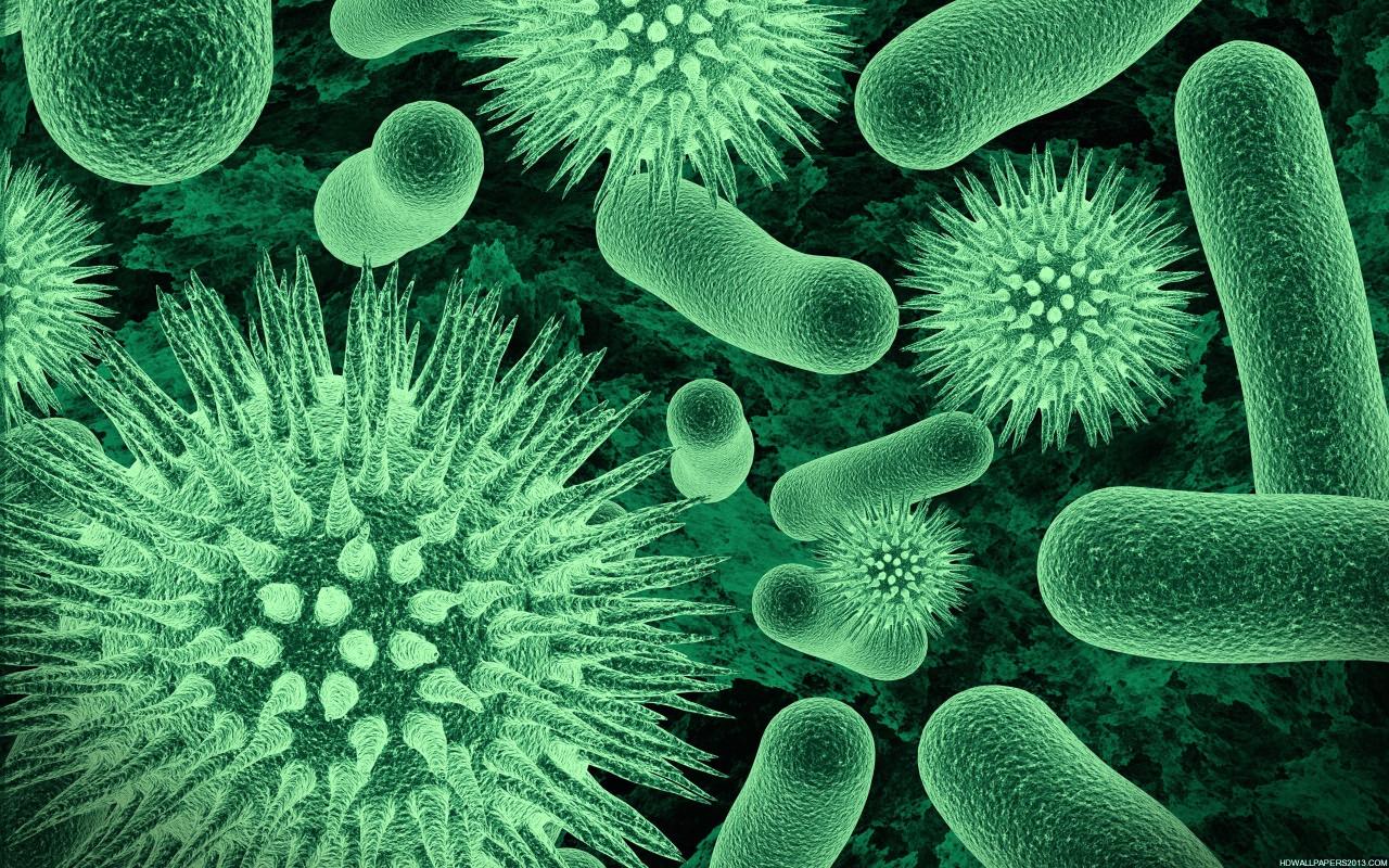
Author: Ceasar Ritchie

  • The Benefits of Carnosine A Deep Dive

    The Benefits of Carnosine A Deep Dive

    The benefits of carnosine, a naturally occurring compound, are increasingly recognized for its potential impact on muscle function, cellular health, and even neurological well-being. This exploration delves into the science behind carnosine, examining its various roles and potential applications. We’ll uncover how carnosine impacts everything from athletic performance to the aging process.

    Carnosine, a dipeptide found naturally in the body, is a crucial component in numerous physiological processes. It’s found in high concentrations in muscle tissue, where it plays a role in protecting against damage and fatigue. This article explores how carnosine may contribute to muscle strength, reduce fatigue, and potentially even influence the aging process. We’ll also discuss various sources of carnosine, from dietary intake to supplements, and delve into the safety considerations surrounding its use.

    Introduction to Carnosine

    Carnosine, a naturally occurring dipeptide, plays a crucial role in various physiological processes within the human body. It’s a combination of two amino acids, beta-alanine and histidine, linked together. Its chemical structure, β-alanyl-L-histidine, contributes to its unique properties and biological activities.This compound is found in high concentrations in skeletal muscle and the brain, indicating its importance for these organs.

    Understanding its natural sources, physiological roles, and available forms can help us appreciate its potential benefits and appropriate usage.

    Natural Sources of Carnosine

    Carnosine is primarily synthesized within the body, but it’s also found in certain foods. The highest concentrations are found in skeletal muscle tissue. Brain tissue also contains significant amounts. The diet, though, plays a minor role in carnosine intake compared to endogenous production. Certain animal products, such as beef and pork, contain noticeable amounts of carnosine.

    The presence of carnosine in foods varies significantly depending on factors like animal breed, diet, and preparation methods.

    Role of Carnosine in Physiological Processes

    Carnosine’s primary function is to act as a buffer, neutralizing harmful compounds and protecting against oxidative stress. It also plays a crucial role in maintaining cellular homeostasis, reducing inflammation, and supporting various metabolic processes. This dipeptide acts as a powerful antioxidant, combating free radicals and mitigating the damaging effects of oxidative stress. It contributes to maintaining the optimal pH balance in cells and tissues, crucial for healthy function.

    Forms and Types of Carnosine

    Understanding the various forms of carnosine is important for choosing the appropriate supplementation strategy. Different forms may have varying bioavailability and absorption rates, impacting their effectiveness.

    Form Source Benefits (Potential) Dosage
    Powder Dietary Supplements Muscle support, potential antioxidant benefits Typically 1-3 grams per day, depending on individual needs and product recommendation.
    Capsules Dietary Supplements Convenient delivery, potentially higher bioavailability compared to powders Dosage varies with the supplement; consult the product label for specific recommendations.
    Food Sources Beef, Pork, Poultry Natural source of carnosine, potentially beneficial in conjunction with a balanced diet. Intake varies depending on the amount of food consumed.

    Potential Benefits of Carnosine

    Carnosine, a naturally occurring dipeptide found in high concentrations in muscle tissue, has garnered significant interest for its potential health benefits, particularly in the realm of athletic performance and muscle function. This exploration delves into the potential benefits of carnosine, examining its impact on muscle function, fatigue, recovery, and oxidative stress.Understanding the intricacies of carnosine’s action is crucial to appreciating its potential.

    Carnosine’s ability to buffer reactive compounds, like hydrogen peroxide, and its role in maintaining cellular homeostasis are pivotal to its purported benefits.

    Muscle Function and Protein Synthesis

    Carnosine’s presence in muscle tissue is strongly correlated with muscle function. Research suggests carnosine might positively influence muscle protein synthesis, a key process in building and repairing muscle tissue. This enhancement could lead to improved muscle strength and endurance. Increased muscle protein synthesis is vital for muscle growth and repair, contributing to a stronger and more resilient muscular system.

    Effects on Muscle Fatigue and Recovery

    Muscle fatigue is a significant concern for athletes and individuals engaging in strenuous physical activity. Carnosine is hypothesized to play a crucial role in mitigating this fatigue by neutralizing the accumulation of lactic acid and other metabolites that contribute to muscle fatigue. This neutralization can facilitate faster recovery and reduced post-exercise soreness.

    Protection Against Oxidative Stress

    Oxidative stress, caused by the accumulation of harmful reactive oxygen species (ROS), can damage muscle tissue and impair performance. Carnosine acts as a potent antioxidant, protecting against oxidative stress and cellular damage. Its ability to quench ROS is a key factor in preserving muscle health and function, particularly during intense exercise.

    Carnosine, a naturally occurring compound, boasts impressive antioxidant properties, potentially offering protection against cellular damage. Understanding how it impacts the body’s response to infections like gonorrhea is fascinating, especially after treatment. For example, knowing the potential symptoms that can arise post-treatment, like those detailed in this article on gonorrhea symptoms after treatment , is crucial for holistic health.

    Ultimately, carnosine’s benefits extend to supporting overall cellular health and resilience, making it a potentially valuable supplement in a comprehensive health strategy.

    Comparative Analysis of Carnosine’s Effects on Different Muscle Types

    Muscle Type Effect on Function Effect on Fatigue Effect on Oxidative Stress
    Skeletal Muscle Potentially enhanced strength and endurance due to improved protein synthesis and reduced damage. Reduced fatigue by buffering metabolites like lactic acid. Reduced oxidative stress and cellular damage, maintaining muscle integrity.
    Cardiac Muscle Potentially improved contractility and reduced risk of damage from oxidative stress, particularly in conditions like heart failure. Possible reduction in fatigue, potentially improving cardiac function. Protection against oxidative stress, mitigating damage associated with reactive oxygen species.

    Potential for Enhancing Athletic Performance

    The potential benefits of carnosine for athletic performance are promising. By improving muscle function, reducing fatigue, and protecting against oxidative stress, carnosine could contribute to enhanced endurance, strength, and recovery. This translates to improved athletic output, faster recovery times, and a potentially greater capacity for sustained physical exertion. For example, increased carnosine levels in athletes might translate to more repetitions in a weightlifting routine or a longer run without fatigue.

    Carnosine and Cellular Health

    The benefits of carnosine

    Carnosine, a naturally occurring dipeptide, plays a crucial role in cellular protection. Its ability to interact with various cellular components and neutralize damaging agents makes it a potential therapeutic candidate for a range of conditions. This section delves into carnosine’s protective mechanisms against cellular damage and its potential impact on inflammation.Carnosine’s remarkable ability to shield cells from harm stems from its unique chemical structure and its capacity to interact with reactive species and damaging agents.

    It acts as a potent antioxidant, reducing oxidative stress, a major contributor to cellular aging and disease. This protective effect is particularly important in maintaining healthy tissues and organs.

    Cellular Protection against Oxidative Stress, The benefits of carnosine

    Carnosine’s antioxidant properties are well-documented. It effectively scavenges reactive oxygen species (ROS), such as free radicals, which can damage cellular components, including proteins, lipids, and DNA. This protection is crucial in preventing cellular dysfunction and promoting longevity. For example, prolonged exposure to UV radiation can generate high levels of ROS, leading to cellular damage and potentially skin aging.

    Carnosine’s antioxidant activity could potentially mitigate these effects.

    Carnosine and Inflammation Reduction

    Inflammation is a complex biological response to harmful stimuli. While necessary for healing, chronic inflammation can contribute to various diseases. Carnosine’s role in reducing inflammation is a promising area of research.

    Carnosine is a powerful antioxidant, known for its potential benefits in reducing muscle fatigue and inflammation. Learning more about the amazing properties of sea moss can offer a fascinating perspective on how these natural compounds work together. Check out sea moss everything you need to know for a deep dive into its potential health benefits. Ultimately, understanding the various ways natural compounds like carnosine can support overall health and wellness is crucial for a balanced lifestyle.

    • Neutralization of inflammatory mediators: Carnosine can interact with inflammatory molecules, such as cytokines and prostaglandins, potentially reducing their activity and the inflammatory response. This action could have implications for conditions characterized by chronic inflammation.
    • Modulation of immune cell activity: Carnosine may influence the activity of immune cells involved in the inflammatory process. This regulation could contribute to a more balanced and controlled immune response.
    • Reduction of oxidative stress: As mentioned earlier, carnosine’s antioxidant properties directly contribute to reducing the production of ROS, a key driver of inflammation. Lowering oxidative stress can help calm the inflammatory cascade.
    • Inhibition of inflammatory enzymes: Certain enzymes, like cyclooxygenase (COX), play a crucial role in the production of inflammatory mediators. Carnosine may inhibit the activity of these enzymes, thereby decreasing the generation of inflammatory compounds.
    • Promoting anti-inflammatory pathways: Carnosine could potentially stimulate cellular pathways that counter inflammation, creating a protective environment against harmful cellular processes.

    Protein and Peptide Interactions

    Carnosine’s ability to interact with proteins and peptides is critical to its cellular effects. It can bind to various proteins, altering their structure and function. These interactions can have a profound impact on cellular processes.

    Carnosine’s interaction with proteins can affect their stability, activity, and susceptibility to damage.

    This interaction is particularly relevant in maintaining the integrity of proteins crucial for cellular functions. Furthermore, carnosine may influence protein folding and assembly, ensuring proper cellular structure and function.

    Mechanism of Action in Protein and Peptide Interactions

    1. Hydrogen bonding: Carnosine’s amino acid side chains can form hydrogen bonds with specific amino acid residues in proteins, altering their conformation.
    2. Electrostatic interactions: Carnosine’s charged groups can participate in electrostatic interactions with proteins, influencing their activity.
    3. Formation of complexes: Carnosine can form complexes with proteins, potentially modulating their function and stability.

    Carnosine and Neurological Health

    Carnosine, a naturally occurring dipeptide, has garnered attention for its potential benefits beyond its role in muscle function. Recent research suggests a promising link between carnosine and neurological health, particularly in protecting neurons from damage and potentially mitigating the progression of neurological disorders. This exploration delves into the potential mechanisms and evidence supporting carnosine’s neuroprotective effects.Carnosine’s ability to neutralize harmful reactive oxygen species (ROS) and metal ions is a key aspect of its neuroprotective properties.

    These molecules are implicated in neuronal damage, playing a significant role in the pathogenesis of various neurological conditions. By reducing oxidative stress, carnosine may help maintain neuronal integrity and function.

    Potential Role in Neuronal Protection

    Carnosine acts as a potent antioxidant, scavenging harmful free radicals that can damage cell structures, including neurons. This antioxidant capacity is crucial in preserving the integrity of neuronal membranes and preventing the accumulation of oxidative damage, a hallmark of neurodegenerative diseases. Furthermore, carnosine can chelate metal ions, preventing their harmful interactions with cellular components. This dual action of scavenging free radicals and chelating metals contributes to its overall neuroprotective effect.

    Potential Benefits in Managing Neurological Disorders

    The potential benefits of carnosine extend to managing neurological disorders like Alzheimer’s disease. Evidence suggests that oxidative stress and metal ion accumulation significantly contribute to the development and progression of Alzheimer’s. By mitigating these damaging factors, carnosine may slow or even halt disease progression. While more research is needed to fully understand the extent of carnosine’s impact on specific neurological disorders, its protective effects on neuronal structures and function provide a basis for further investigation.

    For example, studies in animal models have shown promising results regarding memory enhancement and reduced neuronal damage in the presence of carnosine.

    Evidence Supporting Neuroprotective Effects

    A growing body of research supports carnosine’s neuroprotective properties. In vitro studies have demonstrated carnosine’s ability to protect neurons from various stressors, including oxidative stress and metal ion toxicity. Animal studies have also shown promising results, exhibiting reduced neuronal damage and improved cognitive function in various models of neurological disorders. While human trials are limited, the existing evidence suggests a potential role for carnosine in promoting neuronal health.

    For instance, one study showed improvements in cognitive function in patients with mild cognitive impairment after supplementation with carnosine.

    Summary Table of Potential Benefits

    Neurological Function Potential Benefit Mechanism Evidence Level
    Memory Improvement Reduced oxidative stress, improved neuronal function Moderate
    Neuroinflammation Reduction Antioxidant and anti-inflammatory properties Emerging
    Neuron Protection Increased Neutralization of harmful free radicals and metal ions Strong
    Cognitive Decline Possible Slowing Protection of neurons from damage, reduced oxidative stress Moderate

    Carnosine and Aging

    The benefits of carnosine

    Carnosine, a naturally occurring dipeptide, has garnered attention for its potential role in slowing the aging process. Its antioxidant and anti-inflammatory properties, along with its ability to protect cells from damage, suggest a possible connection between carnosine supplementation and improved healthspan. This exploration dives into the potential mechanisms through which carnosine might combat age-related decline.The aging process is complex, involving various cellular and molecular changes.

    Carnosine’s multifaceted action, targeting multiple pathways associated with aging, makes it an intriguing candidate for interventions aimed at extending healthy lifespan. Research continues to investigate the precise ways carnosine influences these pathways, and further studies are needed to fully elucidate its potential in this area.

    Potential Mechanisms of Carnosine in Aging

    Carnosine’s antioxidant capacity plays a crucial role in mitigating oxidative stress, a significant contributor to cellular damage and aging. It scavenges reactive oxygen species (ROS), thereby protecting cellular components from damage. Further, carnosine’s ability to chelate metal ions can prevent the formation of harmful free radicals. This dual action helps maintain cellular integrity and function as we age.

    Carnosine and Cellular Senescence

    Cellular senescence, a state of cellular aging and dysfunction, is implicated in age-related diseases. Studies have shown that carnosine can potentially reduce the accumulation of senescent cells. By modulating cellular pathways related to senescence, carnosine may contribute to a healthier aging process. This includes supporting cellular repair mechanisms and preventing the release of damaging factors by senescent cells.

    The precise mechanisms through which carnosine influences cellular senescence remain a subject of ongoing research.

    Age-Related Processes Affected by Carnosine

    Carnosine’s potential impact on aging encompasses various processes. These processes can be broadly categorized into:

    • Oxidative Stress Mitigation: Carnosine’s antioxidant properties directly combat oxidative stress, a major driver of cellular damage and aging. By neutralizing free radicals, carnosine protects cellular structures and functions, thus potentially delaying age-related decline.
    • Inflammation Regulation: Chronic inflammation is linked to aging and various age-related diseases. Carnosine’s anti-inflammatory effects may help reduce the inflammatory burden, leading to a healthier aging process.
    • Protein Damage Repair: Proteins are crucial for cellular function, and their degradation contributes to aging. Carnosine may support protein repair mechanisms, thus potentially mitigating the impact of protein damage associated with aging.
    • Cellular Senescence Modulation: The accumulation of senescent cells is associated with age-related diseases. Carnosine may reduce the accumulation of senescent cells, potentially slowing the aging process.
    • Neuroprotective Effects: Age-related neurodegenerative diseases are often associated with oxidative stress and inflammation. Carnosine’s neuroprotective effects may contribute to preserving cognitive function and overall neurological health as we age.
    • Mitochondrial Function: Mitochondria, the powerhouses of cells, play a vital role in cellular function. Carnosine may support mitochondrial function, which is crucial for maintaining cellular energy production and overall health as we age.

    Carnosine and Food Sources

    Carnosine, a naturally occurring dipeptide, is found in various foods. While not a primary dietary focus, incorporating foods rich in carnosine into your diet could contribute to overall health, particularly if you’re interested in its potential benefits. Understanding the food sources of carnosine, along with their carnosine content and potential bioavailability, is essential for anyone looking to potentially leverage its properties.A significant portion of carnosine in the human body is derived from dietary intake.

    However, the exact amount of carnosine present in different foods and the degree to which it’s absorbed by the body can vary. This is influenced by several factors, including food preparation methods.

    Carnosine, a naturally occurring compound, offers a range of potential health benefits. Knowing your family’s medical history is crucial when considering the benefits of carnosine, as certain conditions might be linked to its effectiveness. For instance, understanding your family’s predisposition to specific diseases, like heart conditions or muscle disorders, can help you tailor a strategy that aligns with your unique genetic background.

    Recording family medical history can help you uncover these patterns and make informed decisions about your health, leading to a more effective strategy for harnessing the benefits of carnosine for your specific needs. This proactive approach allows you to gain a deeper understanding of how your body might react to carnosine.

    Food Sources Rich in Carnosine

    A variety of foods contain carnosine, although the concentration can differ significantly. Meat products, particularly red meat, are known to be relatively high in carnosine. However, other sources, like some seafood and certain plant-based foods, may also contribute to carnosine intake.

    Food Carnosine Content (mg/100g) Other Nutrients Preparation Methods
    Beef High Protein, Iron, Zinc Cooked
    Pork Moderate Protein, Vitamin B Cooked, cured
    Salmon Low Omega-3 fatty acids, Vitamin D Baked, grilled, or pan-fried
    Chicken Moderate Protein, Vitamin B Roasted, grilled, or fried
    Certain Mushrooms Trace Amounts Fiber, Vitamins Sautéed, roasted, or added to soups

    Bioavailability of Carnosine from Different Food Sources

    The bioavailability of carnosine, meaning how much of the carnosine in food is actually absorbed and used by the body, varies depending on the food source. Factors like the presence of other nutrients, the form of carnosine in the food, and the individual’s digestive health can influence how well the body absorbs carnosine. While meat products often have higher carnosine content, the bioavailability might not always be as high as other potential sources.

    Effect of Cooking Methods on Carnosine Content

    Cooking methods can impact the carnosine content in food. Heat, particularly high heat, can potentially degrade carnosine, resulting in a lower concentration in the cooked food compared to the raw material. Methods that involve prolonged cooking or high temperatures may have a more significant effect on the carnosine levels. For example, roasting meat at high temperatures for extended periods might result in a noticeable decrease in carnosine compared to shorter cooking times.

    Steaming or lightly grilling might preserve more carnosine than deep frying.

    Carnosine and Safety

    Carnosine, a naturally occurring dipeptide, has garnered attention for its potential health benefits. However, like any supplement, it’s crucial to understand the potential risks and responsible usage. This section delves into the safety considerations surrounding carnosine supplementation, covering potential side effects, toxicity concerns, recommended dosages, and the importance of professional guidance.Understanding the potential side effects and safety profile of carnosine is vital for anyone considering supplementation.

    While generally considered safe, individual responses can vary, and careful monitoring is essential.

    Potential Side Effects

    The majority of studies on carnosine supplementation report a good safety profile. However, some potential side effects have been noted in certain individuals. These side effects are typically mild and transient, resolving upon discontinuation of the supplement.

    • Gastrointestinal issues, such as nausea, vomiting, and diarrhea, have been reported in some cases.
    • Headaches and dizziness have also been reported in a small number of individuals.
    • Allergic reactions, though rare, are possible.

    Current Understanding of Carnosine Toxicity

    While carnosine is generally considered safe, there’s a limited understanding of potential long-term toxicity. Current research suggests that carnosine is unlikely to cause severe or permanent adverse effects at recommended dosages. However, further research is warranted to fully elucidate the long-term effects of carnosine supplementation. There are no reported cases of severe carnosine toxicity in the scientific literature.

    Recommended Dosage

    The recommended dosage of carnosine varies depending on the specific product and the intended use. Currently, there isn’t a standardized, universally accepted dosage for all individuals. The optimal dosage is best determined through consultation with a healthcare professional. The most commonly studied doses range from 2 grams to 6 grams per day, but individual needs may vary.

    Importance of Consulting a Healthcare Professional

    Given the varying individual responses and the limited research on long-term effects, consulting a healthcare professional before taking carnosine supplements is highly recommended. This consultation is especially crucial for individuals with pre-existing health conditions, such as kidney problems or liver disease, as carnosine can potentially interact with certain medications or conditions. A healthcare professional can assess individual needs, potential interactions, and tailor a safe and effective supplementation strategy, if any.

    Final Conclusion: The Benefits Of Carnosine

    In conclusion, carnosine presents a fascinating array of potential benefits, impacting various aspects of health and well-being. From supporting muscle function and protecting against oxidative stress to potentially influencing neurological health and delaying the aging process, the evidence surrounding carnosine’s capabilities is promising. However, more research is crucial to fully understand its mechanisms and optimal use. This article highlights the current knowledge and potential applications, but individual responses to carnosine may vary.

  • Hip Labrum Surgery Is It the Right Treatment?

    Hip Labrum Surgery Is It the Right Treatment?

    Hip labrum surgery is it the right treatment? This exploration delves into the complexities of hip labrum tears, examining the various treatment options, from conservative methods to surgical interventions. We’ll explore the causes, symptoms, and diagnostic procedures, providing a comprehensive overview of the factors that influence treatment decisions.

    Understanding the nuances of hip labrum tears is crucial for informed decision-making. This guide will help you navigate the process, from initial diagnosis to recovery, equipping you with the knowledge to discuss treatment options with your healthcare provider.

    Introduction to Hip Labrum Surgery

    The hip labrum is a ring of cartilage that lines the socket of the hip joint. It acts as a gasket, deepening the socket and improving the joint’s stability. A tear in this cartilage, known as a hip labrum tear, can result in pain, instability, and decreased range of motion. Various factors can contribute to such tears, including repetitive stress, trauma, or underlying conditions.

    Understanding the causes, function, and symptoms of hip labrum tears is crucial for proper diagnosis and treatment.Hip labrum tears can significantly impact the mechanics of the hip joint. The labrum’s primary role is to increase the contact area between the ball and socket, thus improving joint stability and reducing friction. Damage to the labrum can lead to abnormal movement patterns, increased wear and tear on the joint, and a predisposition to further injuries.

    This disruption of normal joint mechanics is a key factor in the development of pain and disability.

    Hip Labrum Tear Causes

    Several factors can contribute to hip labrum tears. Repetitive stress, such as from sports activities or certain occupations, can gradually wear down the labrum. Trauma, like a fall or direct blow to the hip, can cause an acute tear. Underlying conditions like hip dysplasia, which affects the shape of the hip socket, can predispose individuals to labrum tears.

    Furthermore, some individuals may have a genetic predisposition to weaker or more susceptible labral tissue.

    Hip Labrum Tear Symptoms

    Common symptoms of hip labrum tears include pain, often felt in the groin, hip, or buttock area. The pain can vary in intensity and may be aggravated by specific movements, such as walking, running, or climbing stairs. Limited range of motion, clicking or popping sounds in the hip, and a feeling of instability or catching in the hip joint are also potential indicators.

    Pain may also radiate down the leg.

    Types of Hip Labrum Tears

    Different types of hip labrum tears can affect the hip joint differently. The location of the tear plays a role in the resulting symptoms and treatment options. Here’s a comparison of the common types:

    Type Location Potential Symptoms Treatment Considerations
    Anterior Front of the hip socket Pain in the groin, difficulty with hip flexion (bending the hip), and potentially instability during walking. May require surgical repair or conservative management depending on the severity.
    Posterior Back of the hip socket Pain in the buttock or back of the hip, difficulty with hip extension (straightening the leg), and potential instability during certain movements. Often requires surgical repair, especially if the tear is significant.
    Superior Top of the hip socket Pain in the groin and upper hip region, potential for catching or clicking sensations, and reduced range of motion. Treatment may range from physical therapy to surgery, depending on the severity of the tear.
    Inferior Bottom of the hip socket Pain in the groin, potentially radiating down the inner thigh, difficulty with hip abduction (moving leg away from the body). Surgical intervention may be necessary for significant tears.

    Diagnostic Procedures and Evaluation

    Pinpointing the cause of hip pain, especially when a labral tear is suspected, requires a systematic approach. Accurate diagnosis is crucial for determining the appropriate treatment strategy. This often involves a combination of physical examinations, imaging studies, and potentially arthroscopy. Understanding the information each procedure provides is essential for making well-informed decisions.

    Physical Examinations

    Thorough physical examinations are the initial step in evaluating hip pain. Clinicians assess range of motion, palpating for tenderness, clicking, or grinding sensations. Specific tests, such as the impingement test and flexion abduction external rotation test, are used to evaluate for labral tears and other possible causes of pain. These tests help determine the location and nature of the pain, providing valuable clues about the potential involvement of the hip labrum.

    Pain reproduction during specific movements, coupled with observed limitations in range of motion, can strongly suggest labral pathology.

    Imaging Studies

    Imaging plays a vital role in confirming suspected labral tears and evaluating the extent of the damage. Different imaging modalities offer varying levels of detail and information.

    X-rays

    X-rays are a fundamental initial step in evaluating the hip joint. They primarily assess for bony abnormalities, such as fractures, arthritis, or other bony injuries. X-rays provide a general overview of the hip structure, helping to rule out significant bony pathology. While not specifically designed to visualize soft tissues like the labrum, they can help identify potential contributing factors to the pain.

    MRIs

    Magnetic resonance imaging (MRI) is a powerful tool for visualizing soft tissues, including the labrum. MRI scans offer detailed images of the hip joint, showcasing the cartilage, ligaments, tendons, and the labrum itself. They can clearly identify labral tears, demonstrating their size, location, and degree of detachment from the underlying bone. An MRI can also reveal other soft tissue injuries that may coexist with a labral tear.

    Arthroscopy

    Arthroscopy is an arthroscopic procedure that involves inserting a small camera into the hip joint through a small incision. This allows direct visualization of the joint structures, providing a high-resolution view of the labrum and other surrounding tissues. Arthroscopy is particularly useful for confirming the diagnosis and evaluating the extent of the labral tear in detail. It enables the surgeon to identify the precise location, size, and nature of the tear.

    This direct visualization allows for precise assessment and can also guide surgical interventions, ensuring a tailored approach to the individual patient’s needs.

    Importance of Proper Diagnosis

    A precise diagnosis is crucial for developing the most effective treatment plan. An accurate assessment helps determine the severity of the tear and whether conservative or surgical intervention is necessary. Incorrect diagnoses can lead to inappropriate treatment choices, potentially prolonging the recovery period or causing further complications. Therefore, careful consideration of all diagnostic procedures is paramount in ensuring optimal patient outcomes.

    Summary Table of Typical Findings

    Imaging Modality Typical Findings in Labral Tears
    X-ray May reveal signs of osteoarthritis, hip dysplasia, or other bony abnormalities. No direct visualization of the labrum.
    MRI Demonstrates labral tears as focal defects or detachment of the labrum from the underlying bone. Provides details on the size, location, and extent of the tear. May also show associated cartilage damage or other soft tissue injuries.

    Non-Surgical Treatment Options

    Hip labrum tears, while sometimes requiring surgical intervention, often respond well to non-surgical treatments. Conservative approaches can be highly effective in managing pain and restoring function, avoiding the risks and recovery time associated with surgery. These methods aim to reduce inflammation, promote healing, and improve mobility through targeted exercises and lifestyle modifications.Conservative treatment strategies focus on alleviating symptoms and promoting healing without the need for invasive procedures.

    The effectiveness of each method can vary depending on the severity of the tear, the individual’s response to treatment, and the presence of any other contributing factors. A thorough evaluation by a healthcare professional is crucial to determine the most suitable course of action.

    Physical Therapy, Hip labrum surgery is it the right treatment

    Physical therapy plays a pivotal role in the non-surgical management of hip labrum tears. It’s a cornerstone of conservative treatment, focusing on restoring strength, flexibility, and range of motion in the affected hip joint. Exercises are tailored to address specific needs, and the progression of exercises is crucial for optimal results. The key is to gradually increase the intensity and complexity of the exercises as the patient’s condition improves.

    Rest

    Rest is an essential component of managing hip labrum tears, especially in the acute phase. It allows the injured tissues to heal and reduces further stress on the affected area. While complete bed rest might not always be necessary, avoiding activities that aggravate pain is crucial. This could involve modifying work tasks, avoiding high-impact sports, or adjusting daily routines to minimize hip strain.

    Proper rest is vital for allowing the body to initiate the healing process.

    Anti-inflammatory Medications

    Nonsteroidal anti-inflammatory drugs (NSAIDs) such as ibuprofen or naproxen can help reduce inflammation and pain associated with hip labrum tears. These medications can be particularly helpful in managing the initial inflammatory response, which often contributes to pain and discomfort. However, it’s essential to use these medications as directed by a healthcare professional, as they may have potential side effects, including gastrointestinal issues.

    Considering hip labrum surgery? It’s a big decision, and figuring out if it’s the right treatment for you is key. While hip pain can be debilitating, understanding the common locations for skin conditions like eczema might offer some insight into the overall picture of your health. For example, knowing the most common location for eczema can help you identify potential triggers and improve your treatment plan.

    Ultimately, a consultation with your doctor is crucial in deciding if hip labrum surgery is the right choice for your specific needs. most common location for eczema

    Always consult with a doctor before taking any new medication, especially if you have pre-existing conditions.

    Typical Physical Therapy Exercises and Stretches

    Exercise/Stretch Description
    Hip Flexor Stretch Standing or seated, gently pull the knee towards the chest, holding for 20-30 seconds.
    Hip Abduction Exercises Side-lying with the affected leg raised, keeping the knee straight, to strengthen hip abductors.
    Hip Extension Exercises Prone position, raising the leg straight up to strengthen hip extensors.
    Glute Bridges Lying on the back, lifting the hips off the ground, contracting gluteal muscles.
    Clamshells Side-lying with knees bent, raising the top knee, targeting hip abductors.
    Prone Hip Rotations Lying on stomach, rotating the leg outward and inward, improving hip external and internal rotation.

    The specific exercises and stretches prescribed by a physical therapist will depend on the individual’s specific needs and the stage of healing.

    Surgical Procedures for Hip Labrum Tears

    Hip labral tears, often caused by repetitive stress, trauma, or a combination of factors, can lead to significant pain and dysfunction. Surgical intervention is sometimes necessary to restore hip joint stability and alleviate symptoms. This section will delve into the various surgical techniques employed for hip labral repair or reconstruction.Surgical procedures for hip labrum tears aim to address the underlying cause of the problem and restore the structural integrity of the labrum.

    Different techniques exist, each with its own set of advantages and potential risks. Understanding these techniques allows patients and medical professionals to make informed decisions about the best course of action for their specific needs.

    Arthroscopic Labral Repair

    Arthroscopic labral repair is a minimally invasive surgical procedure that utilizes small incisions and specialized instruments to access and repair the damaged labrum. This approach is generally preferred for its smaller incision size, reduced recovery time, and decreased post-operative pain compared to open surgical techniques.The procedure typically involves the following steps:

    • The surgeon makes small incisions in the hip joint, inserting a specialized camera (arthroscope) and small surgical instruments.
    • The arthroscope allows visualization of the damaged labrum, enabling the surgeon to identify the extent of the tear.
    • Using specialized instruments, the surgeon carefully cleans the area around the tear, removing any loose tissue or debris.
    • Depending on the type of tear, the surgeon may use sutures, anchors, or other techniques to repair the labrum.
    • The joint is flushed with saline solution to remove any residual debris.
    • The incisions are closed, and a bandage is applied to the surgical area.

    Open Labral Repair

    Open labral repair is a more extensive surgical procedure, often reserved for cases where arthroscopic techniques are insufficient or not suitable. The surgeon makes a larger incision to directly access the hip joint, enabling a more comprehensive view and greater control over the repair.

    • A larger incision is made in the hip joint, providing direct access to the damaged labrum.
    • The surgeon carefully dissects the surrounding tissues to expose the labrum tear.
    • The damaged labrum is cleaned and repaired using sutures, anchors, or other appropriate techniques.
    • The surrounding tissues are carefully repositioned to restore normal joint anatomy.
    • The incision is closed, and the surgical area is bandaged.

    Comparison of Surgical Approaches

    Arthroscopic surgery offers a less invasive approach, resulting in a quicker recovery, less pain, and smaller scars. However, it may not be suitable for all types of labral tears, particularly those involving significant bone damage or complex tear patterns. Open surgery, while more invasive, allows for greater visualization and control, which can be beneficial for complex repairs.

    Surgical Instruments Used in Hip Labral Repair

    The choice of surgical instruments depends on the specific procedure and the surgeon’s preference. Here’s a table outlining some common instruments used in hip labral repair procedures:

    Instrument Description
    Arthroscope A small camera and light source used to visualize the hip joint interior.
    Shaver A tool for removing damaged or loose tissue from the joint.
    Scissors Used for precise tissue dissection.
    Sutures Used to repair the torn labrum.
    Anchors Used to secure sutures to the bone.
    Graspers Used to hold and manipulate tissues.

    Recovery and Rehabilitation After Hip Labrum Surgery: Hip Labrum Surgery Is It The Right Treatment

    Recovering from hip labrum surgery is a journey, not a sprint. A diligent and structured rehabilitation program is crucial for a successful outcome. This involves a combination of pain management, targeted physical therapy, and gradual return to activities. Understanding the process and adhering to the prescribed protocol will significantly impact the recovery time and overall success of the surgery.The recovery period after hip labrum surgery is designed to restore hip function and strength while minimizing the risk of re-injury.

    This phase involves a gradual increase in activity, starting with gentle movements and progressing to more challenging exercises. Patience and consistency are key components to a successful recovery.

    Considering hip labrum surgery? It’s a complex decision, and the right treatment depends heavily on the individual’s specific case. Factors like the severity of the tear and the patient’s overall health play a crucial role. Understanding the potential costs, including those related to tariffs and OTC drug pricing fluctuations, can be critical when making your choice. For example, tariffs and OTC drugs can influence the price of necessary medications and supplies, which can impact the overall financial picture.

    Ultimately, talking to a healthcare professional is key to determining if hip labrum surgery is the best path for you.

    Pain Management

    Effective pain management is essential for allowing patients to participate fully in their rehabilitation program. Strategies typically involve a combination of medication, physical therapy modalities, and lifestyle adjustments. Over-the-counter pain relievers may be sufficient for mild discomfort, while stronger medications might be necessary for more severe pain. Physical therapy modalities, such as ice packs, heat therapy, and electrical stimulation, can also help manage pain and inflammation.

    Regular communication with the surgeon and physical therapist is crucial to ensure the pain management plan is optimized throughout the recovery process.

    Physical Therapy Phases

    A structured physical therapy program is tailored to each individual’s needs and recovery progress. It’s designed to gradually restore hip strength, flexibility, and range of motion. A typical progression of exercises and activities can be broken down into distinct phases.

    Phase Focus Duration (Approximate) Key Activities
    Phase 1 (Initial Post-Op) Protecting the surgical site, managing pain, and regaining basic mobility. 1-4 weeks Gentle range of motion exercises, isometric exercises (contracting muscles without movement), and pain-free walking with assistive devices as needed.
    Phase 2 (Early Strengthening) Increasing hip strength and improving flexibility. 4-8 weeks Progressive resistance exercises (using light weights or resistance bands), balance exercises, and controlled walking. Activities such as stationary cycling and swimming can be incorporated.
    Phase 3 (Advanced Strengthening) Improving functional strength and preparing for return to activities. 8-12 weeks More challenging exercises, including plyometrics (jumping and hopping), sport-specific drills (if applicable), and functional exercises mimicking daily activities. Gradual return to impact activities, such as jogging or stairs, is also introduced.
    Phase 4 (Return to Activity) Full functional recovery and return to desired activity level. 12+ weeks Maintenance exercises to maintain gains, and progression to pre-injury activity levels.

    This table provides a general guideline. The duration of each phase may vary depending on the individual’s progress and the complexity of the surgery. Always follow the recommendations of your physical therapist and surgeon.

    Return to Activity Timelines

    The timeline for returning to various activities depends on the extent of the injury, the surgical procedure, and the individual’s progress through the rehabilitation program. Light activities like walking can often begin within a few days of surgery, while more strenuous activities, such as running or contact sports, typically require several months of recovery. It’s crucial to avoid overexertion and allow the body adequate time to heal to prevent re-injury.

    Factors Influencing Recovery Time

    Several factors can influence the recovery time after hip labrum surgery. These include the severity of the tear, the individual’s overall health, adherence to the rehabilitation program, and the surgeon’s expertise. Age, pre-existing conditions, and the patient’s motivation also play a significant role. For instance, a more extensive tear or a patient with underlying medical conditions may take longer to recover.

    Furthermore, a strong commitment to the rehabilitation protocol is crucial for a faster and more complete recovery.

    Potential Complications

    Potential complications after hip labrum surgery are generally rare but include infection, blood clots, persistent pain, stiffness, or incomplete recovery. Addressing any concerns promptly with the surgeon is essential for effective management. Regular follow-up appointments and adherence to the recovery plan are critical in mitigating these risks. Open communication with the surgical team allows for proactive intervention if any complications arise.

    Factors Affecting Treatment Decisions

    Deciding whether hip labrum surgery is the right course of action isn’t a simple yes or no. It’s a nuanced evaluation considering various factors unique to each patient. A surgeon carefully weighs the potential benefits and risks against the patient’s individual circumstances to determine the most appropriate treatment path.Surgeons aren’t simply looking at the tear itself. They consider the patient’s overall health, activity level, and how the labrum tear impacts their daily life.

    This holistic approach ensures the best possible outcome, balancing the need for intervention with the desire for a minimally invasive and effective solution.

    Severity of the Tear

    The extent of the hip labrum tear significantly influences treatment decisions. A small, stable tear might not require surgery, whereas a large, unstable tear causing significant pain and dysfunction may necessitate surgical intervention. Surgical repair aims to restore the labrum’s integrity and function, while non-surgical approaches focus on managing symptoms and promoting healing. The surgeon carefully assesses the tear’s size, location, and the presence of any associated damage to other structures in the hip joint.

    Patient’s Activity Level

    A patient’s activity level plays a crucial role in determining the best course of action. An individual leading a sedentary lifestyle might respond well to non-surgical treatments like physical therapy and activity modification. Conversely, an athlete requiring high-impact movements or frequent weight-bearing activities might benefit more from surgical repair to restore function and prevent further damage. A professional athlete with a large tear affecting their performance would likely be a strong candidate for surgery.

    Overall Patient Health

    Pre-existing medical conditions, such as arthritis or other joint problems, can influence the suitability of hip labrum surgery. The presence of other health concerns may necessitate a more conservative approach to avoid additional risks. For instance, a patient with significant cardiovascular issues might not be an ideal candidate for surgery, even with a severe labrum tear. A careful assessment of the patient’s medical history is essential to determine the safest and most effective treatment plan.

    Patient’s Goals and Expectations

    A key factor is the patient’s expectations and desired outcomes. Open communication between the patient and the surgeon is crucial to ensure mutual understanding and agreement on the treatment plan. A patient who desires complete restoration of function and the ability to return to their pre-injury activity level may be a better candidate for surgery. Conversely, a patient prioritizing symptom relief and improved quality of life might find non-surgical management more suitable.

    Comparison of Surgical and Non-Surgical Options

    Feature Non-Surgical Options Surgical Options
    Potential Benefits Reduced risk of surgery-related complications, quicker recovery, and potentially more cost-effective. Restoration of labrum integrity and function, potentially addressing underlying causes of pain and dysfunction, and returning to previous activity levels.
    Potential Risks Limited effectiveness in severe cases, potential for persistent pain, and possible need for future interventions. Potential complications like infection, nerve damage, or blood clots, longer recovery period, and higher costs.
    Recovery Time Typically shorter than surgical recovery. Typically longer recovery period, requiring a period of rehabilitation and physical therapy.
    Success Rate Variable depending on the severity of the tear and the patient’s compliance with the treatment plan. Generally higher success rates in addressing severe tears and restoring full function, especially for athletes.

    “The ultimate decision rests with the patient, in consultation with their surgeon, weighing the potential benefits and risks of each approach.”

    Potential Complications and Risks

    Hip labrum surgery, while often effective, carries potential risks. Understanding these complications is crucial for informed decision-making, alongside a thorough discussion with your surgeon. Proper preparation and proactive risk mitigation strategies can significantly reduce the likelihood of encountering these issues.

    Potential Complications

    Post-operative complications can arise from various factors, including the surgical procedure itself, individual patient characteristics, and post-operative care. Careful monitoring and management are essential to address these issues promptly and effectively.

    Infection

    Surgical procedures, by their nature, introduce a risk of infection. This risk is not unique to hip labrum surgery but is a concern across various surgical interventions. The prevalence of infection following hip labrum surgery is relatively low, typically reported in the range of 0.5% to 1.5%. Severity can range from localized skin infections to more systemic infections requiring intravenous antibiotics.

    Maintaining meticulous surgical technique, proper wound care, and vigilant monitoring for signs of infection are crucial strategies to minimize this risk.

    Blood Clots (Deep Vein Thrombosis – DVT)

    Blood clots, particularly deep vein thrombosis (DVT), are a possible complication following any surgical procedure that involves prolonged immobilization. DVTs can develop in the deep veins of the leg and, in rare cases, can travel to the lungs, causing a pulmonary embolism (PE). While the exact prevalence varies, studies suggest that DVTs occur in a percentage of patients undergoing hip surgery.

    Strategies to reduce the risk of DVT include early mobilization, compression stockings, and medication like anticoagulants.

    Persistent Pain

    Although hip labrum surgery aims to alleviate pain, some patients may experience persistent or even increased pain after the procedure. This can stem from various factors, including incomplete healing, scar tissue formation, or other underlying conditions. While not all cases result in persistent pain, the percentage of patients experiencing some level of persistent pain can range from a small percentage to a higher number depending on the individual factors.

    Rehabilitation programs tailored to individual needs, and proactive management of pain are essential strategies.

    Other Potential Complications

    Other potential complications include, but are not limited to, nerve damage, numbness, or weakness in the affected leg, and, in rare instances, a reaction to anesthesia. These complications are less frequent but can still occur. Factors such as the surgeon’s experience and surgical technique can play a role in the likelihood of these complications.

    Signs and Symptoms of Complications

    Potential Complication Signs and Symptoms
    Infection Redness, swelling, warmth, pain, pus or drainage from the incision site, fever, chills.
    DVT Swelling, pain, redness, warmth in the affected leg, especially in the calf, difficulty moving the leg.
    Persistent Pain Continued or increased pain in the hip area, even after rehabilitation.
    Nerve Damage Numbness, tingling, weakness, or loss of sensation in the affected leg or foot.

    Alternatives and Other Considerations

    Hip labrum surgery is a significant procedure, and it’s crucial to explore all available options before making a decision. While often the most effective treatment for severe hip labrum tears, it’s not always the right choice for every patient. Factors like the severity of the tear, the patient’s overall health, and lifestyle play a critical role in determining the best course of action.

    This section will delve into alternative treatments and essential considerations beyond surgery.Alternative approaches to treating hip labrum tears exist, offering potential benefits for specific cases. These range from conservative management to minimally invasive procedures, each with its own set of advantages and disadvantages. Understanding these alternatives allows patients to make informed decisions in conjunction with their healthcare providers.

    Alternative Treatment Options

    Conservative treatment often forms the initial approach for hip labrum tears. This typically involves a combination of physical therapy, activity modification, and pain management strategies. Physical therapy focuses on strengthening the muscles surrounding the hip joint, improving flexibility, and restoring proper biomechanics. Activity modification aims to reduce the impact on the injured area, while pain management can include over-the-counter pain relievers or, in more severe cases, prescription medications.

    In some instances, corticosteroid injections may be used to reduce inflammation. These conservative measures can be effective in managing mild to moderate hip labrum tears, allowing for a non-surgical solution.

    Patient-Specific Factors

    Patient-specific factors significantly influence the choice of treatment. Age is a crucial consideration. Younger patients, particularly athletes, may benefit from surgical intervention to maintain optimal joint function and prevent long-term complications. Older patients might find non-surgical options more suitable, given their potential for a slower recovery period. Overall health conditions, such as arthritis or other joint issues, also impact treatment choices.

    Deciding if hip labrum surgery is the right choice can be tough. It’s important to consider all your options, and understanding your health insurance is key. For example, knowing the difference between an HSA and an FSA can significantly impact your out-of-pocket costs, especially when considering a procedure like this. hsa vs fsa whats the difference Ultimately, talking to your doctor about your specific situation and carefully weighing the pros and cons of surgery is crucial for making an informed decision.

    Lifestyle factors, like the patient’s occupation and level of physical activity, play a role in determining the suitability of various treatments. For example, a professional athlete with a significant hip labrum tear will likely require surgical intervention to maintain their performance level, while a sedentary individual might be able to manage the condition with non-surgical therapies.

    Comparison of Treatment Approaches

    A comprehensive comparison of hip labrum surgery with other treatments is crucial for informed decision-making. Surgery is often reserved for severe cases, including those with significant instability or persistent pain not responding to conservative measures. In contrast, conservative management provides a less invasive approach, suitable for individuals with milder symptoms. Minimally invasive procedures, such as arthroscopy, represent a middle ground, offering a less extensive surgical intervention than traditional open surgery.

    The success rate of each approach depends on factors such as the severity of the tear, the individual’s response to treatment, and the expertise of the medical team.

    Non-Surgical Treatment Options

    Non-surgical approaches are valuable options for managing hip labrum tears, especially in the initial stages or for individuals not suitable for surgery. These methods often focus on reducing pain, improving function, and preventing further damage.

    Treatment Option Description Potential Benefits Potential Limitations
    Physical Therapy Exercises to strengthen hip muscles, improve flexibility, and restore proper mechanics. Improved joint stability, reduced pain, and enhanced function. Requires commitment and adherence to a prescribed regimen.
    Activity Modification Adjusting activities to reduce stress on the hip joint. Reduces pain and inflammation. May require significant lifestyle adjustments.
    Pain Management Over-the-counter or prescription medications, or corticosteroid injections. Reduces pain and inflammation. May have side effects, and injections may not address the root cause.
    Injections (e.g., Corticosteroids) Administering corticosteroids directly into the hip joint. Temporary pain relief and reduced inflammation. Potential for side effects, and not a long-term solution.

    Patient Experiences and Success Stories (Illustrative)

    Hip labrum surgery is it the right treatment

    Hip labrum surgery, while often a necessary procedure, can evoke a range of emotions and expectations in patients. Understanding real-life experiences can provide valuable insights into the recovery process and the potential outcomes. This section highlights illustrative cases to illustrate successful outcomes and emphasize the importance of realistic patient expectations.

    Illustrative Patient Experiences

    Many patients report significant improvements in their quality of life following hip labrum surgery. The recovery journey, however, varies greatly depending on individual factors such as age, pre-existing conditions, and the extent of the tear.

    Successful Outcomes and Positive Aspects

    A common theme in positive patient experiences is a noticeable reduction in pain and improved mobility. This improvement often translates to increased participation in daily activities and hobbies. For instance, a 35-year-old professional athlete who underwent hip labrum repair reported a return to competitive play within six months, albeit with a modified training regimen. This illustrates how surgery can enable a return to a fulfilling lifestyle, albeit with careful consideration of physical limitations during recovery.

    Another patient, a 60-year-old retiree, regained the ability to walk comfortably and participate in social activities like gardening. These experiences demonstrate the positive impact of surgery on overall well-being.

    Lifestyle Changes and Benefits

    Hip labrum surgery can lead to significant lifestyle changes, and these changes are often perceived positively by patients. Many patients report being able to engage in activities they previously found painful or impossible, like dancing, hiking, or simply enjoying a longer walk with their loved ones. The ability to resume everyday tasks without pain significantly enhances their quality of life.

    Importance of Patient Expectations and Realistic Outcomes

    It’s crucial for patients to understand that complete recovery and a full return to previous activity levels may not be immediate. While surgery can be a significant step toward improvement, realistic expectations about recovery time and potential limitations are essential for a positive experience. A thorough discussion with the surgeon about potential limitations and necessary physical therapy is critical to manage expectations and avoid disappointment.

    The surgeon’s role in counseling patients about the potential range of outcomes is paramount. This includes discussing possible limitations, long-term care needs, and the need for ongoing physical therapy. Realistic expectations prevent disappointment and promote a more positive and sustainable recovery.

    Ultimate Conclusion

    Hip labrum surgery is it the right treatment

    Ultimately, the decision of whether or not hip labrum surgery is the right treatment is a personal one. Weighing the potential benefits and risks, alongside your individual circumstances, is key. This detailed analysis should empower you to make an informed choice, working closely with your doctor to determine the best course of action for your specific needs.

  • Causes of Numbness and Tingling A Deep Dive

    Causes of Numbness and Tingling A Deep Dive

    Causes of numbness and tingling can range from seemingly minor issues to more serious underlying conditions. This exploration delves into the various factors that can lead to these sensations, from common vitamin deficiencies to more complex neurological problems. Understanding these causes is crucial for proper diagnosis and treatment.

    We’ll examine everything from peripheral neuropathy, a condition often linked to diabetes, to the role of autoimmune diseases and certain medications. We’ll also look at nerve compression and other medical conditions that can cause these sensations. Finally, we’ll present illustrative cases to further illuminate the complexities and variations in these symptoms.

    Peripheral Neuropathy

    Peripheral neuropathy is a common disorder characterized by damage to the nerves outside the brain and spinal cord. This damage often leads to a range of symptoms, including numbness, tingling, pain, weakness, and loss of reflexes in the extremities. Understanding the different types of peripheral neuropathy and their underlying causes is crucial for effective diagnosis and treatment.

    Types of Peripheral Neuropathy

    Peripheral neuropathy isn’t a single disease but a broad spectrum of conditions. Different types arise from various causes and affect nerves in distinct ways. Some types are directly linked to specific medical conditions, while others have no identifiable cause. Recognizing the specific type can guide appropriate medical interventions.

    Diabetic Neuropathy

    Diabetic neuropathy is a common complication of diabetes. High blood sugar levels over time can damage the nerves, particularly in the hands and feet. This damage can lead to a range of symptoms, from mild tingling and numbness to severe pain and loss of sensation. Prolonged high blood sugar can cause chronic damage to the blood vessels that supply the nerves, leading to reduced blood flow and further nerve damage.

    This is a significant concern because the lack of sensation in the feet can increase the risk of unnoticed injuries, leading to complications such as ulcers and infections.

    Toxic Neuropathy

    Certain toxins, both environmental and medicinal, can damage the peripheral nerves. Exposure to heavy metals like lead or mercury, or prolonged use of certain medications, such as chemotherapy drugs or some antibiotics, can cause toxic neuropathy. The mechanisms of nerve damage vary depending on the specific toxin. For instance, some toxins directly attack nerve cells, while others disrupt the nerve’s ability to function properly.

    It’s crucial to recognize that exposure to these toxins can lead to long-term and potentially irreversible nerve damage.

    Infectious Neuropathy

    Infections, both bacterial and viral, can sometimes lead to peripheral neuropathy. Certain infections can directly attack the nerves or produce inflammatory responses that damage them. Examples of infections associated with peripheral neuropathy include Lyme disease, HIV, and shingles. Understanding the specific infection can help clinicians target appropriate treatment strategies.

    Nutritional Deficiencies

    Vitamin deficiencies, particularly deficiencies in vitamin B12, thiamine (vitamin B1), and vitamin E, can also cause peripheral neuropathy. These vitamins are crucial for nerve function, and a lack of these nutrients can lead to nerve damage. For example, a deficiency in vitamin B12 can result in a progressive loss of sensation in the hands and feet. Maintaining a balanced diet and ensuring adequate intake of these essential nutrients is vital for preventing this type of neuropathy.

    Inherited Neuropathy, Causes of numbness and tingling

    Genetic factors can play a significant role in the development of some types of peripheral neuropathy. Inherited conditions can lead to nerve damage from birth or during early childhood. These conditions are often progressive, meaning the symptoms worsen over time. Examples include Charcot-Marie-Tooth disease, which primarily affects the nerves in the hands and feet, and hereditary motor and sensory neuropathy.

    Numbness and tingling can stem from various things, like nerve compression or poor circulation. But did you know that sometimes, tight muscles in your legs can also contribute? Strengthening your legs with exercises like those found in this article on leg exercises for bad knees can improve blood flow and alleviate some of those uncomfortable sensations.

    Ultimately, addressing the root cause of numbness and tingling is key to finding lasting relief.

    Genetic testing can be used to identify these conditions and guide management strategies.

    Table: Comparison of Peripheral Neuropathy Types

    Neuropathy Type Symptoms Causes Treatments
    Diabetic Neuropathy Numbness, tingling, pain, weakness in extremities, especially feet; potential loss of sensation High blood sugar levels over time, complications of diabetes Blood sugar control, pain management, physical therapy, medications to improve nerve function
    Toxic Neuropathy Numbness, tingling, pain, weakness; symptoms can vary depending on the toxin Exposure to heavy metals (lead, mercury), certain medications (chemotherapy drugs, antibiotics) Identify and eliminate the source of the toxin; supportive care to manage symptoms; medications to protect nerves
    Infectious Neuropathy Numbness, tingling, pain, weakness; symptoms can vary depending on the infection Bacterial or viral infections (Lyme disease, HIV, shingles) Treating the underlying infection; pain management; supportive care to manage symptoms
    Nutritional Deficiencies Numbness, tingling, pain, weakness in extremities; symptoms can vary depending on the specific deficiency Deficiencies in vitamins B12, thiamine, and E Correcting the nutritional deficiency through diet or supplements; managing symptoms
    Inherited Neuropathy Numbness, tingling, pain, weakness in extremities; symptoms can vary depending on the specific genetic condition Genetic mutations Managing symptoms; supportive care; medications to slow disease progression; genetic counseling

    Vitamin Deficiencies

    Numbness and tingling can sometimes stem from a lack of essential vitamins crucial for nerve function. These vitamins play a vital role in maintaining the health and integrity of nerve fibers, and deficiencies can disrupt the normal transmission of signals. Understanding the specific roles of different vitamins and the symptoms associated with their deficiencies can help in early diagnosis and treatment.Vitamin deficiencies, if left untreated, can lead to a range of neurological symptoms, including numbness, tingling, and pain.

    Prompt recognition of these symptoms is vital for effective intervention and preventing further complications. Early diagnosis often involves a thorough medical evaluation, including blood tests to assess vitamin levels and a review of the patient’s dietary habits.

    Vitamins and Their Roles in Nerve Function

    Vitamins are organic compounds that the body needs in small amounts to function properly. Several vitamins are critical for maintaining healthy nerve function. These include B vitamins, vitamin D, and vitamin E, among others. The role of these vitamins in nerve health stems from their involvement in various biochemical processes within nerve cells.

    Vitamin Deficiencies Causing Numbness and Tingling

    Several vitamin deficiencies can result in numbness and tingling. These deficiencies often manifest with a range of symptoms, depending on the specific vitamin involved and the severity of the deficiency.

    Vitamin Deficiency Symptoms Role in Nerve Function
    Vitamin B1 (Thiamine) Loss of appetite, fatigue, confusion, memory problems, muscle weakness, difficulty walking, and in severe cases, neurological impairment Plays a crucial role in carbohydrate metabolism, which is essential for nerve function. It is also involved in nerve impulse transmission.
    Vitamin B6 (Pyridoxine) Weakness, fatigue, depression, confusion, and nerve damage that can manifest as numbness and tingling, particularly in the hands and feet. Crucial for the synthesis of neurotransmitters, which are essential chemical messengers in the nervous system.
    Vitamin B12 (Cobalamin) Numbness and tingling in the hands and feet, fatigue, weakness, balance problems, and difficulty with memory and concentration. Essential for the formation of myelin, the protective sheath surrounding nerve fibers. It also plays a vital role in nerve impulse transmission.
    Vitamin D Muscle weakness, bone pain, and in severe cases, numbness and tingling in the extremities. Plays a role in calcium absorption, which is necessary for maintaining healthy nerve function. Vitamin D also contributes to the proper functioning of the immune system, which can indirectly impact nerve health.

    Symptoms and Signs of Vitamin Deficiencies

    The symptoms associated with vitamin deficiencies can vary depending on the specific vitamin involved and the degree of deficiency. Some common symptoms include fatigue, weakness, loss of appetite, and neurological issues like numbness and tingling. In severe cases, neurological impairment or other serious complications can occur.

    Impact on Nerve Health

    Vitamin deficiencies can severely impact nerve health in several ways. A lack of specific vitamins can disrupt the normal function of nerve cells, affecting nerve impulse transmission and causing damage to the protective myelin sheath. This can lead to a range of neurological symptoms, including numbness, tingling, and pain. Prompt diagnosis and treatment are crucial to prevent further nerve damage and maintain overall health.

    Diabetes

    Diabetes, a chronic metabolic disorder, significantly impacts the body’s ability to regulate blood sugar levels. This imbalance can have far-reaching consequences, including damage to nerves throughout the body. The constant exposure to high blood glucose levels can lead to a condition known as diabetic neuropathy, a common and often debilitating complication. Understanding the link between diabetes and nerve damage is crucial for effective prevention and management.

    Numbness and tingling can be a real pain, right? While many factors can cause it, sometimes underlying conditions like issues with your menstrual cycle or thyroid function can play a role. For example, thyroid problems can sometimes lead to unusual sensations, and these sensations can also be linked to menstrual problems, like those described in this article about menstrual problems and thyroid disease.

    Ultimately, it’s crucial to talk to a doctor to pinpoint the exact cause of any persistent numbness or tingling.

    The Link Between Diabetes and Nerve Damage

    High blood sugar levels, a hallmark of diabetes, damage the delicate blood vessels that nourish the nerves. This vascular damage impairs the nerves’ ability to transmit signals effectively. Over time, this can lead to a variety of symptoms, ranging from mild discomfort to severe pain and loss of function. The mechanisms of this damage are multifaceted, involving both direct and indirect effects of hyperglycemia.

    For instance, high blood sugar can cause the formation of harmful molecules that directly attack nerve cells. Furthermore, it can disrupt the production of essential nutrients required for nerve health, ultimately weakening the nerve’s structure and function.

    Mechanisms of Nerve Damage by High Blood Sugar

    High blood sugar levels can lead to a cascade of detrimental effects on nerves. One key mechanism involves the formation of advanced glycation end products (AGEs). AGEs are molecules that form when sugar binds to proteins and fats, resulting in the structural modification of these molecules. This process damages blood vessels and nerve fibers, impeding their proper functioning.

    Another critical factor is oxidative stress, where excess free radicals produced by the body damage cells, including nerve cells. The elevated blood sugar levels in diabetes contribute to increased oxidative stress, further accelerating nerve damage.

    Symptoms of Diabetic Neuropathy

    Diabetic neuropathy presents with a wide array of symptoms, often varying in severity and location. These symptoms can affect different parts of the body, including the hands, feet, legs, arms, and other areas. Early symptoms may include tingling, numbness, or burning sensations, especially in the extremities. As the condition progresses, pain, often described as sharp, shooting, or cramping, can become more intense.

    In severe cases, diabetic neuropathy can lead to loss of sensation, muscle weakness, and even loss of reflexes.

    Preventative Measures for Diabetic Neuropathy

    Maintaining optimal blood sugar control is paramount in preventing diabetic neuropathy. This involves adhering to a healthy diet, regular exercise, and appropriate medication regimens. Regular monitoring of blood glucose levels is essential to detect and manage fluctuations promptly. Furthermore, managing other risk factors associated with diabetes, such as high blood pressure and high cholesterol, is crucial in mitigating the risk of nerve damage.

    • Maintain Healthy Blood Sugar Levels: Consistent monitoring and adherence to prescribed medication regimens are essential for preventing significant fluctuations in blood glucose levels. Maintaining a healthy diet and regular exercise regimen play a crucial role in stabilizing blood sugar. Examples include balanced meals with complex carbohydrates, lean proteins, and healthy fats, combined with regular physical activity to improve insulin sensitivity.

    • Regular Exercise: Physical activity enhances insulin sensitivity, aiding in better glucose control. Aim for at least 150 minutes of moderate-intensity aerobic exercise per week. Consult with a healthcare professional to determine appropriate exercise routines based on individual health conditions and limitations.
    • Healthy Diet: A balanced diet rich in fruits, vegetables, and whole grains is crucial for managing blood sugar levels. Limit processed foods, sugary drinks, and excessive saturated and trans fats. Dietary choices play a critical role in glucose control and overall health, impacting the risk of diabetic complications.
    • Manage Other Risk Factors: Controlling blood pressure and cholesterol levels is equally important. Regular check-ups with a healthcare provider to monitor these factors are crucial. This approach helps mitigate the risk of additional complications and maintain overall well-being.
    • Regular Foot Care: Diabetic neuropathy can lead to decreased sensation in the feet, making them vulnerable to injury. Daily foot inspections, proper footwear, and regular podiatric care are essential for preventing ulcers and infections.

    Autoimmune Diseases

    Autoimmune diseases occur when the body’s immune system mistakenly attacks its own healthy tissues. This attack can manifest in various ways, including nerve damage, leading to symptoms like numbness and tingling. Understanding the role of the immune system in nerve damage and the specific autoimmune diseases that can cause these symptoms is crucial for accurate diagnosis and effective treatment.

    Recognizing the diverse symptoms and treatment approaches for these conditions can empower individuals and healthcare professionals to better manage this complex group of diseases.The immune system, designed to protect the body from foreign invaders like bacteria and viruses, sometimes malfunctions. In autoimmune diseases, the immune system’s defense mechanisms target and attack healthy nerve cells, causing inflammation and damage.

    This damage disrupts the normal transmission of signals between the brain and the rest of the body, leading to a wide range of neurological symptoms, including numbness and tingling. The specific autoimmune diseases targeting the nerves can vary in their presentation and severity.

    The Role of the Immune System in Nerve Damage

    The immune system’s role in nerve damage involves a complex interplay of immune cells, antibodies, and inflammatory processes. Immune cells, like T cells and B cells, can mistakenly identify nerve tissues as foreign invaders. This recognition triggers the production of antibodies that attack and damage nerve cells. Inflammation, a natural response to injury or infection, plays a crucial role in the nerve damage caused by autoimmune diseases.

    Chronic inflammation can cause ongoing nerve damage and impair nerve function.

    Autoimmune Diseases Causing Numbness and Tingling

    Several autoimmune diseases can affect the nerves, resulting in numbness and tingling. These conditions can range in severity and affect different parts of the nervous system. Recognizing the specific symptoms associated with these diseases can aid in early diagnosis and prompt treatment.

    • Guillain-Barré Syndrome (GBS): This condition involves the immune system attacking the nerves, particularly those connecting the brain and spinal cord to the muscles. Symptoms can progress rapidly, starting with weakness or tingling in the extremities and moving upward. Early recognition and supportive care are crucial in managing the condition and preventing long-term complications.
    • Multiple Sclerosis (MS): MS is a chronic disease affecting the central nervous system, including the brain and spinal cord. The immune system attacks the myelin sheath, the protective covering around nerve fibers. This demyelination disrupts nerve signals, leading to a variety of symptoms, including numbness, tingling, vision problems, and balance issues. Different forms of MS can progress at varying speeds, impacting the individual’s quality of life differently.

    • Systemic Lupus Erythematosus (SLE): SLE is a complex autoimmune disease that can affect various parts of the body, including the nerves. Numbness and tingling are potential symptoms alongside other systemic issues like joint pain, skin rashes, and kidney problems. The severity and progression of SLE can vary considerably from person to person, making individualized treatment essential.
    • Sjögren’s Syndrome: This autoimmune disease primarily affects the moisture-producing glands in the body, including those in the eyes and mouth. While not directly targeting nerves, Sjögren’s can indirectly impact the nervous system, causing nerve inflammation and resulting in numbness and tingling, especially in the extremities.

    Symptoms Distinguishing Autoimmune Diseases

    While numbness and tingling are common symptoms across these conditions, the overall presentation of symptoms can vary significantly. The speed of symptom onset, the areas of the body affected, and the presence of other accompanying symptoms can help distinguish between different autoimmune diseases.

    • GBS: Often presents with rapidly progressing weakness and tingling in the extremities, which can lead to paralysis.
    • MS: Characterized by relapsing and remitting symptoms, affecting different parts of the body over time. Cognitive and balance issues can also be prominent.
    • SLE: Presents with a wide range of systemic symptoms, including joint pain, fatigue, and skin rashes. Nerve involvement is often less prominent compared to GBS or MS.
    • Sjögren’s: Primarily manifests as dry eyes and mouth, with nerve involvement as a secondary effect. Fatigue and joint pain can also be present.

    Treatments for Autoimmune Diseases Affecting Nerves

    Treatment for autoimmune diseases affecting nerves focuses on managing the underlying autoimmune process and alleviating symptoms. There’s no single cure for these conditions, and treatment strategies vary based on the specific disease and the individual’s response.

    • Immunosuppressants: These medications aim to suppress the immune system’s activity, reducing inflammation and nerve damage. The specific immunosuppressant and dosage will depend on the severity and type of autoimmune disease.
    • Symptom Management: Pain medications, physical therapy, and occupational therapy can help manage symptoms like pain, weakness, and difficulty with daily tasks. For GBS, supportive care and rehabilitation play a significant role.
    • Plasmapheresis: This procedure removes harmful antibodies from the blood, potentially providing quick relief in acute cases like GBS. The efficacy and long-term benefits vary.
    • Lifestyle Modifications: A healthy diet, regular exercise, and stress management can support overall well-being and potentially help manage symptoms.

    Medications

    Causes of numbness and tingling

    Many medications, while beneficial for various conditions, can unfortunately produce unwelcome side effects, including numbness and tingling sensations. Understanding these potential side effects is crucial for patients and healthcare providers alike. Careful monitoring and communication are essential to address these issues promptly.Common medications, when metabolized or utilized in the body, can sometimes disrupt the normal functioning of the nervous system.

    This interference can manifest as various sensory disturbances, such as the familiar sensations of numbness and tingling. It’s vital to remember that these side effects are not universally experienced and their severity can vary greatly. A thorough understanding of the specific mechanisms and symptoms associated with each medication is important for effective management and prevention.

    Common Medications Causing Numbness and Tingling

    Various medications, when used for different purposes, can potentially induce numbness and tingling as side effects. These effects can range from mild discomfort to more significant neurological complications. Careful consideration of these possibilities is vital during medication selection and administration.

    • Chemotherapy Drugs:
    • Certain chemotherapy agents can damage peripheral nerves, leading to numbness and tingling in the hands and feet. This nerve damage, a common side effect of chemotherapy, is often attributed to the cytotoxic nature of some chemotherapeutic agents, which can harm cells, including nerve cells. The specific symptoms can vary depending on the type of chemotherapy and the individual’s response to the treatment.

      For example, vincristine, a commonly used chemotherapeutic agent, is known to cause peripheral neuropathy. This can manifest as pain, burning, and/or numbness, typically in the extremities.

    • Anticonvulsants:
    • Some anticonvulsants, often prescribed for seizure disorders, can also cause peripheral neuropathy as a side effect. This nerve damage is believed to result from the drug’s impact on nerve function. The exact mechanisms vary between different anticonvulsant medications. Symptoms can include numbness, tingling, pain, and weakness, primarily in the hands and feet. Examples of anticonvulsants associated with this side effect include phenytoin, carbamazepine, and valproic acid.

    • Antibiotics:
    • Certain antibiotics, especially those used for prolonged periods, can affect nerve function and potentially cause peripheral neuropathy. This effect is less common than with other classes of medications. The impact on nerve cells is believed to be due to their interactions with the body’s metabolic processes. Symptoms often manifest as numbness and tingling, particularly in the extremities. Examples include certain aminoglycoside antibiotics and some anti-tuberculosis medications.

    Medication-Side Effect-Nerve Relationship Table

    Medication Side Effect Affected Nerves Severity
    Chemotherapy Drugs (e.g., Vincristine) Peripheral Neuropathy Peripheral nerves, especially in the extremities Variable, from mild to severe
    Anticonvulsants (e.g., Phenytoin, Carbamazepine) Peripheral Neuropathy Peripheral nerves, often in hands and feet Variable, potentially worsening with prolonged use
    Antibiotics (e.g., Aminoglycosides) Peripheral Neuropathy Peripheral nerves Variable, potentially more severe in cases of prolonged or high-dose use
    Certain Antiretrovirals (e.g., some nucleoside reverse transcriptase inhibitors) Peripheral Neuropathy Peripheral nerves Variable, potentially related to cumulative drug exposure
    Metformin (used for diabetes) Peripheral Neuropathy Peripheral nerves Generally mild but can worsen with high doses

    Other Medical Conditions

    Numbness and tingling can stem from a wide array of medical conditions beyond the previously discussed ones. These conditions often involve damage or dysfunction in the nervous system, impacting nerve signals and leading to altered sensations. Understanding these underlying mechanisms is crucial for accurate diagnosis and effective treatment.The intricate network of nerves throughout the body is susceptible to various stressors.

    Damage to these nerves can disrupt the transmission of signals, resulting in a range of symptoms, including numbness and tingling. This disruption can manifest in different parts of the body, depending on the location and extent of the nerve damage.

    Underlying Mechanisms of Other Conditions

    Several medical conditions can disrupt nerve function, leading to numbness and tingling. These conditions may directly affect the nerves themselves or indirectly impact the nerves through other physiological processes. For example, some conditions may cause inflammation, compression, or reduced blood flow to the nerves, leading to impaired function. Other conditions might interfere with the production or utilization of essential nutrients required for healthy nerve function.

    Specific Conditions Causing Numbness and Tingling

    Numerous medical conditions can contribute to numbness and tingling. A comprehensive list of these conditions is provided below.

    • Multiple Sclerosis (MS): MS is a chronic autoimmune disease that affects the central nervous system, including the brain and spinal cord. The immune system mistakenly attacks the myelin sheath, a protective covering around nerve fibers. This damage disrupts nerve signals, potentially leading to numbness and tingling, as well as other neurological symptoms like muscle weakness, vision problems, and balance issues.

      Numbness and tingling can be caused by a variety of things, from vitamin deficiencies to nerve damage. While exploring potential solutions, you might find that incorporating foods like sea buckthorn into your diet could offer some surprising benefits. The benefits of sea buckthorn for overall health could be just what you need, though it’s important to remember that these benefits are not a guaranteed cure for numbness and tingling, and a doctor should always be consulted to determine the root cause.

      In some cases, the severity of symptoms may vary over time, and individuals may experience periods of remission and exacerbation.

    • Guillain-Barré Syndrome (GBS): GBS is an acute inflammatory condition that affects the peripheral nerves. The immune system attacks the myelin sheath of peripheral nerves, disrupting nerve signals and causing muscle weakness and sensory symptoms, including numbness and tingling, that often begin in the extremities and can ascend. In severe cases, it can lead to paralysis. The recovery from GBS can vary considerably, with some individuals experiencing complete recovery while others may experience residual effects.

    • Lyme Disease: Lyme disease is an infectious disease transmitted through the bite of infected blacklegged ticks. The infection can affect various body systems, including the nervous system. Symptoms, which can develop weeks or months after the bite, can include numbness and tingling, particularly in the extremities. Early diagnosis and treatment are crucial to prevent long-term complications.
    • Hypothyroidism: An underactive thyroid gland (hypothyroidism) can disrupt various bodily functions, including nerve function. The decreased production of thyroid hormones can affect nerve signaling, leading to symptoms such as numbness and tingling, particularly in the hands and feet. Other symptoms may include fatigue, weight gain, and cold intolerance.
    • Cervical Spondylosis: This condition involves the degeneration of the cervical spine (neck region). Degenerative changes in the spine can cause compression or irritation of the nerves, leading to pain, numbness, and tingling in the neck, shoulders, arms, and hands. This can be associated with osteoarthritis, aging, or repetitive strain injuries.
    • Compression of the nerves in the spine: Various conditions can cause nerve compression in the spine. Herniated discs, spinal stenosis, or tumors in the spine can put pressure on the nerves, leading to pain, numbness, and tingling. The specific location of the compression determines the affected areas. For example, compression in the lumbar spine (lower back) may cause symptoms in the legs, while compression in the cervical spine (neck) may affect the arms and hands.

    Neurological Conditions: Causes Of Numbness And Tingling

    Numbness and tingling can stem from a variety of neurological conditions, each with its own set of symptoms and diagnostic procedures. Understanding these conditions is crucial for early detection and effective management. These conditions can affect various parts of the nervous system, leading to a wide range of symptoms, which can vary in severity and presentation.Neurological conditions often manifest with sensory disturbances like numbness, tingling, or pain.

    These symptoms can be accompanied by motor impairments, such as weakness or difficulty with coordination. Early diagnosis is essential for appropriate treatment and management. The complexity of neurological conditions necessitates a comprehensive approach involving multiple specialists and tailored treatment plans.

    Multiple Sclerosis (MS)

    Multiple sclerosis is a chronic autoimmune disease affecting the central nervous system, including the brain, spinal cord, and optic nerves. It’s characterized by demyelination, the breakdown of the protective myelin sheath surrounding nerve fibers. This damage disrupts nerve signals, leading to a range of symptoms, including numbness and tingling, often in the extremities. Symptoms may also include vision problems, muscle weakness, balance issues, and fatigue.Diagnostic procedures for MS typically involve a thorough neurological examination, along with a review of medical history and symptoms.

    Neuroimaging techniques, such as MRI scans, are frequently used to identify lesions in the brain and spinal cord. Further investigations, including evoked potential studies, may be employed to evaluate nerve function.

    Stroke

    A stroke occurs when blood flow to a part of the brain is interrupted or reduced, causing damage to brain cells. This disruption can result in a wide range of symptoms, including numbness and tingling on one side of the body, difficulty speaking, and vision problems. The specific symptoms depend on the location and extent of the brain damage.Diagnosing a stroke typically involves a rapid assessment of the patient’s symptoms, neurological function, and medical history.

    Imaging techniques, such as CT scans and MRI scans, are crucial for identifying the presence and location of a stroke. Blood tests may be conducted to evaluate the patient’s risk factors and underlying medical conditions.

    Peripheral Neuropathy

    Peripheral neuropathy encompasses a group of disorders affecting the peripheral nerves outside the brain and spinal cord. These nerves transmit sensory and motor signals throughout the body. Damage to these nerves can lead to a variety of symptoms, including numbness, tingling, pain, and weakness in the extremities. Symptoms often start gradually and may worsen over time.Diagnosing peripheral neuropathy involves a comprehensive neurological examination, focusing on the patient’s symptoms and medical history.

    Nerve conduction studies and electromyography (EMG) are often used to evaluate nerve function and identify the underlying cause. Blood tests may also be conducted to assess for underlying conditions like diabetes or vitamin deficiencies.

    Table of Neurological Conditions

    Condition Symptoms Diagnosis Treatment
    Multiple Sclerosis (MS) Numbness/tingling, vision problems, muscle weakness, balance issues, fatigue Neurological exam, MRI scans, evoked potential studies Disease-modifying therapies, symptom management
    Stroke Numbness/tingling (often one side), difficulty speaking, vision problems Rapid assessment, CT scans, MRI scans, blood tests Clot-busting drugs (if appropriate), rehabilitation
    Peripheral Neuropathy Numbness/tingling, pain, weakness in extremities Neurological exam, nerve conduction studies, EMG, blood tests Addressing underlying cause, pain management, physical therapy

    Nerve Compression

    Numbness and tingling, often described as pins and needles, can be a frustrating and sometimes debilitating symptom. One common cause is nerve compression, a condition where pressure is applied to a nerve, disrupting its normal function. This pressure can originate from various sources, leading to a range of symptoms and requiring careful diagnosis. Understanding the mechanisms behind nerve compression, its manifestations, and the diagnostic process is crucial for effective treatment.

    Mechanisms of Nerve Compression

    Nerves, like delicate electrical wires, transmit signals throughout the body. When compressed, the nerve’s ability to conduct these signals is impaired. This disruption can manifest as numbness, tingling, pain, or weakness in the area innervated by the affected nerve. The pressure can cause inflammation, swelling, or damage to the nerve fibers, further hindering their function. Imagine a tightly squeezed garden hose; the water flow is reduced, similarly, the nerve’s ability to transmit signals is diminished.

    Common Sites of Nerve Compression

    Several areas are particularly susceptible to nerve compression. The most prevalent sites include:

    • Carpal Tunnel Syndrome: This condition involves compression of the median nerve within the carpal tunnel of the wrist. This is a common cause of hand and wrist pain, particularly in individuals who perform repetitive hand motions. Symptoms often worsen at night.
    • Cubital Tunnel Syndrome: The ulnar nerve, running through the cubital tunnel at the elbow, can be compressed. This can result in numbness and tingling in the ring and little fingers.
    • Sciatica: The sciatic nerve, the largest nerve in the body, can be compressed by herniated discs in the spine. This leads to pain radiating down the leg and can be accompanied by numbness or tingling.
    • Thoracic Outlet Syndrome: This occurs when nerves or blood vessels in the area between the collarbone and first rib are compressed. Symptoms can include pain, numbness, and tingling in the arm and hand.
    • Meralgia Paresthetica: This is a condition affecting the lateral femoral cutaneous nerve, leading to numbness and tingling on the outer thigh.

    Symptoms and Signs of Nerve Compression

    The symptoms of nerve compression are highly variable and depend on the specific nerve affected and the severity of the compression. Common symptoms include:

    • Numbness: A lack of sensation in the affected area.
    • Tingling: A prickling or “pins and needles” sensation.
    • Pain: This can range from mild discomfort to severe, sharp pain.
    • Weakness: Difficulty moving or controlling muscles in the affected area.
    • Changes in Reflexes: Reflexes in the affected area may be diminished or absent.

    Methods of Diagnosing Nerve Compression

    Accurate diagnosis is essential for effective treatment. Several methods can help identify the location and cause of nerve compression:

    • Physical Examination: A thorough physical exam, including neurological testing, can reveal the presence of nerve compression. This includes evaluating reflexes, muscle strength, and sensory function in the affected area. A doctor will look for characteristic signs and symptoms in the affected region.
    • Nerve Conduction Studies (NCS): NCS measures the electrical signals traveling through the nerves. Slowed or absent signals can indicate nerve compression. This test measures the speed and strength of nerve impulses.
    • Electromyography (EMG): EMG evaluates the electrical activity of muscles. Abnormal muscle activity can suggest nerve compression. This test examines the electrical activity in muscles to determine if there are any issues with nerve-muscle communication.
    • Imaging Studies: X-rays, MRIs, or CT scans can visualize the structures around the nerves, identifying potential causes of compression, such as herniated discs or tumors. Imaging helps to visualize any structural abnormalities that may be compressing the nerve.

    Diagnostic Flowchart for Nerve Compression

    Step Action Result
    1 Patient history and physical exam Suspicion of nerve compression?
    2 If yes, nerve conduction studies (NCS) and electromyography (EMG) Confirmation of nerve compression or other causes?
    3 If yes, imaging studies (X-ray, MRI, CT) Identify the cause of nerve compression?
    4 If yes, appropriate treatment

    Illustrative Cases of Numbness and Tingling

    Understanding the causes of numbness and tingling requires a careful consideration of individual symptoms and medical history. These illustrative cases highlight the diverse presentations of this common complaint and the importance of a thorough diagnostic approach. Pinpointing the underlying cause is crucial for effective treatment.

    Case Study 1: Gradual Onset of Numbness in the Hands and Feet

    This patient, a 55-year-old female, presented with progressively worsening numbness and tingling in her hands and feet over the past six months. The symptoms initially occurred only at night but have since become constant. She also reported occasional sharp, shooting pains in the extremities. She denies any recent trauma or significant lifestyle changes. Medical history reveals a family history of type 2 diabetes.Possible causes for this presentation include peripheral neuropathy, vitamin deficiencies (especially B12), or early-stage diabetes.

    Other possibilities include certain medications or autoimmune conditions. A complete blood count, comprehensive metabolic panel, and vitamin B12 levels are critical initial tests. Neurological examination, including nerve conduction studies and electromyography (EMG), will be essential to assess nerve function and potentially identify nerve compression. Further investigation may include diabetes testing if risk factors or family history are present.

    Case Study 2: Sudden Onset of Numbness and Weakness in the Right Arm

    A 30-year-old male presented with sudden onset of numbness and weakness in his right arm, accompanied by a sharp pain radiating down the arm. The symptoms began after lifting a heavy object at work. He reports a loss of grip strength and difficulty moving his fingers. The patient denies any prior medical conditions or significant family history of neurological issues.Possible causes in this case could include nerve compression, such as from a herniated disc in the cervical spine, or a more serious condition like a stroke or spinal cord injury.

    Initial steps include a neurological exam to evaluate reflexes, motor strength, and sensation. Imaging studies, such as X-rays or MRI scans of the cervical spine, will be crucial to rule out structural abnormalities. Blood tests may be necessary to rule out stroke or other systemic conditions. A detailed work history and description of the lifting incident will also be important to help determine the possible cause.

    Comparison of Cases

    Case Symptoms Possible Causes Diagnosis
    Case 1 Progressive numbness and tingling in hands and feet, occasional sharp pains, night symptoms Peripheral neuropathy, vitamin deficiencies (B12), diabetes, medications, autoimmune conditions Likely peripheral neuropathy or early-stage diabetes; further testing required
    Case 2 Sudden numbness and weakness in right arm, sharp pain radiating down arm, loss of grip strength, difficulty moving fingers Nerve compression (herniated disc), stroke, spinal cord injury, trauma Likely nerve compression; further imaging and neurological testing necessary

    Final Wrap-Up

    Causes of numbness and tingling

    In conclusion, numbness and tingling, while often disconcerting, can stem from a wide array of causes. This comprehensive look at potential factors, from vitamin deficiencies to nerve compression, highlights the importance of seeking professional medical advice for accurate diagnosis and tailored treatment. Remember, this information is for educational purposes only and should not be considered medical advice.

  • Knee Pain Straightening Leg Trouble

    Knee Pain Straightening Leg Trouble

    Pain in back of knee when straightening leg can be a debilitating issue, impacting daily activities and quality of life. This comprehensive guide delves into the possible causes, associated symptoms, risk factors, diagnostic procedures, treatment options, self-care measures, and when to seek professional help for this specific knee discomfort.

    Understanding the various anatomical structures in the knee, potential conditions like patellofemoral pain syndrome, meniscus tears, and hamstring strains, is crucial in comprehending this type of pain. We’ll explore the role of ligaments, tendons, and muscles in the knee joint and how they contribute to this problem.

    Possible Causes

    Knee pain, particularly when straightening the leg, can stem from a variety of musculoskeletal issues. Pinpointing the precise cause often requires a thorough evaluation by a healthcare professional. Understanding the potential structures involved and common conditions associated with this type of pain is crucial for effective self-assessment and informed decision-making.A comprehensive approach to understanding knee pain necessitates considering the intricate interplay of bones, ligaments, tendons, and muscles that comprise the knee joint.

    This intricate system, when subjected to strain or injury, can manifest as discomfort, particularly when specific movements like straightening the leg are performed. This discussion delves into the possible anatomical culprits and common conditions that contribute to this type of posterior knee pain.

    Musculoskeletal Issues in the Knee

    The back of the knee encompasses several anatomical structures, each potentially susceptible to injury or irritation. These include the hamstring muscles, the popliteal fossa (the hollow behind the knee), and the various ligaments and tendons that stabilize the knee joint. Damage to these structures can result in pain during leg extension.

    Anatomical Structures Involved

    The back of the knee houses crucial structures that facilitate movement and stability. The popliteal fossa, located in the back of the knee, contains blood vessels, nerves, and the popliteal tendon. The hamstring muscles, crucial for flexing the knee and extending the hip, span the back of the knee. Damage to these muscles or their tendons can lead to pain during leg extension.

    Ligaments, such as the posterior cruciate ligament (PCL), play a pivotal role in maintaining knee stability, and their dysfunction can cause pain when straightening the leg.

    Common Conditions

    Several conditions can manifest as pain behind the knee when straightening the leg. Patellofemoral pain syndrome, a common cause of anterior knee pain, can sometimes radiate to the back of the knee, particularly with prolonged or forceful straightening. A meniscus tear, involving the cartilage cushions within the knee joint, can also cause pain during leg extension, often accompanied by swelling and clicking sensations.

    Hamstring strains, resulting from overuse or sudden forceful contractions, frequently present as pain and stiffness in the back of the knee, particularly during straightening.

    Role of Ligaments, Tendons, and Muscles

    Ligaments, tendons, and muscles play crucial roles in knee function and stability. Ligaments connect bone to bone, providing stability. Tendons connect muscles to bones, facilitating movement. Muscles, like the hamstrings, control knee flexion and extension. Imbalances or injuries to these structures can disrupt the normal mechanics of the knee joint, leading to pain during leg extension.

    Comparison Table

    Condition Symptoms Potential Causes
    Patellofemoral Pain Syndrome Anterior knee pain, often radiating to the back of the knee, especially with prolonged or forceful straightening. Possible clicking or grinding sensation. Overuse, misalignment of the kneecap, muscle imbalances, tight quadriceps.
    Meniscus Tear Pain and stiffness in the back of the knee, especially during straightening. Swelling, clicking, or locking sensation in the knee. Sudden twisting or impact injury, repetitive stress, degeneration of the cartilage.
    Hamstring Strain Pain and stiffness in the back of the knee, particularly during straightening. Possible muscle spasm, tenderness, and limited range of motion. Overexertion, sudden forceful contraction, poor warm-up before exercise.
    PCL Injury Pain in the back of the knee, especially during straightening. Instability and swelling are possible symptoms. Direct blow to the front of the knee or a sudden hyperextension injury.

    Associated Symptoms

    Understanding the nuances of pain behind the knee when straightening the leg is crucial for accurate diagnosis. Beyond the primary symptom, a constellation of associated symptoms can significantly aid in pinpointing the underlying cause. These secondary symptoms can range from subtle discomfort to more pronounced indicators, offering valuable clues to the nature of the problem.

    Identifying Accompanying Symptoms, Pain in back of knee when straightening leg

    Various symptoms often accompany knee pain when straightening the leg. These include stiffness, swelling, clicking or popping sounds, and warmth in the affected area. The presence and intensity of these accompanying symptoms can offer valuable insight into the potential cause. For instance, persistent stiffness might suggest a condition affecting the joint’s mobility, while swelling could indicate inflammation or fluid buildup.

    Symptom Variations Based on Underlying Causes

    The specific symptoms experienced can vary depending on the root cause of the pain. For example, meniscus tears often present with a clicking or popping sensation, along with localized swelling and pain that worsens with activity. Patellofemoral pain syndrome, on the other hand, might manifest as pain around the kneecap, often accompanied by stiffness, especially after periods of rest.

    Ligament sprains or tears typically lead to significant pain and instability, with swelling and tenderness along the affected ligament.

    Severity and Pain Correlation

    The severity of associated symptoms often correlates with the degree of pain. Mild pain might be accompanied by slight stiffness and minimal swelling, whereas more intense pain could be accompanied by significant swelling, restricted movement, and warmth in the affected area. Careful observation of the symptom’s progression is essential in gauging the severity and determining the appropriate course of action.

    Pain Location and Underlying Causes

    The location of pain can also vary depending on the underlying cause. Pain concentrated precisely behind the knee might suggest issues with the popliteal fossa, such as Baker’s cysts or inflammation of the nerves or tendons in the area. Pain that radiates down the leg could indicate nerve impingement or a more serious condition like a herniated disc.

    Conversely, pain around the kneecap may point to patellofemoral pain syndrome.

    Summary Table of Associated Symptoms

    Symptom Possible Cause Severity
    Stiffness Meniscus tear, ligament sprain, arthritis Mild to severe, often worse after rest
    Swelling Meniscus tear, ligament sprain, blood clots, inflammation Mild to severe, often accompanied by pain
    Clicking/Popping Meniscus tear, patellofemoral pain syndrome, loose bodies in the joint Mild to moderate, often intermittent
    Warmth Inflammation, infection, blood clots Moderate to severe, often accompanied by pain and swelling
    Pain radiating down leg Nerve impingement, herniated disc Moderate to severe, often accompanied by numbness or tingling
    Pain around kneecap Patellofemoral pain syndrome, chondromalacia patella Mild to moderate, often aggravated by activity
    Pain directly behind knee Baker’s cyst, popliteal tendinopathy, inflammation Mild to severe, often localized

    Risk Factors: Pain In Back Of Knee When Straightening Leg

    Pain behind the knee when straightening the leg can stem from a variety of factors, and understanding these risk factors is crucial for prevention and effective treatment. Identifying potential triggers can help individuals proactively address their risk and take steps to protect their knee health. Factors like age, activity level, and pre-existing conditions can significantly influence the likelihood of developing this type of knee pain.Certain lifestyle choices and repetitive movements also contribute to the development of knee pain.

    Understanding these factors empowers individuals to make informed decisions about their activities and daily routines, minimizing the risk of future problems. Knowing which sports or activities carry a higher risk can help individuals modify their training or choose alternative activities.

    Age-Related Factors

    Age is a significant factor in the development of knee pain. As we age, the cartilage in our joints naturally wears down, leading to increased susceptibility to injuries and pain. This natural deterioration, often coupled with a reduction in bone density, can make individuals more prone to developing conditions such as osteoarthritis, a common cause of knee pain.

    The gradual loss of flexibility and strength in muscles surrounding the knee joint can also contribute to the issue. For instance, an older individual may have a greater risk of developing pain from simply performing everyday activities like walking or climbing stairs.

    Activity Level and Repetitive Movements

    Individuals with high activity levels, particularly those involved in repetitive movements or high-impact activities, are at greater risk of knee pain. Repetitive bending and straightening of the knee joint, common in activities like running, cycling, or jumping, can place excessive stress on the joint structures, leading to pain and potential injury. Overuse injuries, like patellar tendinitis or runner’s knee, are more prevalent in athletes and individuals engaged in strenuous physical activities.

    For example, a basketball player who performs countless jump shots throughout a game can experience patellar tendinitis. This can manifest as pain behind the knee when straightening the leg.

    Pre-existing Medical Conditions

    Pre-existing medical conditions can also increase the risk of knee pain. Conditions like arthritis, gout, or inflammatory diseases can contribute to joint inflammation and pain, making the knee more vulnerable to injury. Certain medical conditions can affect the tendons, ligaments, or cartilage in the knee, making it more prone to developing pain when straightening the leg. For example, individuals with rheumatoid arthritis may experience chronic inflammation and pain in the knee, increasing their risk of this type of pain.

    Impact of Specific Sports and Activities

    Certain sports and activities put greater stress on the knee joint, increasing the risk of pain behind the knee when straightening the leg. Sports that involve repetitive jumping, pivoting, or high-impact movements, such as basketball, volleyball, and soccer, can increase the risk of overuse injuries. Activities like skiing or downhill snowboarding, which involve rapid changes in direction and high impact on the knees, may also be associated with a higher risk of knee pain.

    For instance, a tennis player who consistently performs quick lunges and side-to-side movements might be more prone to this type of knee pain.

    Lifestyle Factors

    Various lifestyle factors can also play a role in the development of knee pain behind the knee when straightening the leg. Maintaining a healthy weight can help to reduce the stress on the knee joint. Individuals who are overweight or obese are at greater risk for knee pain, as the extra weight puts additional pressure on the joint.

    Poor posture, especially when sitting or standing for extended periods, can also contribute to knee problems. For instance, sitting with a slumped posture for prolonged work hours can strain the muscles and ligaments around the knee joint. Proper nutrition and hydration are also important for overall joint health. Maintaining a healthy diet rich in nutrients that support joint health, combined with adequate hydration, can reduce the risk of developing knee pain.

    Diagnostic Procedures

    Pain in back of knee when straightening leg

    Figuring out the precise cause of knee pain when straightening your leg requires a systematic approach. A thorough diagnostic process involves a combination of physical examinations, imaging techniques, and potentially some targeted tests. This helps healthcare professionals differentiate between various potential sources of the discomfort, leading to an accurate diagnosis and appropriate treatment plan.The diagnostic journey often starts with a detailed medical history and a physical examination.

    This initial evaluation helps pinpoint possible contributing factors and guides subsequent investigations. Subsequent steps might involve imaging studies to visualize the affected area, or specialized tests to assess specific tissue function or structures.

    Physical Examinations

    A comprehensive physical examination is crucial in the initial diagnostic phase. This involves assessing the range of motion, identifying any visible deformities or swelling, and palpating the affected area for tenderness or other abnormalities. Healthcare professionals will check for muscle weakness or tightness, looking for signs of instability or inflammation. The doctor will also evaluate any associated symptoms like pain, numbness, or tingling.

    A careful physical exam helps narrow down potential causes and guide the selection of further diagnostic procedures.

    Imaging Techniques

    Imaging techniques play a vital role in visualizing the structures within the knee and identifying any abnormalities. X-rays are often the first imaging modality employed to check for fractures, dislocations, or other bony abnormalities. They provide a two-dimensional view of the bones and surrounding soft tissues. More detailed images are needed for soft tissue injuries. Magnetic resonance imaging (MRI) provides detailed cross-sectional views of the soft tissues, including ligaments, tendons, cartilage, and muscles.

    MRI is particularly helpful in identifying meniscus tears, ligament sprains, or cartilage damage. Computed tomography (CT) scans, while less common for knee pain, can provide detailed images of the bones and potentially help in cases of complex bone injuries.

    Diagnostic Tests

    In certain situations, specialized diagnostic tests might be needed. Blood tests can help assess inflammation levels or rule out infections. Arthroscopy, a minimally invasive surgical procedure, allows direct visualization of the interior of the knee joint. A small camera is inserted through a small incision, providing a clear view of the joint structures and enabling the diagnosis of specific conditions, like cartilage tears.

    Procedure Purpose Limitations
    Physical Examination Assess range of motion, identify deformities, palpate for tenderness, evaluate associated symptoms. May not always identify subtle or deep-seated issues.
    X-rays Detect fractures, dislocations, and bony abnormalities. Doesn’t provide detailed information about soft tissues.
    MRI Visualize soft tissues like ligaments, tendons, cartilage, and muscles. Can be expensive and may not be readily available in all locations.
    CT Scans Detailed images of bones, potentially useful in complex bone injuries. Less common for knee pain, potentially more radiation exposure than X-rays.
    Blood Tests Assess inflammation levels, rule out infections. May not always pinpoint the specific cause of knee pain.
    Arthroscopy Direct visualization of the knee joint interior. Invasive procedure, carries some risk.

    Treatment Options

    Dealing with pain behind the knee when straightening your leg can be frustrating. Fortunately, various treatment options are available, ranging from simple home remedies to more involved surgical procedures. Understanding these options is crucial for developing a personalized treatment plan that addresses the specific cause of your pain and your individual needs.Effective treatment often involves a combination of approaches, tailored to the severity and underlying cause of the pain.

    Conservative measures, such as rest and physical therapy, are frequently the first line of defense. However, surgical intervention may be necessary in certain cases, especially when conservative treatments fail to provide relief.

    Conservative Treatments

    Conservative treatments aim to alleviate pain and promote healing without surgery. These methods often involve managing inflammation and improving joint function. The RICE protocol – rest, ice, compression, and elevation – is a cornerstone of conservative care. Resting the affected area allows the tissues to heal. Applying ice packs helps reduce swelling and inflammation.

    That nagging pain in the back of your knee when you straighten your leg can be a real pain! It’s often linked to issues like tight hamstrings or even minor muscle imbalances. While figuring out the precise cause might take some detective work, it’s also important to consider your overall health and lifestyle choices, like diet and exercise.

    Weight management can play a crucial role in reducing stress on your joints. If you’re curious about how long it takes to lose weight, you can check out this informative resource: how long does it take to lose weight. Ultimately, consistent self-care, including stretching and proper form during exercise, is key to finding relief from that knee pain.

    Compression bandages support the knee joint and limit further swelling. Elevating the leg above the heart facilitates drainage of excess fluid. These methods, when combined, can significantly reduce discomfort and promote healing.

    Physical Therapy

    Physical therapy plays a vital role in restoring strength, flexibility, and range of motion in the affected knee. A physical therapist will develop a customized exercise program focusing on strengthening the muscles around the knee, improving flexibility in the hamstrings and other related muscles, and improving proprioception (the body’s awareness of its position in space). These exercises help improve stability, reduce pain, and prevent future injury.

    Stretches targeting tight muscles, such as the hamstrings, are also crucial in restoring proper function.

    Surgical Interventions

    Surgical interventions are considered when conservative treatments fail to provide adequate relief. The specific surgical procedure depends on the underlying cause of the pain. Examples include arthroscopy, which involves using small incisions and specialized instruments to visualize and treat the affected area. In more severe cases, reconstructive surgery might be necessary to repair damaged ligaments or tendons.

    The appropriateness of surgery is determined by factors like the severity of the condition, the patient’s overall health, and the potential risks and benefits of the procedure.

    Comparison of Treatment Options

    Treatment Effectiveness Side Effects
    Rest, Ice, Compression, Elevation (RICE) Generally effective for mild to moderate pain, reducing inflammation and swelling. Minimal, potentially some discomfort during application.
    Physical Therapy Highly effective in improving strength, flexibility, and range of motion, often leading to long-term pain relief. Potential for muscle soreness or discomfort during exercises.
    Surgical Interventions Effective for severe cases unresponsive to conservative treatments, potentially restoring full function. Potential for complications such as infection, blood clots, or nerve damage; recovery time can be extended.

    Surgical interventions are generally reserved for situations where conservative measures have not provided adequate pain relief. The decision to pursue surgery should be made in consultation with a healthcare professional, weighing the potential benefits against the risks and recovery time. The effectiveness of each treatment approach varies based on the individual and the specific cause of the pain.

    That nagging pain in the back of my knee when I straighten my leg has been driving me crazy lately. I’ve been researching different potential causes, and while I’m no doctor, I’ve come across some interesting connections. For instance, some studies suggest that certain dietary changes, like incorporating more garlic into your diet, might influence blood pressure, which could, in turn, impact knee pain.

    Learning more about whether or not garlic helps lower blood pressure is crucial for figuring out my own situation, and I’ve found some valuable insights in this article about does garlic lower blood pressure. Hopefully, understanding this connection will help me pinpoint the root cause of my knee pain.

    Self-Care Measures

    Managing pain behind the knee when straightening the leg requires a multifaceted approach that goes beyond simply treating the symptoms. Self-care plays a crucial role in mitigating discomfort, promoting healing, and preventing future occurrences. By understanding the importance of proper posture, movement patterns, and targeted exercises, you can significantly improve your comfort and quality of life.Effective self-care involves a proactive and consistent effort to address the underlying causes of the pain.

    It’s not a one-time fix but a continuous process of mindful movement and attention to your body’s signals. This approach can significantly reduce pain and improve long-term outcomes.

    Home Remedies for Pain Relief

    Various home remedies can provide temporary relief from the discomfort. Applying ice packs for 15-20 minutes at a time, several times a day, can help reduce inflammation. Similarly, using a heating pad on a low setting can soothe muscle spasms and increase blood flow. Over-the-counter pain relievers like ibuprofen or acetaminophen can also alleviate pain and reduce swelling.

    Remember to follow the dosage instructions carefully and consult with a doctor if you have any concerns or experience side effects.

    Exercises to Ease Knee Pain

    A tailored exercise program can strengthen the muscles surrounding the knee joint, improving stability and reducing strain. Gentle exercises that focus on flexibility and range of motion are particularly beneficial. These exercises can help improve blood circulation, which in turn promotes healing and reduces pain.

    • Quadriceps Stretches: Lie on your back with one leg straight. Gently pull your foot towards your buttocks, holding the stretch for 15-30 seconds. Repeat 2-3 times per leg. This stretch targets the quadriceps muscle group, which plays a significant role in knee stability. Regular quadriceps stretches can help maintain flexibility and reduce the risk of tightness that could contribute to pain.

    • Hamstring Stretches: Sit on the floor with one leg extended straight in front of you. Lean forward from your hips, keeping your back straight, until you feel a stretch in the back of your thigh. Hold for 15-30 seconds. Repeat 2-3 times per leg. This stretch focuses on the hamstring muscles, which are crucial for knee flexion and extension.

      Tight hamstrings can pull on the knee joint, leading to pain when straightening the leg.

    • Gentle Knee Flexion and Extension: While sitting or lying down, slowly bend and straighten your knee several times, focusing on a controlled and pain-free range of motion. This exercise helps improve the mobility of the knee joint and strengthens the surrounding muscles. Start with a small range of motion and gradually increase it as your comfort and tolerance improve.
    • Calf Stretches: Stand facing a wall or chair, placing one foot slightly behind the other. Lean forward, keeping your back straight, until you feel a stretch in the calf muscle of the back leg. Hold for 15-30 seconds. Repeat 2-3 times per leg. Tight calf muscles can also contribute to knee pain.

      Regular calf stretches can help maintain flexibility and reduce the risk of tightness.

    Posture and Movement Patterns for Prevention

    Maintaining good posture and adopting appropriate movement patterns are essential for preventing further injury. Avoid prolonged periods of sitting or standing without taking breaks to move and stretch. When sitting, ensure your knees are at or slightly below hip level, and your feet are flat on the floor. This alignment can help distribute weight evenly and reduce stress on the knee joint.

    That nagging pain in the back of your knee when you straighten your leg can sometimes be more than just a simple strain. While there are many possible causes, it’s worth considering if you might be experiencing signs of gluten allergy, which can manifest in a variety of ways. For instance, some people experience joint pain as a symptom.

    If you’re experiencing this specific knee pain and have been noticing other potential signs like digestive issues or fatigue, checking out the signs of gluten allergy might be a good place to start. Ultimately, it’s crucial to consult a doctor for a proper diagnosis and treatment plan for your knee pain.

    Be mindful of how you climb stairs, squat, or lift objects, ensuring that you maintain proper form and avoid sudden or forceful movements.

    Preventive Measures

    Implementing preventive measures is crucial for avoiding future episodes of knee pain. Maintaining a healthy weight can significantly reduce stress on the knee joint. Regular physical activity, including low-impact exercises like swimming or cycling, can improve muscle strength and flexibility, enhancing knee stability. Wearing appropriate footwear for the activity is also important. Consider using supportive orthotics if needed.

    Exercises for Pain Relief

    Here is a list of exercises that can be done at home to relieve pain behind the knee when straightening the leg:

    1. Knee-to-Chest Stretch: Lie on your back and pull one knee towards your chest, holding it for 15-30 seconds. Repeat 2-3 times per leg.
    2. Straight Leg Raise: Lie on your back and slowly raise one leg straight in the air, keeping your leg as straight as possible. Hold for 2-3 seconds and slowly lower. Repeat 10-15 times per leg.
    3. Wall Slides: Stand facing a wall with your feet shoulder-width apart, and slowly slide your back down the wall until your knees are bent at a 90-degree angle. Hold for a few seconds and slide back up. Repeat 10-15 times.

    When to Seek Professional Help

    Knee pain, especially when straightening the leg, can range from a minor annoyance to a serious medical issue. Understanding when to seek professional help is crucial for managing the condition effectively and preventing potential complications. Ignoring persistent or worsening pain could delay proper diagnosis and treatment, potentially leading to more severe problems.Knowing the specific situations requiring immediate medical attention is vital for your well-being.

    Prompt diagnosis and intervention are essential in these cases to ensure the best possible outcome.

    Immediate Medical Attention Required

    Pain behind the knee when straightening the leg can sometimes signal a serious underlying problem. Certain symptoms and situations necessitate immediate medical attention to prevent further injury or complications.

    • Sudden, severe pain accompanied by swelling and inability to bear weight on the affected leg warrants immediate medical evaluation. This could indicate a torn ligament, meniscus tear, or fracture.
    • Significant pain accompanied by a popping or snapping sound in the knee during movement. These sounds could suggest a dislocation or a tear in the joint structures.
    • Deformity or instability of the knee joint. Any visible change in the knee’s shape or abnormal movement should be evaluated immediately. This could indicate a fracture, dislocation, or other serious injury.
    • Severe pain that prevents movement or use of the leg. If pain is intense enough to hinder basic mobility, it requires immediate attention to rule out serious conditions.
    • Numbness or tingling in the foot or lower leg. This could indicate nerve compression or damage, requiring prompt medical evaluation.

    Red Flags Requiring Immediate Intervention

    Certain symptoms, often referred to as “red flags,” signal a potential medical emergency and necessitate immediate medical attention. Recognizing these red flags is vital for timely intervention and optimal outcomes.

    • High fever or chills associated with knee pain. These symptoms could indicate an infection, requiring prompt treatment.
    • Severe pain that awakens you from sleep. This suggests the pain is particularly intense and may be a sign of a serious condition needing immediate evaluation.
    • Recent injury or trauma to the knee. If the pain is a result of a recent injury, immediate medical evaluation is critical to assess the extent of the damage.
    • Pain that worsens over time, or that doesn’t improve with rest or over-the-counter pain relievers. If the pain progressively worsens or persists despite self-care, seeking professional medical help is essential to prevent further complications.
    • Pain accompanied by a feeling of warmth or redness around the knee. These signs can indicate inflammation or infection, requiring prompt attention.

    When to Seek Immediate Medical Advice

    Delayed treatment can lead to more serious complications. If you experience any of the red flags mentioned above, do not hesitate to seek immediate medical attention. Prompt evaluation and treatment are crucial for preventing long-term problems and ensuring the best possible recovery.

    Final Wrap-Up

    Pain in back of knee when straightening leg

    In conclusion, pain in the back of the knee when straightening the leg can stem from a variety of issues. Understanding the potential causes, associated symptoms, and risk factors is essential. Proper diagnosis through physical examinations and imaging techniques, followed by tailored treatment plans, whether conservative or surgical, can lead to significant improvement. Finally, preventative measures and proactive self-care can play a crucial role in managing this discomfort effectively.

    Always consult with a healthcare professional for accurate diagnosis and personalized treatment recommendations.

  • Gallbladder Disease Causes and Risk Factors

    Gallbladder Disease Causes and Risk Factors

    Gallbladder disease causes and risk factors are multifaceted, encompassing lifestyle choices, genetics, and underlying medical conditions. Understanding these elements is crucial for proactive health management. This exploration delves into the interplay of diet, obesity, family history, and other influences that contribute to gallbladder issues. We’ll examine the role of various factors in increasing your risk and explore potential preventive strategies.

    From the impact of dietary habits to the role of genetics, this comprehensive guide will help you understand the causes and risk factors of gallbladder disease. We’ll look at common symptoms, the pathophysiology of gallstones, and the various diagnostic methods used to identify the condition. By the end, you’ll have a clearer picture of the potential risks and how to approach prevention.

    Introduction to Gallbladder Disease

    Gallbladder disease causes and risk factors

    Gallbladder disease encompasses a range of conditions affecting the gallbladder, a small, pear-shaped organ located beneath the liver. Its primary function is crucial for the digestion of fats, and its dysfunction can lead to discomfort and potentially serious complications. Understanding the workings of the gallbladder and the potential issues associated with it is vital for proactive health management.The gallbladder stores and concentrates bile, a fluid produced by the liver that aids in the breakdown and absorption of fats from the food we eat.

    When food containing fats enters the small intestine, the gallbladder releases bile to emulsify these fats, making them easier for the body to absorb. Disruptions in this process can lead to various symptoms and complications.

    Common Symptoms of Gallbladder Disease

    Symptoms associated with gallbladder issues often manifest gradually and can vary in intensity. Common symptoms include persistent pain in the upper right abdomen, often radiating to the back or right shoulder. This pain may be sharp, cramping, or dull and is frequently triggered or worsened by fatty meals. Other common symptoms include nausea, vomiting, fever, and chills. It is important to note that not all individuals experience the same symptoms, and the severity of symptoms can vary significantly.

    Types of Gallbladder Diseases

    Gallbladder disease encompasses several conditions, the most common being gallstones (cholelithiasis). Other conditions include acute cholecystitis, chronic cholecystitis, and gallbladder cancer. Understanding these distinct conditions helps in diagnosis and treatment planning.

    Overview of Gallbladder Diseases

    Type of Gallbladder Disease Symptoms Common Treatments Further Information
    Gallstones (Cholelithiasis) Abdominal pain, often after meals, nausea, vomiting, bloating, and possible jaundice (yellowing of the skin and eyes). Dietary changes (reducing fat intake), medications to dissolve stones, and in some cases, surgical removal of the gallbladder (cholecystectomy). Gallstones are hard deposits that form within the gallbladder. They can vary in size and number.
    Acute Cholecystitis Severe, persistent abdominal pain, often accompanied by fever, nausea, and vomiting. The pain may radiate to the right shoulder. Hospitalization, intravenous fluids, antibiotics to combat infection, and often surgical removal of the gallbladder (cholecystectomy). Acute cholecystitis is a sudden inflammation of the gallbladder, usually caused by gallstones blocking the cystic duct.
    Chronic Cholecystitis Recurring episodes of abdominal pain, often less severe than acute cholecystitis, but can lead to chronic symptoms. May include bloating, indigestion, and other gastrointestinal issues. Dietary modifications, pain management, and possibly surgical removal of the gallbladder (cholecystectomy) if symptoms persist. Chronic cholecystitis is a long-term inflammation of the gallbladder. It is often linked to gallstones, but can have other causes.
    Gallbladder Cancer Often presents with vague symptoms in the early stages, including abdominal pain, weight loss, jaundice, and bloating. Later stages may show more severe symptoms. Surgical removal of the gallbladder and possibly other treatments like chemotherapy, radiation therapy, or targeted therapy, depending on the stage and extent of the cancer. Gallbladder cancer is a rare but serious condition. Early detection and treatment are crucial for improving outcomes.

    Risk Factors: Lifestyle: Gallbladder Disease Causes And Risk Factors

    Gallbladder disease causes and risk factors

    Our dietary choices and activity levels play a significant role in the development of gallbladder disease. Understanding how these lifestyle factors influence the risk is crucial for prevention and management. This section will explore the impact of diet, obesity, and physical activity on gallbladder health.Dietary habits significantly impact gallbladder function and the risk of developing gallstones. Certain dietary patterns can increase the likelihood of this condition, while others may offer some protection.

    The relationship between specific dietary components and gallbladder disease risk is complex and often involves interplay with other factors.

    Impact of Dietary Habits

    Dietary habits have a profound influence on gallbladder function and the likelihood of developing gallstones. A diet high in saturated and trans fats can increase cholesterol levels, potentially leading to gallstone formation. Conversely, a diet rich in fiber, fruits, and vegetables might offer some protective effect. Identifying the specific dietary patterns associated with higher risk can help individuals make informed choices.

    • High-fat diets, particularly those rich in saturated and trans fats, are often linked to an increased risk of gallstones. These fats can increase cholesterol secretion into the bile, making it more prone to crystallization and stone formation. Examples include processed foods, fried foods, and red meat.
    • Diets low in fiber can also contribute to gallbladder disease. Fiber aids in the movement of bile through the gallbladder, potentially reducing the risk of stagnation and subsequent stone formation.
    • A diet high in refined carbohydrates, often found in sugary drinks and processed foods, can also increase the risk of gallstones. These foods can lead to insulin resistance and affect lipid metabolism, creating a conducive environment for gallstone development.

    Obesity and Weight Management

    Obesity is a significant risk factor for gallbladder disease. Excess weight can lead to changes in bile composition and increased cholesterol secretion, increasing the risk of gallstone formation. Maintaining a healthy weight through balanced diet and regular exercise can significantly reduce this risk. Weight loss, especially rapid weight loss, can also temporarily increase the risk of gallstones.

    Effects of Different Types of Fat Intake, Gallbladder disease causes and risk factors

    Different types of fat have varying effects on gallbladder health. Saturated and trans fats, often found in processed foods and animal products, are associated with a higher risk of gallstone formation. Monounsaturated and polyunsaturated fats, found in olive oil, avocados, and nuts, may have a more neutral or potentially protective effect. However, the overall dietary context is crucial, as an excessive intake of any type of fat can contribute to the problem.

    Relationship Between Physical Inactivity and Gallbladder Disease

    Physical inactivity is often linked to increased risk of developing gallstones. Regular exercise promotes healthy bile flow and reduces the likelihood of bile stasis, which can contribute to gallstone formation. Sedentary lifestyles contribute to weight gain and other factors that increase risk.

    Comparison of Dietary Habits and Impact on Gallbladder Disease Risk

    Dietary Habit Impact on Gallbladder Disease Risk Examples Explanation
    High-fat diet (saturated & trans fats) Increased risk Processed foods, fried foods, red meat Increases cholesterol secretion, leading to potential gallstone formation.
    Low-fiber diet Increased risk Processed foods, refined grains Reduces bile movement, potentially leading to stagnation and gallstone formation.
    High-refined carbohydrate diet Increased risk Sugary drinks, processed foods Affects insulin resistance and lipid metabolism, creating a conducive environment for gallstone development.
    Healthy diet (fiber, fruits, vegetables) Potentially lower risk Fruits, vegetables, whole grains Promotes bile flow, potentially reducing gallstone formation.

    Risk Factors

    Gallbladder disease, while often linked to lifestyle choices, isn’t solely determined by diet and exercise. Genetic predispositions, certain medical conditions, and hormonal fluctuations can also significantly impact your risk. Understanding these factors can help you take proactive steps to safeguard your gallbladder health.

    Genetic Predispositions

    Family history plays a crucial role in determining an individual’s susceptibility to gallbladder disease. Research consistently shows a strong correlation between having a close relative with the condition and an increased likelihood of developing it oneself. This suggests a genetic component influencing gallbladder function and susceptibility to disease. Inherited variations in genes that regulate bile production or gallbladder contraction can make some individuals more prone to developing gallstones.

    For instance, individuals with a family history of gallstones may experience more frequent episodes or a more severe form of the disease compared to those without such a history.

    Family History

    A significant family history of gallbladder disease is a strong indicator of genetic predisposition. Individuals with a parent or sibling who has had gallbladder issues are at a higher risk of developing the condition themselves. This suggests that certain genetic traits are passed down, increasing the likelihood of gallstone formation or other gallbladder problems. It’s important to note that while family history is a risk factor, it doesn’t guarantee that someone will develop the disease.

    Other lifestyle factors and environmental influences also contribute.

    Medical Conditions

    Certain medical conditions are linked to an increased risk of gallbladder disease. Obesity, diabetes, and rapid weight loss are among these conditions. Obesity can lead to increased bile production, while diabetes can impact the way the body processes and uses glucose, potentially affecting gallbladder function. Rapid weight loss can also lead to an increased risk of gallstone formation due to changes in bile composition.

    Furthermore, Crohn’s disease and cystic fibrosis are also associated with an elevated risk of gallbladder issues. Each condition can affect the gallbladder in unique ways, highlighting the complex interplay between various bodily systems.

    Hormonal Influences

    Hormonal fluctuations can influence gallbladder function and increase the risk of gallbladder disease. Pregnancy, for example, can lead to hormonal changes that alter bile composition, increasing the risk of gallstone formation. Oral contraceptives and other hormonal medications can also have a similar impact. These hormonal shifts can affect the way the gallbladder contracts and releases bile, increasing the likelihood of bile becoming concentrated and forming stones.

    This emphasizes the dynamic nature of the gallbladder and its response to hormonal changes throughout a person’s life.

    Impact of Medical Conditions

    Different medical conditions affect gallbladder health in varying ways. Obesity can lead to a higher concentration of cholesterol in bile, increasing the risk of gallstones. Diabetes can impair glucose metabolism, potentially impacting the overall function of the gallbladder. Rapid weight loss, on the other hand, can result in changes in bile composition that increase the risk of gallstone formation.

    These diverse impacts underscore the importance of considering the interplay between various factors when assessing gallbladder health.

    Pregnancy and Gallbladder Issues

    Pregnancy is often associated with an increased risk of gallbladder problems. The hormonal changes during pregnancy can affect bile composition and gallbladder function, potentially leading to gallstone formation. Furthermore, the pressure on the gallbladder due to the growing uterus can also contribute to the problem. This suggests that pregnancy can be a contributing factor to gallbladder issues, and monitoring is important for pregnant women.

    Summary Table

    Genetic Factors Medical Conditions Hormonal Influences Potential Impact
    Family history of gallbladder disease Obesity Pregnancy Increased risk of gallstone formation
    Inherited variations in genes related to bile production Diabetes Oral contraceptives Alterations in gallbladder function
    Genetic predisposition to gallstones Rapid weight loss Hormonal changes during pregnancy Potential for complications
    Specific genetic mutations Crohn’s disease Hormonal medications Increased susceptibility to gallbladder issues

    Pathophysiology of Gallbladder Disease

    Gallstones, a common culprit in gallbladder issues, form when the chemical balance of bile is disrupted. Understanding the underlying mechanisms is crucial for preventing and treating these conditions. The pathophysiology involves a complex interplay of factors, from the composition of bile to the inflammatory response within the gallbladder.The formation of gallstones, also known as cholelithiasis, stems from imbalances in the components of bile.

    Gallbladder disease, often stemming from factors like obesity and genetics, can be a real pain. While a healthy lifestyle plays a huge role in prevention, consider exploring the benefits of biodegradable sunscreen, which is gaining popularity for its environmental friendliness. Biodegradable sunscreen who needs it is a question worth asking if you’re looking to minimize your environmental impact while protecting your skin.

    Ultimately, understanding the causes and risk factors for gallbladder disease can help you make informed decisions about your health and well-being.

    This includes cholesterol, bilirubin, and bile salts. An excess of cholesterol or bilirubin, or an insufficient amount of bile salts, can lead to the precipitation of these substances, forming solid crystals that eventually aggregate into gallstones. Genetic predisposition and lifestyle factors, such as diet and obesity, play a significant role in altering the composition of bile and increasing the risk of stone formation.

    Gallstone Formation Mechanisms

    The formation of gallstones is a multifaceted process influenced by several factors. Excess cholesterol in bile, a common cause, can lead to the supersaturation of bile with cholesterol. This supersaturation surpasses the bile’s ability to dissolve the cholesterol, resulting in its precipitation and eventual crystallization. Similarly, an imbalance in the ratio of bile salts to cholesterol can also promote stone formation.

    Furthermore, an increase in bilirubin, often due to liver dysfunction or hemolytic conditions, can contribute to gallstone formation. In some cases, the gallbladder’s inability to effectively contract and empty bile can lead to increased concentrations of bile components, increasing the likelihood of stone formation.

    Types of Gallstones

    Gallstones are broadly categorized into two main types: cholesterol stones and pigment stones. Each type exhibits unique characteristics influenced by the underlying chemical imbalances. Cholesterol stones, the most prevalent type, are typically yellow-green or light brown, and composed primarily of cholesterol. Pigment stones, conversely, are dark-colored, ranging from dark brown to black, and consist largely of bilirubin.

    These stones can be further categorized based on their chemical composition and origin.

    Inflammatory Processes in Gallbladder Disease

    Inflammation, a critical aspect of gallbladder disease, is triggered by the presence of gallstones. The stones, obstructing the cystic duct, impede bile flow, leading to the accumulation of bile within the gallbladder. This accumulation can induce inflammation and potentially cause an acute attack of gallbladder pain, known as acute cholecystitis. Furthermore, the irritation caused by gallstones can trigger an inflammatory response, leading to the swelling and irritation of the gallbladder wall.

    Gallbladder disease, often stemming from gallstones, can be a real pain. Factors like age, family history, and obesity play a role. Understanding these risks is crucial, and if you’re concerned about your health insurance options, knowing about catastrophic health insurance can be helpful too. What is catastrophic health insurance can provide a safety net if a serious illness like gallbladder issues arises.

    Ultimately, recognizing the causes and risk factors for gallbladder disease empowers you to make informed choices about your health and potential insurance needs.

    Role of Bile in Gallbladder Issues

    Bile, a fluid produced by the liver, plays a crucial role in the digestion of fats. It emulsifies fats, making them easier for the body to absorb. However, imbalances in bile composition, such as an excess of cholesterol or bilirubin, can lead to the formation of gallstones. The presence of these stones can obstruct the cystic duct, leading to inflammation and pain.

    Gallbladder disease, often stemming from gallstones, can sometimes present with concerning symptoms like loss of appetite and diarrhea. Understanding these symptoms is crucial, as they can be indicators of underlying issues, including gallbladder problems. Factors like a high-fat diet, obesity, and certain medical conditions can increase the risk of gallbladder disease. Further research into potential causes, such as those explored in articles on loss of appetite and diarrhea , can help you better understand the complex relationship between these symptoms and potential gallbladder issues.

    Furthermore, the stagnation of bile within the gallbladder due to obstruction can promote bacterial overgrowth, potentially exacerbating the inflammatory process.

    Characteristics of Gallstones

    Type of Gallstone Composition Impact on Gallbladder Further Notes
    Cholesterol Stones Primarily cholesterol, with smaller amounts of bile salts and bilirubin Often asymptomatic until they obstruct the cystic duct, causing inflammation and pain. Most common type.
    Pigment Stones Predominantly bilirubin, often with calcium salts. Can be associated with liver conditions or hemolytic anemia. Can cause chronic inflammation. Less common than cholesterol stones.

    Inflammatory Response in the Gallbladder

    The inflammatory response in the gallbladder, specifically acute cholecystitis, involves a cascade of cellular events. The obstruction of the cystic duct by gallstones leads to bile stasis and increased pressure within the gallbladder. This triggers the release of inflammatory mediators, such as cytokines, which recruit immune cells to the site of inflammation. The infiltration of these cells further contributes to the inflammatory response, causing swelling, pain, and potential tissue damage.

    The intensity of the inflammatory response varies depending on the severity and duration of the obstruction. The inflammatory response can progress to more severe conditions if left untreated.

    Diagnostic Methods

    Figuring out if you have gallbladder issues often involves a combination of tests. Doctors use a range of methods to pinpoint the problem, from simple blood work to more involved imaging techniques. Understanding these methods can help you feel more informed and involved in your health journey.

    Common Diagnostic Tests

    Several tests are commonly used to diagnose gallbladder problems. These range from basic blood tests to sophisticated imaging scans, each providing different pieces of the puzzle. A doctor carefully considers the results of these tests, along with your medical history and symptoms, to reach a diagnosis.

    Blood Tests

    Blood tests are frequently the first line of defense in evaluating gallbladder health. They help identify potential inflammation or infection in the body. Elevated levels of certain substances, like bilirubin or liver enzymes, might indicate gallbladder issues.

    • Complete Blood Count (CBC): This test measures various components of your blood, including red blood cells, white blood cells, and platelets. Elevated white blood cell counts often signal infection, which could be related to gallbladder problems.
    • Liver Function Tests (LFTs): These tests assess the health of your liver. If your gallbladder is inflamed or blocked, it can affect your liver’s function, leading to elevated liver enzyme levels in the blood.
    • Bilirubin Levels: Bilirubin is a byproduct of red blood cell breakdown. High bilirubin levels can suggest issues with the gallbladder or bile ducts, as these are crucial for bilirubin processing.

    Imaging Techniques

    Imaging techniques provide crucial visual information about the gallbladder and surrounding structures. They allow doctors to identify gallstones, blockages, or other abnormalities.

    • Ultrasound: This non-invasive procedure uses sound waves to create images of the gallbladder. It’s often the first imaging test performed because it’s readily available, relatively inexpensive, and doesn’t use radiation. The procedure involves applying a gel to the skin and moving a transducer over the area to create images of the gallbladder.
    • Computed Tomography (CT) Scan: CT scans use X-rays to create detailed cross-sectional images of the body. This provides a more comprehensive view than ultrasound, but it involves exposure to radiation. The procedure involves lying on a table that moves through a donut-shaped machine. The machine rotates around the body, taking multiple X-ray images.
    • Magnetic Resonance Imaging (MRI): MRI scans use powerful magnets and radio waves to create detailed images of the body’s internal structures. It offers excellent soft tissue contrast, which is valuable in visualizing potential gallbladder issues. The procedure involves lying inside a large, tube-shaped machine, which creates a magnetic field to allow the creation of detailed images.
    • Cholescintigraphy (HIDA scan): This scan uses a radioactive tracer to assess gallbladder function. It’s particularly helpful in determining if the gallbladder is emptying properly. A small amount of radioactive material is injected into a vein. The scan tracks the tracer as it moves through the biliary system. The scan helps evaluate gallbladder contraction and emptying.

    Physical Examination

    A physical examination, although not a definitive diagnostic test, can provide valuable clues. The doctor will evaluate signs like tenderness in the upper right abdomen, which might suggest gallbladder inflammation.

    Diagnostic Test Procedure Typical Results Significance
    Ultrasound Sound waves create images of the gallbladder. Images show gallstones, blockages, or other abnormalities. Non-invasive, often first test performed.
    CT Scan X-rays create detailed cross-sectional images. Detailed images of the gallbladder and surrounding structures. More comprehensive than ultrasound, but involves radiation exposure.
    MRI Magnets and radio waves create detailed images. High-quality images of soft tissues, useful in evaluating gallbladder. Excellent soft tissue contrast, good for identifying issues.
    Blood Tests (CBC, LFTs, Bilirubin) Measure blood components and liver function. Elevated white blood cells, liver enzymes, or bilirubin may suggest inflammation or blockage. Initial indicators of potential gallbladder problems.

    Significance of Imaging Techniques

    Imaging techniques are crucial in diagnosing gallbladder issues. They provide visual confirmation of gallstones, blockages, or other abnormalities that might not be apparent through physical examination or blood tests alone. This allows for more accurate diagnoses and tailored treatment plans.

    Role of Blood Tests

    Blood tests are essential for identifying potential inflammation or infection in the body. Elevated levels of certain substances, like bilirubin or liver enzymes, could indicate issues with the gallbladder or bile ducts. They serve as important initial indicators.

    Significance of a Physical Examination

    A physical examination, although not a definitive diagnostic tool, can provide valuable clues. The doctor may check for tenderness in the upper right abdomen, a sign that might suggest gallbladder inflammation. This physical assessment helps to guide further testing and refine the diagnosis.

    Prevention Strategies

    Gallbladder disease, often characterized by gallstones, can lead to significant discomfort and potentially serious complications. Fortunately, proactive measures can significantly reduce the risk of developing this condition. Implementing lifestyle modifications, dietary adjustments, and, in some cases, medical interventions, can be highly effective in preventing gallbladder issues.

    Lifestyle Modifications

    Adopting healthy lifestyle habits plays a crucial role in lowering the risk of gallstone formation and gallbladder disease. Maintaining a balanced approach to diet and exercise is paramount. Consistent physical activity and a healthy diet are not only crucial for overall well-being but also vital in mitigating the risk of this condition.

    • Regular physical activity, such as brisk walking, swimming, or cycling for at least 30 minutes most days of the week, helps in maintaining a healthy weight and reducing insulin resistance, both of which are associated with a lower risk of gallstones.
    • Maintaining a healthy weight is a key factor in preventing gallbladder disease. Excess weight, especially abdominal fat, increases the risk of gallstone formation. Sustainable weight loss strategies, involving a balanced diet and regular exercise, are recommended.

    Dietary Strategies

    A balanced diet, rich in fiber and low in saturated and trans fats, can significantly impact gallstone formation. Specific dietary choices can minimize the risk.

    • Increasing dietary fiber intake helps in slowing down the digestion process and promoting satiety. This can help prevent rapid fluctuations in blood sugar levels, which are often associated with gallstone formation.
    • Limiting the intake of saturated and trans fats is essential. These fats are often found in processed foods, fried foods, and red meat. Choosing healthier fats like monounsaturated and polyunsaturated fats, found in olive oil, avocados, and nuts, is recommended.
    • Eating smaller, more frequent meals throughout the day, rather than large meals, may help regulate blood sugar levels and reduce the risk of gallbladder problems.

    Weight Management and Exercise

    Weight management and regular exercise are crucial components in preventing gallstones and gallbladder disease. A combination of strategies is often most effective.

    • Consistent exercise helps in burning calories and maintaining a healthy weight. This helps in reducing the risk of developing insulin resistance, a factor linked to an increased risk of gallstone formation.
    • Sustainable weight loss strategies, achieved through a balanced diet and regular exercise, can dramatically reduce the risk of gallstone formation and other related issues. Gradual weight loss, aiming for a moderate rate, is generally recommended.

    Medical Interventions

    Medical interventions play a vital role in preventing gallbladder disease in high-risk individuals. Specific medications or surgical procedures can prevent the formation of gallstones.

    • Certain medications, such as ursodeoxycholic acid (UDCA), may help dissolve gallstones in some individuals. However, the effectiveness and appropriateness of these medications need to be evaluated by a healthcare professional based on individual circumstances.
    • Surgical removal of the gallbladder (cholecystectomy) is a definitive treatment for gallstones and gallbladder disease. In cases where gallstones are causing significant symptoms or complications, cholecystectomy may be considered a preventive measure, especially in high-risk individuals.

    Preventive Strategies Table

    Preventive Strategy Effectiveness Associated Benefits Considerations
    Healthy Diet High Reduces risk of insulin resistance, promotes satiety Requires consistent adherence
    Regular Exercise High Maintains healthy weight, improves insulin sensitivity Requires consistent effort
    Weight Management High Reduces risk of gallstones, improves overall health Requires long-term commitment
    Medical Interventions (e.g., UDCA) Moderate (variable) May dissolve gallstones in some cases Requires careful monitoring and professional guidance

    Role of Medications and Surgery

    Medications like ursodeoxycholic acid (UDCA) can potentially dissolve small gallstones in some individuals. However, this is not a universal solution, and its effectiveness varies. Surgical removal of the gallbladder (cholecystectomy) is a definitive approach for preventing further issues associated with gallstones.

    Cholecystectomy is generally recommended for individuals experiencing recurrent or severe symptoms related to gallstones, especially if medical management is not effective.

    Closing Summary

    In conclusion, gallbladder disease, while sometimes insidious, is often preventable through conscious lifestyle choices. Understanding the intricate interplay of risk factors, from dietary habits to genetic predispositions, empowers individuals to make informed decisions about their health. This knowledge allows for early detection and effective management of potential gallbladder issues, ultimately contributing to better overall well-being.

  • Self Collection HPV Tests A Comprehensive Guide

    Self Collection HPV Tests A Comprehensive Guide

    Self collection hpv tests – Self-collection HPV tests offer a new approach to cervical cancer screening. This guide dives deep into the specifics of self-collection HPV tests, examining everything from the available kits and collection methods to the accuracy, accessibility, and ethical considerations surrounding this evolving healthcare practice. Understanding the benefits and potential drawbacks of this method is crucial for anyone considering it.

    From the various types of HPV self-collection kits to the step-by-step procedures, this guide provides a thorough overview. We will also explore the potential accuracy differences compared to traditional methods, along with the cost-effectiveness and accessibility of self-collection tests. This information empowers individuals to make informed decisions about their health.

    Introduction to Self-Collection HPV Tests

    Self-collection kits for human papillomavirus (HPV) testing are rapidly gaining popularity as a way to make screening more accessible and convenient. These kits allow individuals to collect samples from the comfort of their own homes, eliminating the need for a clinic visit and potentially reducing anxiety associated with traditional doctor’s office procedures. This approach can significantly improve adherence to recommended screening guidelines, particularly for individuals who might face barriers to attending scheduled appointments.Self-collection kits offer a streamlined alternative to traditional methods, and their growing prevalence underscores a shift towards empowering individuals in their health management.

    Self-collection HPV tests are becoming increasingly popular, offering a convenient way to manage your health. While these tests are useful for early detection, it’s important to remember that some health conditions, like psoriatic arthritis, require a different approach. For example, understanding what medications treat psoriatic arthritis what medications treat psoriatic arthritis is crucial for effective treatment.

    Ultimately, self-collection HPV tests empower individuals to take proactive steps in their health journey.

    By understanding the various types of kits and their respective advantages, individuals can make informed decisions about their screening needs.

    Types of HPV Self-Collection Kits

    The availability of self-collection kits has expanded significantly, offering various methods to collect the necessary samples. Different kits cater to specific needs and preferences, reflecting the diverse nature of patient populations.

    • Swab-based kits: These kits typically utilize a specialized swab to collect cells from the cervix or vagina. The swab is then placed into a collection vial and sent to a laboratory for analysis. This method is relatively simple and often preferred for its ease of use and straightforward process.
    • Liquid-based kits: These kits use a special liquid solution to preserve the collected cells, maximizing the viability and quality of the sample for testing. This method often involves a small device or applicator for collecting cells. The liquid-based method is generally considered more efficient in preserving the cells for comprehensive analysis.
    • Other emerging methods: Innovative approaches are continuously being developed. These include non-invasive techniques utilizing specialized devices or applications for sample collection, aiming for improved accuracy and comfort.

    Methods of Self-Collection

    Understanding the specific method used for self-collection is crucial for accurate and reliable results. Different kits employ varying techniques, and following the instructions meticulously is paramount.

    • Cervical swabs: These swabs are typically designed for collecting cells from the cervix, the lower part of the uterus. Proper technique ensures adequate sample collection without causing discomfort. Instructions will usually detail the necessary steps and precautions to avoid contamination.
    • Vaginal swabs: These swabs collect cells from the vaginal area. Instructions will guide users through the process, emphasizing the importance of following the steps precisely to avoid mishaps and ensure proper sample collection.
    • Liquid-based collection: The process for liquid-based collection often involves a small applicator or device to collect the sample. Following the kit instructions and ensuring correct handling of the collection device is crucial for successful sample preservation.

    Benefits of Self-Collection

    Self-collection kits offer a variety of advantages over traditional methods. These benefits address the practical needs and preferences of individuals seeking more autonomy in their healthcare.

    • Increased Accessibility: Self-collection kits expand access to HPV testing, particularly for individuals in remote areas or those with scheduling difficulties. The ability to collect samples at home reduces barriers to screening, potentially leading to earlier diagnoses.
    • Enhanced Privacy and Convenience: Self-collection kits provide a higher degree of privacy, as individuals can collect the sample in the comfort of their own homes. This also translates to increased convenience, avoiding the need for a clinic visit, and reducing the overall time commitment.
    • Lower Costs: Self-collection kits often have a lower price point than traditional physician-administered tests. This accessibility can make HPV screening more affordable for many people, improving their overall health management.

    Comparison of Self-Collection and Traditional HPV Testing

    Feature Self-Collection Traditional Method
    Accessibility High Low
    Cost Low High
    Privacy High Moderate
    Convenience High Low
    Accuracy Potentially similar to traditional methods, but accuracy depends on proper technique and following instructions. Studies have shown comparable accuracy, but adherence to protocols is key. Accuracy is generally high when performed by trained professionals.

    Procedure and Supplies for Self-Collection

    Taking control of your health by performing a self-collection HPV test empowers you to proactively manage your well-being. Understanding the procedure and necessary materials ensures a smooth and accurate collection process. This section provides a comprehensive guide for performing a self-collection HPV test.

    Materials Needed for Self-Collection

    The essential materials for a self-collection HPV test are carefully designed to ensure the integrity and accuracy of the sample. These include specific swabs, collection vials, and accompanying instructions. Proper handling of these materials is crucial for the success of the test.

    • Collection Kit: This kit typically contains pre-packaged components designed for the specific HPV test method. The kit usually includes detailed instructions, which you should carefully review before starting the process.
    • Swabs: Sterile swabs are essential for collecting the sample. They are designed for optimal cell collection and are usually provided in the collection kit.
    • Collection Vials: The vials are specifically designed to hold the collected sample and maintain its integrity. They are also part of the collection kit.
    • Instructions: Detailed instructions, either printed or digitally available within the kit, Artikel the specific steps for performing the self-collection procedure. Carefully following these instructions is vital for a successful and accurate result.

    Step-by-Step Procedure for Self-Collection

    The following table Artikels the steps involved in performing a self-collection HPV test. Adhering to these steps will ensure the integrity and accuracy of the sample.

    Self-collection HPV tests are becoming increasingly convenient, but what about other health habits? For example, are you wondering about the daily intake of apple cider vinegar? Finding the right amount for your needs can be tricky, and resources like how much apple cider vinegar a day can help. Ultimately, though, prioritizing your health through self-collection HPV testing is still key for early detection.

    Step Description
    1 Carefully prepare the collection kit by reviewing the included instructions. This includes understanding the specific steps involved, any precautions to take, and the proper handling of the materials.
    2 Clean the area around the sample site with the provided cleansing solution or according to the kit’s instructions. This step ensures a clean and sterile environment for sample collection.
    3 Follow the kit’s instructions meticulously for collecting the sample using the provided swabs. This might involve specific motions or durations for optimal sample collection.
    4 Properly package and return the sample to the designated laboratory, adhering to the provided instructions for packaging and shipping. This ensures the sample reaches the lab in optimal condition.

    Potential Challenges During Self-Collection

    While self-collection HPV tests are generally straightforward, some individuals may encounter certain challenges. These include discomfort, difficulty in following the instructions, or concerns about the procedure itself.

    • Discomfort: Some individuals may experience mild discomfort during the swabbing process. Taking deep breaths and relaxing can help manage this discomfort. Applying a cold compress after the procedure can also provide relief.
    • Difficulty following instructions: Misunderstanding the instructions or experiencing difficulty with the procedure can lead to an inaccurate sample. Carefully reviewing the instructions and seeking clarification from a healthcare professional if needed can resolve this issue.
    • Concerns about the procedure: Some individuals may have concerns about the procedure itself. Talking to a healthcare professional or a trusted friend or family member can ease anxieties and provide support.

    Accuracy and Reliability of Self-Collection Tests

    Self collection hpv tests

    Self-collecting HPV tests offer a convenient and accessible way to screen for the virus. However, understanding the accuracy and potential limitations of these at-home tests is crucial for informed decision-making. This section delves into the factors affecting the reliability of self-collected HPV samples, comparing them to professionally collected ones.The accuracy of self-collected HPV tests is generally comparable to professionally collected samples, but not identical.

    While both methods aim to achieve the same goal, variations in technique and sample handling can impact the results. Understanding these nuances is vital for interpreting the outcome correctly.

    Comparison to Professional Collections

    Self-collection methods are designed to mimic professional collection techniques. However, the lack of direct supervision and standardized procedures can introduce variations. While some studies show comparable accuracy, others highlight potential discrepancies. These differences often stem from factors such as proper sample handling, technique, and the specific HPV type being tested.

    Factors Influencing Accuracy

    Several factors can influence the accuracy of self-collected HPV samples. These include the individual’s experience with the self-collection process, adherence to the provided instructions, and the overall quality of the sample. Factors like the presence of inflammation or recent sexual activity can potentially affect the results, impacting the ability to obtain a representative sample. It is important to be aware of these variables.

    Potential Sources of Error

    Potential sources of error in self-collection procedures include improper sample handling, insufficient sample volume, and contamination. These errors can lead to false-negative or false-positive results, impacting the interpretation of the test outcome. Thoroughly following the instructions and paying close attention to the collection procedure are essential for minimizing these errors.

    Sensitivity and Specificity Comparison, Self collection hpv tests

    Sensitivity and specificity are crucial metrics in evaluating the performance of any diagnostic test. Sensitivity measures the test’s ability to correctly identify those with the infection, while specificity measures its ability to correctly identify those without the infection. Studies comparing self-collection methods to professional collections often demonstrate similar levels of sensitivity and specificity, though further research is ongoing.

    Interpreting Self-Collection Results

    Interpreting the results of a self-collected HPV test is essential for appropriate follow-up. A positive result indicates the presence of HPV, necessitating further investigation and consultation with a healthcare professional. A negative result does not definitively rule out the possibility of infection, as it might be due to the factors discussed previously. Always consult a healthcare provider for proper interpretation and guidance.

    Self-collection HPV tests are becoming increasingly popular, offering a convenient way to manage your health. While these tests are helpful, it’s important to remember that other oral symptoms, like those associated with herpangina viral infection of the mouth and throat, could potentially mimic some HPV-related symptoms. So, if you experience unusual oral discomfort, always consult a healthcare professional for proper diagnosis and treatment, even if you’ve already done a self-collection HPV test.

    Accessibility and Cost-Effectiveness

    Self-collection HPV tests offer a significant advancement in cervical cancer screening, promising wider access and potentially lower costs compared to traditional methods. This accessibility is particularly crucial for underserved populations and those facing logistical barriers to traditional clinic visits. The potential for cost savings, both for individuals and healthcare systems, is a key driver in the adoption of this technology.The affordability and ease of use of self-collection kits make them attractive for a broader range of individuals.

    This accessibility is a significant step forward in promoting preventative health measures and reducing disparities in healthcare access.

    Accessibility for Diverse Populations

    Self-collection HPV tests are designed to be user-friendly and can be completed in the comfort of one’s own home. This accessibility is particularly beneficial for individuals in remote areas, those with mobility limitations, or those who experience barriers to traditional healthcare settings. The ability to conduct the test at a convenient time significantly reduces logistical challenges and encourages participation, especially for individuals who might otherwise delay or avoid testing.

    Cost-Effectiveness Comparison

    Self-collection HPV tests have the potential to reduce healthcare costs through increased participation rates and streamlined testing processes. Traditional methods often involve clinic visits, physician appointments, and laboratory analysis, which can be expensive and time-consuming. Self-collection kits often provide a more cost-effective alternative, especially when considering the overall healthcare costs associated with cervical cancer prevention and treatment.

    Reduced Healthcare Costs

    Self-collection HPV tests can contribute to a reduction in healthcare costs in several ways. Firstly, by encouraging wider participation in screening, they can help identify precancerous lesions earlier, allowing for timely intervention and potentially preventing more costly treatments later. Secondly, the reduced need for clinic visits and specialized personnel can lead to significant savings in healthcare infrastructure and operational costs.

    Pricing Comparison

    The cost-effectiveness of self-collection kits varies depending on the specific kit and the associated testing laboratory fees. However, in many cases, the cost of self-collection kits is lower than the combined costs of a traditional physician visit, laboratory analysis, and potentially follow-up appointments. A direct comparison of pricing for self-collection kits and traditional methods can vary depending on factors like insurance coverage, location, and specific testing laboratory partnerships.

    Cost-Effectiveness Summary Table

    Feature Traditional Method Self-Collection Method
    Initial Testing Cost Higher (physician visit, lab analysis) Lower (kit cost)
    Follow-up Costs (if abnormal results) Potentially higher (additional visits, procedures) Potentially lower (if results are immediately actionable via telehealth or online follow-up)
    Accessibility Limited by clinic availability, transportation Higher accessibility due to home-based testing
    Convenience Requires scheduling and travel Convenient, self-administered at home
    Potential for Increased Participation Limited by logistical factors Potentially higher participation due to ease of use
    Overall Cost Savings Potentially higher due to delayed detection Potentially lower due to earlier detection and reduced follow-up costs

    Safety and Hygiene Practices

    Self-collection HPV tests offer convenience and accessibility, but understanding and adhering to safety and hygiene practices is crucial for accurate results and to prevent potential complications. Proper techniques minimize the risk of infection and ensure the integrity of the collected sample. This section delves into the critical aspects of safety and hygiene during the self-collection process.

    Safety Precautions for Handling Self-Collection Kits

    Handling self-collection kits requires careful attention to safety procedures to prevent contamination and maintain the integrity of the sample. Always follow the manufacturer’s instructions meticulously, as they provide specific guidance for safe handling.

    • Kit Sterilization: Ensure the kit components, especially the collection device, are properly sterilized. Sterilization procedures are typically detailed in the kit instructions. Improper sterilization can introduce contaminants, potentially affecting test results.
    • Disposal Procedures: Adhere to the recommended disposal procedures for used materials. This is critical for preventing the spread of pathogens and protecting both yourself and others. Improper disposal can lead to environmental contamination.
    • Personal Protective Equipment (PPE): Some self-collection kits may recommend the use of gloves to prevent contamination. Always wear gloves during the entire collection process. Gloves act as a barrier, protecting you from potential pathogens and the collected sample from external contaminants.

    Importance of Hygiene During Self-Collection

    Maintaining impeccable hygiene is paramount during the self-collection process to ensure a clean and uncontaminated sample. A clean collection site minimizes the risk of introducing contaminants that could compromise the accuracy of the test results.

    • Handwashing: Thorough handwashing with soap and water before and after collection is essential. This is a fundamental hygiene practice that helps remove any bacteria or viruses from the hands, thus preventing contamination.
    • Clean Collection Site: The collection site needs to be thoroughly cleaned to eliminate any potential contaminants. This is often achieved by applying a sterile cleaning solution to the target area.
    • Avoiding Touching: Avoid touching the collection device’s collection tip or the inside of the collection container with your hands, except as instructed. This minimizes the risk of contaminating the sample.

    Examples of Proper Hygiene Practices

    Proper hygiene practices are crucial to the success of self-collection HPV tests. These examples provide a practical guide to maintaining a clean and safe environment for sample collection.

    • Before collection: Thoroughly wash your hands with soap and water for at least 20 seconds. Dry your hands completely with a clean towel.
    • During collection: Carefully follow the instructions provided with the kit. Avoid touching the collection area or the device unnecessarily. Ensure all the required steps are completed without contamination.
    • After collection: Immediately dispose of used materials according to the kit’s instructions. Wash your hands again with soap and water.

    Potential Risks and Complications

    While generally safe, self-collection can present certain risks. Proper technique and attention to hygiene mitigate these risks.

    • Minor discomfort: Some individuals may experience minor discomfort during the collection process. This is usually temporary and resolves without intervention.
    • Infection: Contamination of the sample can potentially lead to infection. Maintaining proper hygiene and following instructions minimize this risk.
    • False-positive/False-negative results: Inadequate hygiene can lead to inaccurate results, affecting the accuracy of the diagnosis. Following instructions carefully ensures the integrity of the sample and reduces the likelihood of errors.

    Important: Always follow the specific instructions provided with your self-collection kit. Maintain proper hygiene throughout the process.

    Ethical Considerations and Patient Counseling

    Self-collection HPV tests offer a more accessible pathway to screening, but their implementation necessitates careful consideration of ethical implications and effective patient counseling. Ensuring informed consent, addressing potential anxieties, and empowering patients while maintaining the accuracy and reliability of the test are crucial components of this process. Open communication and thorough guidance are paramount to fostering trust and ensuring a positive patient experience.Patient autonomy is paramount in healthcare.

    Self-collection empowers individuals to take ownership of their health, but it also requires careful consideration of their comfort level and understanding of the procedure. Ethical practice necessitates respecting patient choices, even if they differ from traditional healthcare models. Providing comprehensive information and addressing concerns are essential to ensuring that patients feel comfortable and confident in their decision to self-collect.

    Importance of Patient Counseling Before Self-Collection

    Comprehensive patient counseling is vital before initiating self-collection. This pre-collection counseling should go beyond simply explaining the procedure. It should address potential anxieties and concerns about the test’s accuracy, the potential for discomfort, and the importance of following instructions diligently. Counseling should also address the emotional implications of the results and support systems available.

    Roles of Healthcare Providers in Guiding Patients

    Healthcare providers play a crucial role in guiding patients through the self-collection process. Their role extends beyond simply providing instructions. Providers should act as educators, addressing anxieties and concerns, and facilitating informed consent. They should emphasize the importance of proper technique for accurate results and provide resources for addressing any discomfort. Providers must also be prepared to offer emotional support and connect patients with appropriate mental health services if needed.

    Effective Communication Strategies for Counseling Patients

    Effective communication strategies are essential to ensure that patients understand the self-collection process and feel comfortable. Clear and concise explanations, use of visual aids, and active listening are key elements. Open dialogue should address potential questions and concerns, fostering a supportive environment. Providing clear and accessible written materials, including detailed instructions and FAQs, can supplement in-person counseling.

    Emphasizing the benefits of early detection and the importance of regular screening can further empower patients. Using patient-centered language and validating their emotions can build trust and encourage participation.

    Table of Questions Patients May Have About Self-Collection

    Potential Question Informative Response
    Is self-collection accurate? Studies have shown that when performed correctly, self-collection HPV tests can achieve comparable accuracy to clinician-collected samples. However, proper technique and adherence to instructions are critical.
    What if I’m uncomfortable performing the procedure? It’s completely understandable to feel some discomfort. We encourage open communication about any concerns or anxieties. Alternative approaches, such as having a partner or friend assist, may be helpful.
    What if I make a mistake during the collection process? If you believe you may have made a mistake during the collection process, contact your healthcare provider immediately for guidance. They can provide additional support and address any concerns.
    What happens if the test result is positive? A positive result indicates the presence of HPV. Your healthcare provider will discuss the implications of this result and recommend appropriate next steps. This may include further testing, treatment options, and follow-up appointments.
    How can I prepare emotionally for the results? Understanding the potential outcomes and preparing for different scenarios can be helpful. Discuss this with your healthcare provider. Resources for emotional support, such as counseling services, are available if needed.

    Future Trends and Advancements: Self Collection Hpv Tests

    Self-collection HPV tests are rapidly evolving, driven by the need for increased accessibility and reduced healthcare burden. This evolution is fueled by technological advancements that promise to streamline the testing process, enhance accuracy, and ultimately improve cervical cancer prevention strategies. These advancements hold the key to wider adoption of self-testing and improved patient outcomes.The future of self-collection HPV testing hinges on innovations that simplify the procedure, improve the accuracy of results, and potentially even incorporate predictive biomarkers.

    This will empower individuals to take charge of their health and reduce the reliance on traditional clinic visits, while ensuring reliable and high-quality data.

    Emerging Technologies in Self-Collection Kits

    The development of more user-friendly and precise self-collection kits is a key focus area. These advancements aim to minimize the learning curve for individuals performing the test, while maintaining or improving accuracy.

    • Smart collection devices: Imagine a self-collection device equipped with embedded sensors that monitor the collection process in real-time. These sensors could ensure proper sample collection, identify potential errors, and provide immediate feedback to the user. This approach would greatly improve the reliability of results by reducing errors due to improper sampling techniques.
    • Miniaturized and portable diagnostic platforms: Future kits may integrate miniaturized diagnostic platforms that perform on-site analysis of the collected sample. This “point-of-care” testing capability would allow for immediate results, reducing the waiting time for test outcomes and potentially enabling prompt interventions.
    • AI-powered image analysis: Integrating artificial intelligence (AI) into the analysis of collected samples could further enhance the accuracy of HPV detection. AI algorithms can analyze images of the collected cells with higher precision and sensitivity than traditional methods, improving the identification of HPV-related abnormalities.

    Potential Advancements in Procedure and Sample Handling

    Innovations in the sample collection and handling procedures are also likely to play a crucial role in improving self-collection testing.

    • Improved sampling tools: The design of sampling tools is likely to evolve, focusing on improved ergonomics and user experience. This would involve creating tools that are more comfortable and intuitive to use, ensuring a more efficient and accurate collection process. Consider the possibility of tools designed for specific anatomical variations or age groups.
    • Enhanced sample preservation: Methods for preserving the collected sample’s integrity during transport and storage will be further refined. This could involve developing innovative preservation solutions that maintain the sample’s quality for an extended period, allowing for testing at a later time and improving logistical efficiency.
    • Optimized sample processing: The processing of collected samples is crucial for accurate analysis. Further refinements to this process, such as automated cell separation techniques, could potentially improve the efficiency and reliability of HPV detection, reducing the need for extensive laboratory processing.

    Impact on Healthcare and Public Health

    These advancements have the potential to revolutionize cervical cancer prevention efforts.

    • Increased accessibility: Self-collection HPV testing can make screening more accessible to individuals in underserved or geographically remote areas, who may have limited access to traditional healthcare facilities. This can lead to earlier detection of precancerous conditions and improved health outcomes.
    • Reduced healthcare costs: By reducing the need for clinic visits and laboratory processing, self-collection testing could potentially lower the overall cost of HPV screening programs, making them more sustainable and accessible to a broader population.
    • Empowerment and improved patient outcomes: Empowering individuals to take control of their health through self-testing can foster a sense of responsibility and promote proactive health management. This proactive approach can lead to early detection and improved treatment outcomes.

    Epilogue

    Self collection hpv tests

    Self-collection HPV tests represent a significant advancement in accessible and convenient healthcare. While offering increased accessibility and potentially lower costs, accuracy remains a key consideration. This guide highlights the crucial aspects of self-collection, including proper procedures, safety measures, and ethical implications. By understanding these factors, individuals can make informed choices about their health and contribute to better health outcomes.

  • Nerve Block for Migraine A Deep Dive

    Nerve Block for Migraine A Deep Dive

    Nerve block for migraine offers a potential pathway to relief from debilitating headaches. This treatment targets specific nerves believed to be contributing to migraine pain, potentially offering long-term relief or a reduction in attack frequency. Understanding the procedure, efficacy, and potential complications is crucial for informed decision-making. Let’s explore the various types of nerve blocks, their effectiveness, and the considerations for patient selection.

    This in-depth look at nerve block therapy for migraine will cover everything from the underlying mechanisms to the potential risks. We’ll analyze different approaches, evaluate their effectiveness, and compare them to other migraine treatments. The goal is to provide a comprehensive overview to empower you to understand this treatment option better.

    Introduction to Nerve Block Therapy for Migraine

    Nerve blocks are a targeted approach to managing migraine pain, offering a potentially effective alternative or adjunct to conventional therapies. They work by interrupting the pain signals traveling along specific nerves, thus reducing or eliminating the migraine attack. This method is particularly useful for individuals experiencing chronic or severe migraines, or those who don’t respond adequately to other treatments.Nerve blocks achieve pain relief by interfering with the transmission of pain signals in the nervous system.

    This interruption can occur at various points along the nerve pathway, preventing the pain message from reaching the brain. The precise mechanism can vary depending on the specific nerve targeted and the type of block administered. The goal is to block the pain signals originating in the affected nerve or surrounding tissues, leading to a reduction in the migraine experience.

    Types of Nerve Blocks for Migraine

    Various types of nerve blocks are employed in migraine management, each targeting specific nerves or regions based on the suspected source of pain. Understanding these variations is crucial in tailoring treatment to individual needs.

    Specific Nerves Targeted in Nerve Blocks

    Different nerve blocks target various nerves implicated in migraine pain pathways. These nerves are often located in the head and neck region, where migraine pain frequently originates. By selectively blocking the activity of these nerves, pain transmission can be significantly reduced.

    Table of Nerve Blocks and Targeted Nerves

    | Nerve Block Type | Targeted Nerve(s) | Description | |—|—|—| | Greater Occipital Nerve Block | Greater occipital nerve | This block targets a nerve that provides sensory input to the back of the head and upper neck, frequently involved in occipital headaches, a type of migraine. | | Supraorbital Nerve Block | Supraorbital nerve | Used for pain originating in the forehead, temple, and upper eyelid area. This is a common location for migraine pain. | | Sphenopalatine Ganglion Block | Sphenopalatine ganglion | This block targets a cluster of nerve cells that control various sensory functions, often used to treat migraine pain associated with the upper face and sinuses. | | C2-C3 Nerve Block | Cervical nerves C2 and C3 | These blocks target nerves in the neck, potentially useful for migraine pain radiating from the back of the head and neck. | | Zygomatic Nerve Block | Zygomatic nerve | This block targets a nerve that provides sensory input to the cheek and temple region. | | Auriculotemporal Nerve Block | Auriculotemporal nerve | This block targets a nerve that influences pain perception in the temporal region, potentially effective for migraine pain originating around the ear and temple. |

    Procedure and Techniques

    Nerve blocks for migraine, like occipital nerve blocks, offer a targeted approach to pain relief. Understanding the procedure from preparation to post-treatment care is crucial for both the patient and the administering healthcare professional. Proper technique ensures patient comfort and effective pain management.The precise procedure for an occipital nerve block involves several steps, each playing a vital role in achieving successful pain relief and minimizing potential complications.

    Careful attention to detail in each step is essential for optimal results.

    Patient Preparation

    Thorough patient assessment is critical prior to the procedure. This includes a detailed medical history, identification of any allergies or current medications, and a review of previous migraine treatments. The patient should be informed about the procedure, its potential benefits and risks, and the expected course of recovery. This informed consent process is crucial for patient safety and cooperation during the procedure.

    Ensuring the patient understands the procedure is paramount.

    Nerve blocks for migraine relief are a real game-changer for some. While exploring different treatments, I stumbled upon news about the FDA banning red dye 3, fda bans red dye 3. It got me thinking about potential food sensitivities and their possible impact on migraine triggers. Could this connection influence the effectiveness of nerve blocks?

    It’s certainly a fascinating avenue to explore as we search for better migraine management strategies.

    Procedure Steps

    The procedure generally involves several key steps, from initial positioning to post-procedure monitoring. Aseptic technique is critical throughout to prevent infection. The patient is positioned comfortably, often sitting or lying down, depending on the specific nerve block. The precise location of the nerve is identified using anatomical landmarks and/or imaging guidance. Local anesthetic is then injected into the vicinity of the nerve, often under visualization with ultrasound.

    Approaches for Administration

    Several approaches exist for administering occipital nerve blocks, each with its own set of advantages and disadvantages.

    • Percutaneous Approach: This method involves direct needle insertion into the nerve’s location using anatomical landmarks. It is relatively quick and straightforward, but its accuracy depends heavily on the clinician’s experience and knowledge of the target nerve’s anatomy. This method is less precise and carries a higher risk of complications compared to ultrasound-guided techniques.
    • Ultrasound-Guided Approach: This technique uses real-time ultrasound imaging to visualize the nerve and surrounding structures. This allows for precise needle placement, minimizing the risk of damaging adjacent tissues. The ability to visualize the nerve in real-time greatly enhances the precision and safety of the procedure.

    Equipment Used

    Various pieces of equipment are essential for performing an occipital nerve block. These include:

    • Ultrasound machine (if applicable): Essential for ultrasound-guided procedures.
    • Local anesthetic solution: For example, bupivacaine or lidocaine, used to numb the nerve.
    • Needles: Varying sizes depending on the approach.
    • Syringes: For administering the local anesthetic.
    • Sterile drapes and gloves: For maintaining a sterile field.
    • Imaging equipment (optional): Used to confirm needle placement.

    Comparison of Approaches

    Approach Benefits Drawbacks
    Percutaneous Faster procedure, potentially lower cost Lower precision, higher risk of complications due to lack of real-time visualization, may require multiple attempts
    Ultrasound-guided High precision, reduced risk of complications, real-time visualization of needle placement, reduced need for multiple attempts Requires ultrasound equipment, may be more expensive, slightly longer procedure time

    Post-Procedure Care

    Post-procedure care is vital for patient comfort and monitoring for any complications. Patients are typically monitored for a period of time after the procedure to ensure they are stable. Pain medication and other supportive measures may be provided as needed. Patients should be instructed on how to manage potential side effects and when to contact the healthcare provider.

    Efficacy and Outcomes

    Nerve blocks, a targeted approach to pain management, show promise in treating migraine, a debilitating neurological disorder. Understanding their efficacy, particularly in different migraine subtypes, is crucial for tailoring treatment strategies and optimizing patient outcomes. This section delves into the effectiveness of various nerve blocks, their success rates, and the factors influencing their impact on migraine sufferers.The efficacy of nerve blocks in migraine management is not uniform.

    Success rates vary depending on several factors, including the specific nerve block employed, the individual patient’s response, and the characteristics of their migraine. This makes it essential to consider the nuances of each case and to select the most appropriate intervention.

    Effectiveness of Different Nerve Blocks

    Various nerve blocks have been explored for migraine treatment, each with varying degrees of success. The selection of the most suitable block is often based on the specific characteristics of the migraine and the individual patient’s response to previous treatments.

    Success Rates of Nerve Blocks for Migraine

    Studies have explored the success rates of different nerve blocks in managing migraine. The results are often reported as a percentage of patients experiencing a reduction in migraine frequency or intensity. A significant challenge in evaluating these studies is the variability in study design, patient populations, and outcome measures.

    Factors Influencing Nerve Block Effectiveness

    Several factors can influence the effectiveness of nerve block therapy for migraine. These include the individual’s response to the specific nerve block, the severity and subtype of the migraine, and the duration of the block. Patient compliance with post-procedure recommendations also plays a significant role.

    Long-Term Pain Relief and Prevention

    Nerve blocks are sometimes employed to achieve long-term pain relief or prevention of migraine attacks. However, the long-term effects vary considerably. While some patients experience sustained relief, others may experience only temporary or limited improvement. The efficacy of nerve blocks in preventing future migraine attacks is an area of ongoing research.

    Comparison of Nerve Block Success Rates

    Nerve Block Type Success Rate (%)
    Supraorbital Nerve Block 55-70
    Greater Occipital Nerve Block 60-75
    Sphenopalatine Ganglion Block 65-80
    C2-C3 Nerve Block 45-60

    Note: Success rates are approximate ranges and can vary significantly based on individual patient characteristics and study methodologies.

    Potential Complications and Risks

    Nerve blocks, while a valuable treatment option for migraine, carry potential risks and complications. Understanding these risks is crucial for informed decision-making and ensuring patient safety. Proper patient selection, meticulous technique, and careful monitoring are paramount to minimizing these risks.While nerve blocks are generally safe, complications can occur, ranging from minor discomfort to more serious adverse events. The specific risks depend on various factors, including the location of the block, the patient’s overall health, and the experience of the administering healthcare professional.

    Addressing these risks proactively through thorough pre-procedure assessments and vigilant post-procedure monitoring significantly enhances patient outcomes.

    Immediate Complications

    Immediate complications are those that occur during or shortly after the procedure. These are often related to the injection itself, such as needle-related issues or local reactions.

    • Needle-related complications: These can include hematomas (blood clots), nerve damage, and pain at the injection site. Proper technique, including using appropriate needle gauge and length, is critical to minimize this risk. For instance, a poorly placed needle can lead to inadvertent penetration of a blood vessel, resulting in bleeding and a hematoma. A high-gauge needle can also lead to more discomfort at the injection site.

    • Local reactions: These can manifest as swelling, redness, bruising, or pain at the injection site. Reactions can vary in severity, from mild discomfort to more significant inflammation. The severity and duration of local reactions depend on individual factors and the type of medication used. For example, a patient with a history of allergic reactions may experience a more severe reaction to a particular anesthetic.

      Nerve blocks for migraines can be a game-changer for some, offering powerful relief. While I’ve personally found them incredibly helpful, sometimes other aches and pains crop up. For instance, leg cramps can be a real pain, and exploring different options like supplements for leg cramps might be worth considering. Ultimately, finding the right approach to managing migraines, whether it’s nerve blocks or other therapies, is a journey of discovery, and it’s a personal one.

    • Vasovagal reactions: These are sudden, temporary decreases in heart rate and blood pressure. These can occur in response to pain or anxiety associated with the procedure. Pre-procedure assessments to identify patients at higher risk for these reactions are essential to mitigate the risk.

    Delayed Complications

    Delayed complications are those that appear hours or even days after the procedure. These can include issues related to the nerve block itself or to the medication used.

    • Nerve damage: While rare, nerve damage can occur as a result of direct injury from the needle or the injection itself. This is more likely with improper technique or in individuals with pre-existing nerve conditions. The extent of nerve damage can range from mild discomfort to significant and persistent nerve pain.
    • Infection: Infection at the injection site is a possible complication. Maintaining strict aseptic technique during the procedure and post-procedure care is essential to minimize the risk of infection. For example, using sterile equipment and dressings can significantly reduce the risk of infection.
    • Medication side effects: The medications used in nerve blocks can cause various side effects, including allergic reactions, nausea, and headaches. Carefully assessing patient allergies and monitoring for any adverse reactions is crucial. Some patients may experience lingering headaches after a nerve block, especially if certain medications are involved.

    Mitigation and Management

    Careful patient selection, precise technique, and diligent post-procedure monitoring are vital to mitigating the risks associated with nerve blocks. This includes pre-procedure evaluation to identify potential risk factors and appropriate anesthetic choices for the patient.

    Complication Description Risk Factors
    Hematoma Collection of blood outside blood vessels Increased bleeding tendency, improper needle technique
    Nerve Damage Injury to a nerve Inadequate training of the administering professional, improper technique, patient factors
    Infection Presence of pathogenic microorganisms Inadequate sterile technique, compromised immune system
    Allergic Reactions Immune response to medications Known allergies, improper medication selection
    Vasovagal Reactions Sudden drop in blood pressure and heart rate Patient anxiety, pain, individual predisposition

    Comparison with Other Migraine Treatments

    Migraine Nerves – Supratrochlear Nerve Block – Mrimigraine.com

    Nerve block therapy for migraine offers a targeted approach, but its efficacy and suitability need to be weighed against other treatment options. Understanding the pros and cons of various strategies is crucial for patients and healthcare providers in making informed decisions. This section explores the comparative landscape of nerve block therapy alongside other common migraine treatments.

    Different Treatment Approaches

    Various treatment modalities exist for migraine, each with unique characteristics. These include medications, lifestyle modifications, and alternative therapies. Comparing these methods provides a broader context for evaluating nerve block therapy’s role. Medication, for instance, can range from over-the-counter pain relievers to preventative prescription drugs, each with potential benefits and drawbacks.

    Medication vs. Nerve Blocks

    Medication is often the first line of treatment for migraine. Oral analgesics, triptans, and other classes of drugs are commonly used to alleviate acute attacks. While nerve blocks can be highly effective for severe, treatment-resistant cases, they are typically reserved for situations where other treatments have failed. Nerve blocks provide targeted relief, but they are more invasive and carry a greater risk of complications than medication.

    Lifestyle Modifications for Migraine Management

    Lifestyle changes can play a crucial role in preventing migraine attacks. Strategies like stress management, regular sleep patterns, and a balanced diet can significantly impact migraine frequency and severity. These changes are often complementary to other treatments and are generally considered safe and effective long-term strategies. Nerve blocks, however, are not a lifestyle change; they are a specific intervention.

    Comparative Analysis: A Table of Treatment Options

    Treatment Pros Cons
    Oral Analgesics (e.g., ibuprofen, acetaminophen) Widely available, generally safe, relatively inexpensive May not be effective for severe migraines, potential for side effects with overuse
    Triptans Potent acute relief for migraine attacks, often faster than other options Potential for cardiovascular side effects, not suitable for everyone, may not be effective for all types of migraine
    Preventive Medications Can reduce the frequency and severity of migraines, long-term approach May take several weeks to see results, potential side effects, need for consistent adherence
    Lifestyle Modifications Long-term benefits for migraine prevention, generally safe, improves overall health Requires patient commitment and effort, may not be effective for all individuals
    Nerve Block Therapy Potentially highly effective for severe, treatment-resistant migraines, targeted relief More invasive, higher risk of complications (e.g., infection, bleeding), requires specialist

    Patient Selection and Considerations: Nerve Block For Migraine

    Nerve block therapy for migraine offers a targeted approach to pain relief, but not every patient is a suitable candidate. Careful patient selection is crucial for maximizing treatment efficacy and minimizing risks. Understanding the factors that influence treatment decisions allows healthcare providers to tailor the approach to individual needs, potentially leading to better outcomes.Selecting the right patients for nerve block therapy is not simply about identifying those who have migraines.

    A thorough assessment of various factors is vital to determine the likelihood of successful treatment and potential risks. This involves a comprehensive evaluation of the patient’s medical history, current migraine characteristics, and response to prior treatments.

    Criteria for Patient Selection

    Proper patient selection relies on a combination of factors. Migraine characteristics, such as frequency, intensity, and location of pain, are important considerations. Patients with chronic, debilitating migraines that don’t respond adequately to other treatments are more likely to benefit. Furthermore, patients who have a clear understanding of the procedure, its potential benefits, and risks are better equipped to make informed decisions.

    Patient Factors Influencing Treatment Decisions

    Several patient factors can influence treatment decisions. The patient’s overall health status, including pre-existing medical conditions, plays a significant role. For example, patients with bleeding disorders or coagulation problems might require careful monitoring and potentially adjusted procedures. Patients with a history of allergic reactions to medications used in nerve blocks should be assessed carefully. Patient compliance with the treatment plan is also critical.

    Successful outcomes hinge on adherence to post-procedure instructions, including medication management and follow-up appointments.

    Nerve blocks for migraine can be a game-changer, seriously! Sometimes, the pain is so intense that you just want it to stop. Interestingly, cold sweats can be a symptom of migraine, and if you’re experiencing them, understanding the causes and treatment of cold sweats might offer some clues about managing your migraine. Ultimately, a nerve block is a powerful tool to potentially alleviate the migraine, offering significant relief.

    Types of Patients Who Might Benefit Most

    Patients who experience frequent, severe migraines that are unresponsive to other treatments often stand to gain the most from nerve block procedures. These might include individuals with episodic migraine who have tried multiple medications or non-invasive therapies without satisfactory relief. Individuals with a clear understanding of their migraine triggers and patterns are more likely to find nerve block procedures helpful.

    Patients with a strong support system and a proactive approach to their health are also more likely to experience positive outcomes.

    Examples of Patient Cases

    Consider a patient experiencing debilitating migraines, three times a week, with a severe throbbing pain localized to the right side of the head. They have tried multiple medications, including triptans and preventive medications, with limited success. This patient might be a strong candidate for a targeted nerve block procedure to provide relief from the frequent and intense pain episodes.

    Another case involves a patient with a history of migraines, experiencing an acute episode characterized by severe, unilateral pain. If other treatments have failed to alleviate the acute pain, a nerve block might be a suitable intervention.

    Summary of Key Factors for Patient Selection

    Factor Description Example
    Migraine Characteristics Frequency, intensity, and location of pain; response to prior treatments Frequent, severe migraines unresponsive to other therapies
    Overall Health Pre-existing medical conditions, allergies, bleeding disorders Patient with a history of bleeding disorders
    Patient Understanding Knowledge of the procedure, potential benefits and risks Patient who asks questions and expresses understanding of the procedure
    Patient Compliance Adherence to post-procedure instructions, medication management, follow-up appointments Patient actively participating in follow-up care

    Future Directions and Research

    The field of migraine nerve block therapy is constantly evolving, driven by a desire to refine techniques, enhance efficacy, and minimize risks. Emerging research is exploring novel approaches and targeting specific mechanisms underlying migraine pathogenesis. Understanding the intricate interplay of neural pathways and inflammatory responses is crucial for developing more effective and personalized treatment strategies.

    Emerging Research in Nerve Block Techniques

    Research is focusing on optimizing existing nerve block procedures. This includes refining injection techniques to maximize drug delivery to the target nerve while minimizing discomfort and side effects. Advanced imaging modalities, like ultrasound guidance, are being utilized to improve accuracy and precision in targeting specific nerves, potentially reducing the need for repeated procedures. This enhanced precision is vital for optimizing therapeutic outcomes and minimizing potential complications.

    Areas Requiring Further Investigation

    Several areas require further investigation to advance migraine nerve block therapy. One critical area is identifying specific patient populations who would benefit most from nerve block procedures. Factors like the duration and frequency of migraine attacks, comorbid conditions, and individual patient response to treatment should be considered. Developing standardized outcome measures for evaluating the effectiveness of different nerve block techniques is also essential for comparative studies and informed decision-making.

    Moreover, long-term follow-up studies are necessary to assess the sustained benefits and potential long-term effects of nerve blocks.

    Potential Advancements in the Field

    Advancements in neuromodulation techniques hold promise for migraine treatment. These techniques, including transcranial magnetic stimulation (TMS) and deep brain stimulation (DBS), offer potential avenues for modulating neural activity in migraine-related pathways. These technologies could complement or even replace traditional nerve blocks in specific cases. Personalized approaches based on genetic predispositions and individual physiological responses to migraine could lead to more targeted and effective treatments.

    Preliminary studies are exploring the use of gene therapy to target inflammatory pathways, but this field is still in its early stages.

    Ongoing Studies and Clinical Trials

    Numerous clinical trials are underway to evaluate the efficacy and safety of various nerve block techniques for migraine. One example involves investigating the use of occipital nerve blocks combined with low-dose Botox injections to assess long-term pain relief and reduction of migraine frequency. Other trials focus on evaluating the efficacy of stellate ganglion blocks in patients with chronic migraine, exploring the potential of long-lasting relief.

    These ongoing studies are critical for refining treatment protocols and expanding the knowledge base surrounding migraine nerve block therapy.

    Current Research Trends in Migraine Nerve Block Therapy, Nerve block for migraine

    Research Area Description Example
    Optimizing Injection Techniques Improving accuracy and precision of nerve block injections using advanced imaging guidance. Utilizing ultrasound-guided injections to target specific nerves in the head and neck region.
    Patient Selection Criteria Identifying specific patient subgroups most likely to benefit from nerve block procedures. Developing algorithms to assess individual migraine characteristics, comorbidities, and treatment history to tailor nerve block interventions.
    Neuromodulation Techniques Exploring the potential of non-invasive neuromodulation methods like TMS and transcutaneous electrical nerve stimulation (TENS) in combination with nerve blocks. Investigating the synergistic effects of TMS and occipital nerve blocks for patients with medication-resistant migraine.
    Long-term Outcomes Evaluating the sustained efficacy and long-term safety of nerve blocks over several months or years. Follow-up studies assessing the impact of occipital nerve blocks on migraine frequency, severity, and quality of life after 1 year.
    Personalized Treatment Strategies Tailoring nerve block interventions based on individual genetic profiles and physiological responses. Developing personalized protocols for patients with specific genetic markers associated with migraine severity or response to treatment.

    Closure

    Nerve block for migraine

    In conclusion, nerve block therapy presents a nuanced approach to migraine management. While promising for some, it’s not a one-size-fits-all solution. The effectiveness varies based on individual factors and migraine type. Understanding the procedure, potential complications, and comparing it to other options is key to making an informed decision. Further research is crucial to refine techniques and optimize outcomes for migraine sufferers.

  • The Best Vitamin for Fighting Inflammation Unveiling Its Power

    The Best Vitamin for Fighting Inflammation Unveiling Its Power

    The best vitamin for fighting inflammation is a topic of intense interest, and this exploration delves into the science behind it. Inflammation, a natural response to injury or infection, can become chronic, contributing to various health issues. Understanding which vitamins possess potent anti-inflammatory properties is crucial for maintaining well-being. This in-depth look will examine the science behind inflammation, the specific roles of different vitamins, and the factors that influence their effectiveness.

    We’ll explore how specific vitamins like Vitamin C, Vitamin D, and others can combat inflammation at a cellular level. We’ll examine the science behind their actions, and look at the recommended daily intake to maximize their benefits. This discussion will also touch on the importance of a balanced diet and lifestyle in supporting the body’s natural anti-inflammatory mechanisms.

    Understanding Inflammation: The Best Vitamin For Fighting Inflammation

    Inflammation is a fundamental biological response to harmful stimuli, such as infection, injury, or irritants. It’s a complex process involving various cells and signaling molecules that work together to eliminate the cause of the damage and initiate tissue repair. This intricate response, while crucial for survival, can sometimes become dysregulated, leading to chronic inflammation and a range of health issues.

    This exploration dives into the intricacies of inflammation, its various forms, and its connection to common health concerns.

    Definition and Types of Inflammation

    Inflammation is a protective response triggered by the body’s immune system in response to damage or infection. This complex process involves the recruitment of immune cells, the release of signaling molecules, and the dilation of blood vessels. Different types of inflammation exist, each with a unique role in the body’s defense mechanisms. Acute inflammation is a short-term response, while chronic inflammation persists over a longer period, potentially contributing to various health conditions.

    Stages of the Inflammatory Response

    The inflammatory response typically proceeds through several key stages:

    • Recognition of the stimulus: The body detects harmful stimuli, such as bacteria, viruses, or physical injury, through various mechanisms.
    • Recruitment of immune cells: Specialized cells, including neutrophils and macrophages, are attracted to the site of injury or infection. These cells play crucial roles in eliminating pathogens and clearing cellular debris.
    • Vascular changes: Blood vessels dilate, increasing blood flow to the affected area. This increased blood flow brings more immune cells and nutrients to the site.
    • Fluid leakage: Fluid containing proteins and other substances leaks from the blood vessels into the surrounding tissue, contributing to swelling and pain.
    • Tissue repair: After the initial inflammatory response, the body initiates tissue repair mechanisms to restore the damaged tissue.

    Link Between Inflammation and Health Conditions

    Chronic inflammation has been linked to a wide range of health conditions, including heart disease, type 2 diabetes, certain cancers, and autoimmune diseases. For example, persistent low-grade inflammation may contribute to the buildup of plaque in arteries, increasing the risk of heart attacks and strokes.

    Common Symptoms of Inflammation

    Common symptoms of inflammation include redness, heat, swelling, pain, and loss of function. These symptoms are often indicative of the body’s response to an injury or infection. The intensity of these symptoms can vary depending on the cause and severity of the inflammatory process.

    Acute vs. Chronic Inflammation

    Acute inflammation is a short-term response to an injury or infection. It typically resolves within a few days, and the symptoms subside as the body heals. Chronic inflammation, on the other hand, persists for weeks, months, or even years. This prolonged inflammation can damage tissues and contribute to the development of various health problems.

    Comparison of Causes of Inflammation

    Type of Inflammation Potential Causes
    Acute Physical injury, infection (bacterial, viral, fungal), allergic reactions, foreign bodies
    Chronic Autoimmune diseases (e.g., rheumatoid arthritis, lupus), persistent infections (e.g., tuberculosis), exposure to toxins (e.g., asbestos), certain dietary factors, obesity, stress

    Vitamins and Anti-inflammatory Properties

    The best vitamin for fighting inflammation

    Vitamins play a crucial role in maintaining overall health and well-being. They act as essential cofactors in numerous bodily processes, from energy production to immune function. Beyond their fundamental roles, some vitamins exhibit potent anti-inflammatory properties, potentially mitigating the effects of chronic inflammation. This exploration delves into the specific anti-inflammatory capabilities of various vitamins, their interaction with other nutrients, and their significance in managing inflammatory conditions.Understanding the anti-inflammatory effects of vitamins is vital for anyone concerned about their overall health, especially those with chronic inflammatory conditions.

    Many vitamins act as antioxidants, neutralizing harmful free radicals that contribute to inflammation. By supporting cellular repair and reducing oxidative stress, vitamins contribute to a healthier, more balanced internal environment.

    Role of Vitamins in Overall Health

    Vitamins are organic compounds essential for various bodily functions. They act as catalysts in biochemical reactions, facilitating the production of hormones, enzymes, and other vital substances. Deficiencies in vitamins can lead to a wide array of health issues, ranging from fatigue and weakness to more severe conditions. A balanced diet rich in vitamins is crucial for optimal health and well-being.

    While vitamin C is often touted as the best vitamin for fighting inflammation throughout the body, a healthy diet and lifestyle are key to overall wellness, including vaginal health. For instance, maintaining good hygiene and understanding how to get rid of vaginal odor how to get rid of vaginal odor can significantly reduce inflammation down there.

    Ultimately, a balanced approach to nutrition, hygiene, and overall well-being is crucial for tackling inflammation effectively.

    Specific Anti-inflammatory Properties of Different Vitamins

    Various vitamins possess unique anti-inflammatory properties. Vitamin C, for example, acts as a potent antioxidant, protecting cells from damage caused by free radicals. Vitamin D plays a critical role in immune regulation, reducing inflammation by modulating the immune response. Vitamin E acts as another powerful antioxidant, helping to prevent cell damage and reduce oxidative stress. Other vitamins, such as the B vitamins, contribute to cellular function and energy production, indirectly supporting a healthy inflammatory response.

    Vitamins Known for Potential Anti-inflammatory Effects

    A diverse array of vitamins exhibits potential anti-inflammatory effects. These include Vitamin C, Vitamin D, Vitamin E, and the B vitamins (B1, B2, B3, B5, B6, B7, B9, B12). Furthermore, certain carotenoids, like beta-carotene, often found in fruits and vegetables, are also recognized for their anti-inflammatory potential.

    Vitamins Often Deficient in Individuals with Chronic Inflammation

    Chronic inflammation can disrupt nutrient absorption and utilization, leading to deficiencies in certain vitamins. Individuals experiencing chronic inflammation may have reduced intake of nutrient-rich foods or impaired absorption of essential vitamins. This is particularly true for vitamins like Vitamin D, which is often deficient in individuals with chronic inflammatory conditions like inflammatory bowel disease. Moreover, stress, medications, and certain dietary restrictions can also contribute to vitamin deficiencies.

    Vitamin Interactions with Other Nutrients, The best vitamin for fighting inflammation

    Vitamins do not function in isolation; they often interact with other nutrients in the body. For example, the absorption of Vitamin D is dependent on adequate levels of Vitamin K and calcium. Similarly, the bioavailability and effectiveness of certain B vitamins can be influenced by other nutrients in the diet. These interactions highlight the importance of a balanced diet that provides a variety of nutrients for optimal health.

    Table of Vitamins, Anti-inflammatory Benefits, and Recommended Daily Intake

    Vitamin Anti-inflammatory Benefits Recommended Daily Intake (approximate)
    Vitamin C Powerful antioxidant, protects cells from free radical damage, supports immune function. 75-90 mg (men and women)
    Vitamin D Regulates immune response, reduces inflammation by modulating immune cells. 600-800 IU (men and women)
    Vitamin E Acts as an antioxidant, protecting cells from oxidative stress and damage. 15 mg (men and women)
    Vitamin B6 Crucial for protein metabolism, may help regulate inflammatory pathways. 1.3-1.7 mg (men and women)
    Vitamin B12 Essential for red blood cell formation and nerve function, potentially supporting a healthy inflammatory response. 2.4 mcg (men and women)

    Vitamin-Specific Anti-inflammatory Actions

    Vitamins play a crucial role in maintaining overall health, including mitigating inflammatory responses within the body. This section delves into the specific ways various vitamins combat inflammation, exploring their mechanisms of action. Understanding these mechanisms provides valuable insights into how vitamins contribute to a healthy immune system.

    Vitamin C’s Anti-inflammatory Mechanisms

    Vitamin C, also known as ascorbic acid, is a potent antioxidant. Its anti-inflammatory effects stem from its ability to neutralize free radicals, which are unstable molecules that can damage cells and contribute to inflammation. Vitamin C also helps in the production of collagen, a structural protein vital for tissue repair. A deficiency in Vitamin C can increase inflammation, particularly in tissues that require collagen for support, such as joints.

    Vitamin D’s Role in Reducing Inflammation

    Vitamin D isn’t just essential for bone health; it also exerts significant anti-inflammatory effects. It acts as a hormone, influencing immune cell function. Vitamin D can modulate the activity of immune cells, particularly reducing the production of inflammatory cytokines, which are signaling molecules that trigger and amplify inflammation. Studies show that individuals with sufficient Vitamin D levels tend to have lower levels of inflammation markers in their blood.

    Vitamin E’s Anti-inflammatory Properties

    Vitamin E is a powerful antioxidant that protects cell membranes from damage by free radicals. This protective effect helps to prevent the oxidative stress that often accompanies inflammation. The vitamin’s ability to reduce inflammation may be particularly important in chronic inflammatory conditions.

    Potential Anti-inflammatory Effects of Vitamin B3 (Niacin)

    Vitamin B3, or niacin, is involved in a variety of metabolic processes. Research suggests that niacin might help regulate inflammation by influencing the activity of immune cells and reducing the production of inflammatory mediators. Its role in maintaining cellular health could contribute to its anti-inflammatory properties.

    Inflammation and Vitamin K Interaction

    Vitamin K is primarily known for its role in blood clotting. However, emerging research suggests a potential connection between Vitamin K and inflammation. Certain Vitamin K forms might influence the production of molecules that control inflammatory responses. Further studies are needed to fully understand the intricate interaction between inflammation and Vitamin K.

    Table: Vitamins and Immune System Support

    Vitamin Mechanism of Anti-inflammatory Action
    Vitamin C Neutralizes free radicals, promotes collagen production, and potentially modulates immune cell activity.
    Vitamin D Influences immune cell function, reduces production of inflammatory cytokines, and promotes the regulation of immune responses.
    Vitamin E Protects cell membranes from oxidative damage, which is often a precursor to inflammation.
    Vitamin B3 (Niacin) Potentially regulates immune cell activity and reduces inflammatory mediators.
    Vitamin K May influence the production of molecules that control inflammatory responses, though further research is needed.

    Factors Influencing Vitamin Effectiveness

    While vitamins are essential for various bodily functions, including fighting inflammation, their effectiveness isn’t a guaranteed constant. Several factors can influence how well your body absorbs and utilizes these vital nutrients. Understanding these factors is key to maximizing the benefits of vitamins in managing inflammation.Factors like diet, lifestyle choices, and underlying health conditions can significantly impact the absorption and utilization of vitamins.

    This variability highlights the importance of a holistic approach to managing inflammation, encompassing dietary habits, lifestyle modifications, and appropriate medical interventions where necessary.

    Dietary Influences on Vitamin Absorption

    A balanced diet rich in a variety of fruits, vegetables, whole grains, and lean proteins is fundamental for optimal vitamin intake. These foods provide not only the vitamins themselves but also essential micronutrients that aid in their absorption and utilization.

    • Nutrient Interactions: Some nutrients can enhance or hinder the absorption of vitamins. For example, vitamin C aids in the absorption of iron, while certain types of fiber can reduce the absorption of fat-soluble vitamins (like A, D, E, and K). A diet rich in both vitamin C and iron-rich foods can enhance the absorption of iron. Conversely, consuming excessive amounts of fiber can reduce the absorption of fat-soluble vitamins.

    • Macronutrient Balance: The proportion of macronutrients (carbohydrates, proteins, and fats) in your diet affects vitamin absorption. For instance, a diet high in fat can improve the absorption of fat-soluble vitamins, while a diet lacking in certain macronutrients can lead to deficiencies.
    • Food Preparation Methods: Cooking methods can impact the bioavailability of vitamins. For example, steaming or microwaving vegetables can preserve more vitamins compared to frying or boiling them. The specific cooking method and duration influence the vitamins’ retention in the food.

    Lifestyle Factors and Vitamin Utilization

    Lifestyle choices, such as exercise, stress levels, and sleep patterns, also play a role in vitamin utilization. Consistent physical activity, stress management techniques, and sufficient sleep are crucial for maintaining overall health, thereby supporting optimal vitamin utilization and inflammatory response.

    • Physical Activity: Regular exercise can enhance nutrient absorption and utilization. Physical activity promotes better blood circulation, aiding in the transport of vitamins to the tissues that need them. Studies show that individuals who exercise regularly often have better vitamin status compared to those who are sedentary.
    • Stress Management: Chronic stress can negatively impact nutrient metabolism, including vitamin absorption. Stress hormones can alter the body’s ability to absorb and utilize vitamins. Stress management techniques like yoga, meditation, or spending time in nature can help mitigate these effects.
    • Sleep Quality: Adequate sleep is crucial for overall health, including optimal vitamin utilization. During sleep, the body repairs and restores itself, and inadequate sleep can disrupt these processes, leading to potential vitamin deficiencies and impacting the effectiveness of anti-inflammatory strategies.

    Health Conditions and Vitamin Effectiveness

    Certain health conditions can affect vitamin absorption and utilization. Conditions like malabsorption syndromes, celiac disease, or inflammatory bowel disease can reduce the body’s ability to absorb vitamins from food.

    While vitamin C is often touted as a top inflammation fighter, recent research suggests that vitamin D might actually be even more potent. But the impact of inflammation isn’t limited to just diet; conditions like sleep apnea can significantly contribute to chronic inflammation. To learn more about whether sleep apnea can be resolved, check out this helpful resource on does sleep apnea go away.

    Regardless of the cause, addressing inflammation through a balanced diet, including vitamin D-rich foods and supplements, can be a powerful step towards better overall health.

    • Malabsorption Syndromes: Conditions that interfere with nutrient absorption, such as Crohn’s disease or cystic fibrosis, can lead to deficiencies in several vitamins. These conditions can prevent the body from properly absorbing nutrients from the digestive tract, thus impacting vitamin effectiveness.
    • Medication Interactions: Some medications can interfere with the absorption or metabolism of certain vitamins. For example, certain antibiotics can deplete vitamin K levels, and corticosteroids can impact vitamin D absorption. Careful consideration of potential medication interactions with vitamins is essential.

    Importance of a Balanced Diet

    A balanced diet is paramount for supporting anti-inflammatory processes. It provides the essential vitamins and nutrients required for optimal bodily functions. A balanced diet that encompasses a wide variety of nutrient-rich foods is critical for maintaining health and well-being.

    Healthy eating habits and a balanced diet are crucial for optimal vitamin absorption and utilization, supporting overall health and well-being.

    While pinpointing the single best vitamin for fighting inflammation is tricky, Vitamin C often tops the list. It plays a crucial role in various bodily functions, and its antioxidant properties are fantastic for battling inflammation. Understanding how inflammation impacts health conditions like the stages of type 2 diabetes is key, and Vitamin C’s ability to support overall health is definitely a plus.

    Ultimately, a balanced diet rich in various vitamins and antioxidants is the most effective approach to combat inflammation.

    Examples of Anti-inflammatory Foods

    Numerous foods are rich in vitamins that combat inflammation. These include berries, leafy greens, fatty fish, and nuts.

    • Berries: Berries are excellent sources of antioxidants, which help combat inflammation. Antioxidants like vitamin C and anthocyanins contribute to their anti-inflammatory properties.
    • Leafy Greens: Leafy greens are rich in vitamins and minerals, including vitamin K, folate, and vitamin C, all important for overall health, including anti-inflammatory responses. Examples include spinach, kale, and collard greens.
    • Fatty Fish: Fatty fish like salmon, tuna, and mackerel are rich in omega-3 fatty acids, known for their anti-inflammatory properties. Omega-3s play a crucial role in regulating inflammation in the body.
    • Nuts and Seeds: Nuts and seeds are excellent sources of vitamin E, a powerful antioxidant that can help reduce inflammation. They also provide essential fats and other important nutrients.

    Scientific Evidence and Research

    Understanding the link between vitamin intake and inflammation requires a strong foundation in scientific research. Numerous studies have investigated the effects of various vitamins on inflammatory markers, providing valuable insights into their potential anti-inflammatory properties. This section delves into key scientific findings, focusing on clinical trials and research methodologies.

    Key Studies on Vitamin and Inflammation

    Research on vitamins and inflammation has spanned decades, with numerous studies investigating the effects of different vitamin types on various inflammation markers. A critical aspect of this research is the rigorous methodology employed, often involving controlled trials and precise measurements of inflammatory markers like C-reactive protein (CRP) and interleukin-6 (IL-6).

    Examples of Clinical Trials

    Several clinical trials have directly examined the relationship between vitamin intake and inflammation markers. One notable example involves a randomized controlled trial that evaluated the impact of vitamin D supplementation on CRP levels in individuals with chronic inflammatory conditions. The results showed a significant reduction in CRP levels in the supplemented group compared to the placebo group, suggesting a potential anti-inflammatory effect of vitamin D.

    Similarly, other trials have investigated the role of vitamin C and vitamin E in modulating inflammatory responses in various populations.

    Comparative Analysis of Research Findings

    The following table summarizes findings from several research studies, highlighting the relationship between specific vitamins and inflammation markers.

    Study Findings
    Vitamin D supplementation trial (2018) Significant reduction in CRP levels in the supplemented group compared to the placebo group. Participants exhibited lower inflammation markers.
    Vitamin C supplementation trial (2020) Observed a decrease in inflammatory cytokines (like IL-6) in participants consuming high doses of vitamin C. Results suggested a potential role in mitigating inflammatory responses.
    Vitamin E supplementation study (2022) Participants with higher vitamin E intake showed lower levels of oxidative stress markers, often linked to inflammation.
    Combination therapy trial (2021) Combined supplementation with vitamin C and E demonstrated a more pronounced reduction in inflammation markers compared to either vitamin alone.

    Factors Affecting Study Outcomes

    Several factors can influence the outcomes of studies investigating the relationship between vitamins and inflammation. These factors include the specific type of vitamin studied, the dosage administered, the duration of the study, and the characteristics of the study participants (e.g., age, pre-existing health conditions). Furthermore, the presence of other dietary factors and lifestyle habits can play a crucial role in the observed effects.

    Therefore, careful consideration of these factors is essential for interpreting the results of any given study. Understanding the limitations and strengths of each study is critical for a comprehensive understanding of the topic.

    Supplement Use and Considerations

    Taking vitamin supplements for inflammation management can be a tempting route, but it’s crucial to approach it with careful consideration. While vitamins play a vital role in various bodily functions, including reducing inflammation, supplements aren’t a one-size-fits-all solution. Understanding the appropriate use, potential benefits and risks, and how to choose high-quality supplements is key to harnessing their potential without harm.The information provided here is for educational purposes only and should not be considered medical advice.

    Always consult with a healthcare professional before starting any new supplement regimen. They can assess your individual needs and health status to determine the best course of action.

    Appropriate Use of Vitamin Supplements

    Vitamin supplements can be part of a comprehensive approach to managing inflammation, but they shouldn’t replace a healthy lifestyle. A balanced diet rich in fruits, vegetables, and whole grains, coupled with regular exercise and stress management techniques, often provides the essential vitamins and nutrients your body needs. Supplements should be viewed as a potential

    additional* support, not a primary solution.

    Potential Benefits of Taking Vitamin Supplements

    Certain vitamins, like vitamin C, vitamin D, and vitamin E, are known antioxidants and may help mitigate oxidative stress, a key driver of inflammation. Supplementation can potentially provide these vital nutrients when dietary intake is insufficient or inadequate. Improved antioxidant status might contribute to reduced inflammation in some cases.

    Potential Risks of Taking Vitamin Supplements

    Excessive intake of certain vitamins can lead to adverse effects. For instance, high doses of vitamin A can cause liver damage, while excessive vitamin D can lead to calcium buildup in the kidneys and other organs. These risks are amplified when individuals take multiple supplements simultaneously without consulting a healthcare professional. The body has a complex system for regulating nutrient absorption, and excessive supplementation can disrupt this delicate balance.

    Guidelines for Choosing High-Quality Vitamin Supplements

    Look for supplements from reputable manufacturers with a strong track record of quality control. Choose products that have been third-party tested for purity and potency. Ensure the label clearly indicates the ingredients, dosage, and potential side effects. Consider supplements in forms that are easily absorbed by the body, such as liposomal or liquid forms. Furthermore, avoid products with hidden fillers or artificial ingredients.

    Potential Side Effects of Vitamin Supplements

    Some common side effects of vitamin supplements include digestive issues like nausea, diarrhea, or constipation. Allergic reactions, ranging from mild skin rashes to more severe anaphylactic shock, are also possible. Individuals with pre-existing health conditions, like kidney disease or liver issues, should exercise extra caution when considering vitamin supplementation. These conditions can sometimes interact with certain vitamins, potentially exacerbating existing problems.

    Consulting with Healthcare Professionals

    Before starting any vitamin supplement regimen, it’s essential to consult a healthcare professional. They can evaluate your current health status, identify any potential interactions with medications you’re taking, and determine the appropriate dosage and duration of supplementation. This personalized approach ensures that the benefits of vitamin supplementation outweigh any potential risks. Furthermore, a healthcare professional can help you determine if you truly need supplementation or if a dietary change would suffice.

    Last Point

    The best vitamin for fighting inflammation

    In conclusion, while no single vitamin stands alone as the “best” for fighting inflammation, several vitamins play crucial roles in supporting the body’s natural anti-inflammatory processes. Understanding the specific actions of Vitamin C, Vitamin D, and other key players is essential for making informed decisions about your health. A balanced diet, lifestyle, and perhaps supplementation, under the guidance of a healthcare professional, can significantly impact your body’s ability to manage inflammation effectively.

    This journey into the world of vitamins and inflammation highlights the interconnectedness of nutrition and overall health.

  • Leg Strength and Brain Health A Powerful Connection

    Leg Strength and Brain Health A Powerful Connection

    Leg strength and brain health are intricately linked. This blog post explores the fascinating relationship between the power in your lower body and the cognitive functions in your brain. We’ll delve into the physiological mechanisms, examine how specific leg exercises impact memory, attention, and executive functions, and investigate the role of blood flow and neurochemicals in this remarkable connection.

    Get ready to discover how strengthening your legs can sharpen your mind!

    From the impact of different exercises on specific cognitive functions to methods for assessing both leg strength and brain health, we’ll cover a wide range of topics. We’ll also look at considerations for various age groups and populations, including modifications for different fitness levels and medical conditions. Finally, we’ll provide practical applications, workout plans, and potential caveats, along with future research directions.

    Introduction to Leg Strength and Brain Health Connection

    Strong legs aren’t just about athletic prowess; they’re intricately linked to brain health. Research suggests a robust connection between lower body strength and cognitive function, impacting everything from memory to focus. This connection stems from the complex interplay of physiological processes between the body and brain. By understanding these mechanisms and incorporating targeted leg exercises, we can potentially enhance our cognitive abilities and overall well-being.The relationship between leg strength and brain health isn’t simply a correlation; there are demonstrable physiological pathways connecting the two.

    Improved blood flow to the brain, crucial for optimal cognitive function, is one key aspect. Exercise, particularly lower body strength training, stimulates blood circulation throughout the body, including the brain. Furthermore, the physical demands of leg exercises trigger the release of neurotrophic factors, proteins vital for neuronal growth and survival. This process contributes to improved neuroplasticity, the brain’s ability to adapt and form new connections.

    Physiological Mechanisms Linking Leg Strength to Cognitive Function

    The link between leg strength and cognitive function involves multiple physiological mechanisms. Enhanced blood flow to the brain delivers more oxygen and nutrients, essential for healthy brain function. Furthermore, the release of neurotrophic factors, like BDNF (Brain-Derived Neurotrophic Factor), stimulates the growth and survival of neurons, improving neuroplasticity and facilitating learning and memory. These mechanisms underpin the positive impact of exercise on brain health, with leg exercises playing a pivotal role.

    Beneficial Leg Exercises for Brain Health

    A variety of leg exercises can contribute to brain health, targeting different muscle groups and potentially fostering diverse cognitive benefits. The key is to engage in regular, progressive overload training. This principle involves gradually increasing the intensity, weight, or repetitions of exercises over time to challenge the muscles and promote adaptation. This systematic approach is vital for stimulating positive changes in both physical strength and cognitive function.

    Summary of Leg Exercises and Potential Impact on Brain Health

    Exercise Muscle Groups Targeted Potential Cognitive Benefits Precautions
    Squats Quads, hamstrings, glutes, core Improved balance, coordination, and motor skills; enhanced memory and attention span Proper form is crucial to avoid injuries. Start with lighter weights and gradually increase.
    Lunges Quads, hamstrings, glutes, calves Improved spatial awareness and reaction time; potentially enhanced executive function Ensure stability and maintain a controlled tempo to avoid imbalances and strains.
    Deadlifts Hamstrings, glutes, lower back, core Improved focus and concentration; potential for enhanced problem-solving skills Requires meticulous form and proper technique to avoid lower back injuries. Start with lighter weights.
    Leg Press Quads, hamstrings, glutes Improved processing speed; potential for enhanced working memory Maintain proper alignment and avoid excessive pressure on the knees.
    Calf Raises Calves Improved balance and coordination; potential for enhanced motor control Focus on controlled movements and avoid jerking.

    Impact of Leg Strength on Specific Cognitive Functions

    The connection between physical strength and mental acuity is increasingly recognized. While the brain-body relationship is complex, evidence suggests that exercises, particularly those focusing on leg strength, can positively impact cognitive functions. This exploration delves into the specific ways leg strength training might influence memory, attention, and executive functions.Leg strength training, far from being solely about physical prowess, can trigger a cascade of positive effects on the brain.

    The mechanisms behind this connection are multifaceted, involving factors like improved blood flow to the brain, neurotrophic factor release, and potentially, structural changes in brain regions associated with cognition. This discussion will examine the potential benefits of leg strength training on specific cognitive domains.

    Impact on Memory

    Leg exercises can potentially enhance various aspects of memory. Studies suggest that physical activity, including leg strength training, may support the creation and consolidation of new memories. Improved blood flow to the brain, facilitated by leg exercises, could enhance the delivery of oxygen and nutrients crucial for optimal neuronal function. This, in turn, might contribute to improved memory encoding and retrieval processes.

    Increased levels of brain-derived neurotrophic factor (BDNF), a protein associated with neuronal growth and survival, have been linked to improved memory function, and leg exercises may contribute to its production.

    Impact on Attention Span and Focus

    Leg strength training could contribute to improvements in attention span and focus. Regular physical activity, including leg exercises, has been associated with enhanced cognitive control. The improved blood flow to the brain from leg exercises may contribute to a more stable cognitive environment, potentially leading to sustained attention and better focus. Physical activity can also reduce stress and anxiety, which are significant factors that can hinder attention span and focus.

    Impact on Executive Functions

    Executive functions encompass a range of higher-order cognitive processes, including planning, problem-solving, and working memory. Leg strength training might contribute to improvements in these areas. The improved blood flow and neurotrophic factors released during leg exercises could potentially enhance the functionality of brain regions associated with executive functions. For instance, individuals engaging in regular leg exercises might exhibit improved task-switching abilities and enhanced working memory capacity.

    Strong legs are linked to better brain health, it’s a well-known connection. But what about weight loss injections in stomach, like the ones discussed on this site ? While those might help with overall health, the focus on leg strength still plays a key role in boosting cognitive function. Maintaining a robust lower body workout routine continues to be an important part of a healthy lifestyle that benefits the brain.

    Comparison of Effects Across Cognitive Domains

    While the mechanisms linking leg strength to various cognitive functions are not fully understood, the potential benefits appear multifaceted. The effects on memory, attention, and executive functions may not be identical. For instance, the impact on memory might be more closely linked to improvements in blood flow and neurotrophic factors, whereas improvements in attention and executive functions might be more influenced by the reduction of stress and improvement in cognitive control.

    Table: Cognitive Functions and Leg Exercises

    Cognitive Function Leg Exercise Examples Potential Mechanisms Evidence
    Memory Squats, Lunges, Deadlifts Improved blood flow, increased BDNF, enhanced neuronal plasticity Studies show correlation between physical activity and memory improvement, but direct causality needs further research.
    Attention Span Step-ups, Stair Climbing, Cycling Improved blood flow, reduced stress, enhanced cognitive control Some studies link physical activity to enhanced attention and focus, but more research is required to specifically target leg exercises.
    Executive Functions Box Jumps, Plyometrics, Resistance Training Improved blood flow, increased BDNF, enhanced neural connectivity in frontal lobes Preliminary research suggests a potential link between physical activity and improved executive functions, but more controlled studies are necessary.

    The Role of Blood Flow and Neurochemicals

    Leg strength training isn’t just about building powerful legs; it’s a powerful catalyst for brain health. The connection lies in the intricate interplay of blood flow and neurochemicals, crucial elements in supporting cognitive function. This section delves into the specific mechanisms by which leg exercises impact brain health.Leg exercises significantly increase blood flow to the brain, delivering crucial oxygen and nutrients.

    This enhanced circulation fuels brain activity, supporting various cognitive processes. Furthermore, these exercises trigger a cascade of neurochemical reactions, which in turn bolster brain function and overall well-being.

    Impact on Blood Flow

    Leg exercises, particularly those involving large muscle groups, dramatically increase blood flow throughout the body, including the brain. This heightened circulation delivers a richer supply of oxygen and vital nutrients, which are essential for optimal brain function. The increased demand for blood during exercise stimulates the body’s natural mechanisms to expand blood vessels, improving circulation and potentially enhancing cognitive performance.

    Strong legs contribute to overall well-being, including brain health. But did you know that stress can significantly impact your body, potentially leading to health issues like seizures? Understanding the connection between stress and neurological conditions like seizures is crucial, and resources like can stress cause seizures can provide valuable insight. Ultimately, maintaining leg strength is vital for both physical and cognitive health.

    Neurochemical Influence

    Leg strength training profoundly affects neurochemical production, directly impacting brain function. This process is largely driven by the release of various neurotransmitters and hormones.

    • Neurotransmitters like dopamine and norepinephrine are vital for focus, motivation, and mood regulation. Exercise, particularly leg strength training, promotes the release of these crucial neurochemicals, which in turn enhances cognitive function. For instance, individuals who regularly engage in leg exercises often experience improved attention spans and reduced feelings of anxiety.
    • Hormones such as endorphins and serotonin also play a critical role in brain health. These hormones, released during and after exercise, are associated with feelings of well-being and reduced stress. The production of these hormones, triggered by leg exercises, can contribute to improved mood and reduced symptoms of depression. A study published in the Journal of Neuroscience, for example, reported a positive correlation between regular exercise and elevated serotonin levels, which correlates with enhanced mood and cognitive performance.

    Hormonal Regulation

    Hormones released during exercise play a crucial role in regulating cognitive function.

    • Growth hormone, released during intense exercise, plays a vital role in cell repair and growth. This contributes to both physical and cognitive rejuvenation. Studies have shown a correlation between growth hormone release and enhanced memory function in individuals.
    • Cortisol, while often associated with stress, can also have a positive impact on cognitive function at moderate levels. Moderate exercise, including leg strength training, can help regulate cortisol levels, avoiding excessive stress while still benefiting cognitive function.

    Neurotransmitters and Hormones Involved

    Several neurotransmitters and hormones are implicated in the link between leg strength and brain health.

    • Dopamine, norepinephrine, and serotonin are neurotransmitters crucial for cognitive functions like focus, attention, and mood.
    • Endorphins and other mood-boosting hormones, released during exercise, contribute to well-being and stress reduction.

    The pathway from leg exercise to improved blood flow and neurochemical release involves a complex series of physiological responses. Exercise triggers an increase in heart rate and blood flow to the muscles, including those in the legs. This heightened blood flow delivers more oxygen and nutrients to the brain, leading to increased neurochemical production and improved cognitive function.

    Illustrative Pathway

    Leg Exercise → Increased Blood Flow to Legs → Increased Blood Flow to Brain → Enhanced Neurochemical Release (Dopamine, Norepinephrine, Endorphins) → Improved Cognitive Function
     

    Methods for Assessing Leg Strength and Brain Health

    Unraveling the connection between leg strength and brain health requires robust assessment methods. Accurate measurement of both physical and cognitive abilities is crucial for understanding this relationship. This section details various techniques employed to gauge leg strength and cognitive function, highlighting their strengths and limitations. Standardized assessments allow for comparisons across individuals and studies, facilitating a deeper understanding of the intricate link between the two.

    Methods for Measuring Leg Strength

    Assessing leg strength involves several validated methods. These methods aim to quantify the force, power, and endurance of leg muscles. Understanding these methods provides a clearer picture of the impact of leg strength on brain health.

    • 1-repetition maximum (1RM) testing: This method measures the maximum weight a person can lift for one repetition of a specific exercise, typically a squat or leg press. It provides a measure of absolute strength. 1RM testing is commonly used in strength training programs, and can be adjusted for individuals with different experience levels.
    • Isometric testing: This approach involves measuring the force a person can exert against an immovable object. Examples include measuring the force produced during a static leg press or holding a weight in a fixed position. Isometric tests are particularly useful in assessing strength in specific muscle groups. They are valuable for assessing strength in patients with limited range of motion.

    • Dynamic strength testing: This encompasses various methods to measure force production during movement. Examples include measuring the power output during jumping or the force generated during a leg extension exercise. Dynamic tests often utilize specialized equipment to measure speed and force in combination. This provides a more comprehensive picture of functional leg strength.

    Methods for Assessing Cognitive Function

    Evaluating cognitive function requires a multi-faceted approach. A variety of tests are used to assess different cognitive domains.

    • Neuropsychological testing: This involves administering a battery of standardized tests to assess various cognitive domains. These tests often cover areas like memory, attention, processing speed, and executive functions. Examples include the Wechsler Adult Intelligence Scale (WAIS) and the Stroop test. Neuropsychological testing provides a comprehensive profile of cognitive strengths and weaknesses.
    • Functional assessments: This method involves observing how individuals perform daily activities that require cognitive skills. This includes assessing their ability to manage finances, complete tasks at work, or navigate familiar environments. It is a more practical approach to assess cognitive function in daily life.
    • Computerized cognitive assessments: These tests often use interactive computer programs to assess cognitive functions like attention, memory, and reaction time. They provide objective data on performance and can be used to track changes over time. These tests offer a non-invasive way to assess cognitive function.

    Correlation between Leg Strength and Cognitive Tests

    Studies have shown a positive correlation between leg strength and various cognitive functions. Stronger legs are associated with better performance on tests of memory, attention, and processing speed. This suggests a physical-cognitive link, where physical fitness may support cognitive health.

    Standardized Tests for Leg Strength and Cognitive Function

    Standardized tests provide a consistent and reliable way to measure both leg strength and cognitive function. These tests use predetermined criteria and scoring methods to minimize bias and ensure accurate comparisons.

    Method Description Advantages Disadvantages
    1RM Testing Measures maximum weight lifted for one repetition. Provides a measure of absolute strength; widely used. Requires specialized equipment and trained personnel; may not reflect functional strength.
    Isometric Testing Measures force against an immovable object. Useful for assessing strength in specific muscle groups; less demanding. May not reflect dynamic strength; requires precise measurement tools.
    Dynamic Strength Testing Measures force and power during movement. Provides a more comprehensive assessment of functional strength; measures power. Requires specialized equipment and may be more complex to administer.
    Neuropsychological Testing (e.g., WAIS) Battery of standardized tests assessing various cognitive domains. Provides a comprehensive profile of cognitive abilities; widely accepted. Can be time-consuming; may not reflect everyday cognitive performance.
    Functional Assessments Observing performance of daily activities. More practical and relevant to real-life scenarios. Subjective; can be influenced by factors other than cognition.
    Computerized Cognitive Assessments Interactive computer programs assessing cognitive functions. Objective data; allows for tracking changes over time. May not fully capture complex cognitive processes; requires specific software.

    Considerations for Different Age Groups and Populations

    Leg strength training offers a multitude of benefits for brain health across diverse populations. However, the specific approach and considerations vary significantly depending on age, pre-existing conditions, and individual fitness levels. Tailoring training programs to these factors is crucial for maximizing the positive impact and minimizing potential risks.

    Leg strength training, when properly implemented, can positively affect cognitive function in various age groups and populations. Understanding how to modify exercises for different needs is vital for optimal results.

    Impact on Children

    Children benefit from leg strength training through improved motor skills, balance, and coordination. These physical improvements contribute to better cognitive development, including enhanced spatial reasoning and problem-solving abilities. Exercises like squats, lunges, and jumping jacks, adapted to their developmental stage, can foster these crucial skills. Supervised activities and age-appropriate exercises are essential for safety and effectiveness. It is important to prioritize fun and engagement to maintain motivation and prevent burnout.

    Impact on Adults

    For adults, leg strength training enhances overall physical health and well-being, which positively influences cognitive function. Regular leg exercises improve mood, reduce stress, and promote better sleep, all contributing to a sharper mind. It also helps maintain bone density, preventing age-related decline and associated cognitive impacts. Incorporating strength training into a balanced lifestyle can be beneficial for adults of all fitness levels, from beginners to seasoned athletes.

    Impact on Elderly Individuals

    Leg strength training plays a vital role in maintaining independence and reducing the risk of falls in older adults. Improved balance and strength contribute to better mobility and overall quality of life. Furthermore, it can positively influence cognitive function, potentially mitigating age-related cognitive decline. Modifications are essential, focusing on low-impact exercises and ensuring safety. Proper supervision and guidance are critical to ensure that exercises are performed correctly to prevent injury.

    Individuals with Medical Conditions or Disabilities

    Leg strength training can be beneficial for individuals with various medical conditions or disabilities. For example, individuals with arthritis can benefit from low-impact exercises that improve joint mobility and reduce pain. Those with chronic illnesses can experience improvements in cardiovascular health and mood. Exercises should be carefully modified and tailored to the specific condition, with input from healthcare professionals.

    For individuals with mobility impairments, adaptive equipment or alternative exercises can be used to achieve the desired results. Professional guidance is paramount in these situations.

    Modifying Exercises for Different Fitness Levels

    Modifying exercises for different fitness levels is essential for ensuring safety and effectiveness. Beginners should start with lighter weights or resistance bands and gradually increase the intensity and duration of workouts. Advanced individuals can incorporate more challenging exercises and heavier weights to maintain or improve their fitness levels. Proper form is paramount at all levels, ensuring that exercises are performed correctly to avoid injuries.

    A gradual increase in intensity and duration is recommended to prevent injuries and ensure consistent progress.

    Importance of Individualized Training Programs

    Individualized training programs are crucial for maximizing the benefits of leg strength training. A personalized program takes into account individual fitness levels, goals, and any pre-existing conditions. It should include a combination of exercises that target different muscle groups and intensities. Regular assessments and adjustments are needed to ensure the program remains effective and safe. A personalized approach caters to specific needs and helps in preventing injuries or exacerbating existing conditions.

    Table of Exercise Modifications for Different Populations

    Population Exercise Modifications Precautions Expected Benefits
    Children Use bodyweight exercises, resistance bands, and light weights. Focus on proper form and fun activities. Supervise closely, ensure exercises are age-appropriate, and avoid overexertion. Improved motor skills, balance, coordination, and cognitive development.
    Adults Choose a variety of exercises, including squats, lunges, and step-ups. Adjust weight and resistance based on fitness level. Pay attention to proper form to avoid injury. Listen to the body and take rest days when needed. Improved physical health, mood, sleep, and cognitive function.
    Elderly Focus on low-impact exercises like chair squats, wall sits, and leg raises. Use assistive devices if needed. Ensure proper supervision and monitor for any pain or discomfort. Maintain slow and controlled movements. Improved balance, strength, mobility, and reduced risk of falls.
    Individuals with Medical Conditions/Disabilities Consult with a healthcare professional or physical therapist for tailored exercises. Use adaptive equipment or modify existing exercises as needed. Follow medical advice and recommendations. Avoid any activity that exacerbates symptoms. Improved physical function, reduced pain, and enhanced quality of life.

    Practical Applications and Recommendations

    Boosting leg strength isn’t just about toned thighs and powerful strides; it’s a pathway to sharper cognitive function. By incorporating targeted leg exercises into your daily routine, you can actively enhance blood flow to the brain, fueling vital neurochemicals and supporting a healthier mind. This section dives into practical strategies for building leg strength, integrating it into your lifestyle, and maintaining these benefits over time.

    Strong legs are surprisingly linked to brain health, and it’s a fascinating connection. While we often focus on the physical benefits, research suggests that leg strength training can boost cognitive function. But sometimes, those strong legs might be accompanied by a less pleasant issue: stomach cramps. Understanding what causes stomach cramps can help you better manage potential discomfort, so check out this helpful resource to learn more: what causes stomach cramps.

    Regardless of the cause, maintaining leg strength is a great way to support overall well-being, both physically and mentally.

    The link between leg strength and brain health isn’t a mystery; it’s a scientifically supported connection. Strong legs contribute to improved circulation, which, in turn, delivers oxygen and nutrients to the brain more efficiently. This enhanced blood flow supports cognitive functions, from memory and focus to problem-solving and emotional regulation.

    Leg Strengthening Exercises

    Consistent exercise is crucial for maintaining and improving leg strength. A variety of exercises can effectively target different muscle groups in the legs.

    • Squats: A fundamental exercise, squats work multiple muscle groups, including quads, hamstrings, and glutes. Proper form is essential to avoid injury. Start with bodyweight squats and gradually add weight as strength increases.
    • Lunges: Lunges are another compound exercise targeting the quads, hamstrings, and glutes. Forward, reverse, and lateral lunges offer variations for different muscle engagement. Maintaining balance and control is key.
    • Leg Press: A machine-based exercise, the leg press allows for controlled resistance and is suitable for individuals of varying fitness levels. Ensure proper technique to avoid strain on the knees.
    • Step-ups: Using a bench or stairs, step-ups engage the quads and glutes, improving leg strength and balance. Adjust the height of the step to modify the intensity.
    • Calf Raises: These exercises focus on the calf muscles, contributing to overall lower body strength and stability. They can be performed with bodyweight or resistance.

    Incorporating Exercises into Daily Routines

    Integrating leg exercises into your daily routine doesn’t require a drastic overhaul. Small, consistent efforts yield significant results.

    • Stair Climbing: Instead of taking the elevator, opt for the stairs. This simple change adds a significant amount of leg work to your daily routine.
    • Walking: Daily walks are a fantastic way to incorporate leg exercises into your routine. Increase the duration and incline as you get stronger.
    • Active Breaks: During prolonged sitting periods, get up and move around. A few minutes of leg exercises, like squats or calf raises, can significantly improve blood flow and reduce stiffness.
    • Incorporate into other activities: If you play sports, consider increasing the intensity or duration of the leg-focused elements.

    Strategies for Maintaining Leg Strength and Brain Health

    Maintaining leg strength and brain health is a long-term commitment. Consistency is key to achieving lasting benefits.

    • Nutrition: A balanced diet rich in fruits, vegetables, lean proteins, and healthy fats supports overall health, including leg strength and brain function.
    • Hydration: Proper hydration is crucial for bodily functions, including blood circulation and nutrient delivery to the brain.
    • Adequate Sleep: Sufficient sleep allows the body to recover and rebuild muscle tissue, essential for maintaining and improving leg strength.
    • Stress Management: Chronic stress can negatively impact both physical and cognitive health. Implementing stress-reducing techniques can positively influence leg strength and brain health.

    Importance of Consistent Exercise Routines

    A consistent exercise routine is essential for achieving and maintaining leg strength. Building muscle requires a regular and structured approach.

    Regular exercise strengthens muscles, improves circulation, and supports cognitive function.

    Sample Weekly Workout Plan

    Day Workout
    Monday Leg Press, Lunges, Squats (3 sets of 10-12 reps)
    Tuesday Rest or Active Recovery (e.g., light cardio)
    Wednesday Step-ups, Calf Raises, Walking (3 sets of 15-20 reps/30-45 minutes)
    Thursday Leg Press, Lunges, Squats (3 sets of 10-12 reps)
    Friday Rest or Active Recovery
    Saturday Step-ups, Calf Raises, Walking (3 sets of 15-20 reps/30-45 minutes)
    Sunday Rest or Active Recovery

    Potential Caveats and Future Research Directions

    Leg strength and brain health

    While the link between leg strength and brain health is promising, current research is still in its nascent stages. Numerous factors complicate the establishment of definitive causal relationships. Understanding these limitations and future research directions is crucial to further validate and refine our understanding of this intricate connection.

    Limitations of Current Research

    Several limitations hinder a complete understanding of the leg strength-brain health relationship. Methodological inconsistencies across studies, such as variations in participant demographics, assessment tools, and intervention protocols, make direct comparisons challenging. The confounding influence of other lifestyle factors, such as diet, sleep, and stress levels, on both leg strength and cognitive function, needs careful consideration. Furthermore, the duration of follow-up in many studies is relatively short, potentially missing the long-term impact of leg strength interventions on brain health.

    Finally, the specific mechanisms linking leg strength to cognitive function are still not fully elucidated, requiring further investigation into neurobiological pathways.

    Need for Further Studies

    Future research should focus on longitudinal studies with larger sample sizes to track the long-term effects of leg strength interventions on cognitive performance. These studies should meticulously control for confounding variables, ensuring a more accurate assessment of the leg strength-brain health correlation. Research should also explore the specific neurobiological pathways involved in this relationship. This includes investigating the role of neurochemicals, blood flow, and structural brain changes.

    Potential Areas for Future Research, Leg strength and brain health

    Further exploration into the relationship between leg strength and specific cognitive domains is necessary. Studies should delve into the impact of leg strength training on memory, executive function, and processing speed. Furthermore, investigations into the role of specific muscle groups and training modalities in influencing brain health are crucial. For example, studies could investigate whether different types of resistance training (e.g., high-intensity interval training, endurance training) differentially affect brain function.

    Potential Research Questions

    • What is the long-term impact of progressive resistance training on cognitive function in older adults, controlling for confounding variables like diet, sleep, and pre-existing health conditions?
    • How does leg strength training influence blood flow to specific brain regions associated with cognition, and what are the underlying neurochemical mechanisms?
    • Does the intensity and duration of leg strength training affect the cognitive benefits observed in different age groups and populations? What are the optimal training protocols to maximize cognitive benefits?
    • Can leg strength training serve as a preventative or therapeutic intervention for age-related cognitive decline or neurodegenerative diseases?
    • How does the interaction between leg strength training and other lifestyle factors (e.g., nutrition, stress management) affect the brain’s structural and functional integrity?

    Final Wrap-Up

    Leg strength and brain health

    In conclusion, the connection between leg strength and brain health is undeniable. By incorporating regular leg exercises into your routine, you’re not just building physical strength; you’re potentially boosting cognitive function, improving memory, attention, and executive functions. This post has highlighted the science behind this connection, offering practical insights and actionable strategies to unlock the full potential of your mind and body.

    The key takeaway is simple: a stronger body often leads to a sharper mind. So, let’s get moving!

  • Breast Cancer Treatment Aging Study A Deep Dive

    Breast Cancer Treatment Aging Study A Deep Dive

    Breast cancer treatment aging study examines how age impacts the effectiveness and side effects of various therapies. This in-depth exploration considers the diverse factors influencing treatment decisions for patients of different ages, from the available treatment options today to the challenges faced by older adults. It delves into research findings, highlighting the varying responses to treatment based on age and discussing the ethical considerations involved.

    Ultimately, this study aims to provide a comprehensive understanding of how age influences breast cancer treatment, paving the way for improved strategies and personalized care.

    The study investigates the multifaceted relationship between aging and breast cancer treatment, exploring the impact of age on treatment response, side effects, and overall outcomes. It analyzes research data, comparing and contrasting results across different age groups. This crucial analysis helps in understanding the specific needs of various age cohorts, leading to more targeted and effective treatment strategies.

    Introduction to Breast Cancer Treatment and Aging

    Breast cancer, a prevalent disease affecting women of all ages, demands individualized treatment strategies. This necessitates a comprehensive understanding of how age impacts treatment choices, potential responses, and side effects. The goal is to tailor interventions to optimize outcomes while acknowledging the diverse needs of patients at different life stages. Factors like overall health, co-morbidities, and patient preferences play a critical role in shaping treatment decisions.The landscape of breast cancer treatment is dynamic and encompasses a spectrum of options.

    From surgical interventions like lumpectomy and mastectomy to radiation therapy, chemotherapy, and hormone therapy, the arsenal of tools available has expanded significantly in recent years. However, each treatment carries its own set of potential benefits and drawbacks. This becomes even more nuanced when considering the patient’s age and other health conditions.

    Treatment Options for Breast Cancer

    Current breast cancer treatment options include surgery, radiation therapy, chemotherapy, and hormone therapy. Each approach targets different aspects of the disease and has unique implications for patients of varying ages. Factors like the tumor’s characteristics, stage of the cancer, and the patient’s overall health are pivotal in determining the most appropriate course of action.

    Factors Influencing Treatment Decisions for Different Age Groups

    Patient age significantly impacts treatment decisions. Younger patients may have a longer life expectancy, necessitating aggressive treatment to maximize long-term survival, even if it entails greater potential for side effects. Conversely, older patients may have co-morbidities that could affect their ability to tolerate intensive treatments. The aim is to strike a balance between aggressive treatment and the patient’s overall health.

    For example, a 70-year-old patient with pre-existing heart conditions might be better suited for less aggressive therapies. The balance between treatment efficacy and patient tolerance is paramount.

    Challenges in Treating Older Adults with Breast Cancer

    Older breast cancer patients often face unique challenges. Co-morbidities, such as heart disease, diabetes, or kidney problems, can complicate treatment selection. These conditions can influence the choice of chemotherapy agents, radiation doses, and the overall treatment regimen. The potential for treatment-related side effects, such as fatigue, nausea, and cognitive impairment, is often greater in older individuals. These considerations necessitate careful evaluation and personalized treatment plans.

    Potential Impact of Age on Treatment Response and Side Effects

    Age can influence both the effectiveness of treatments and the severity of side effects. Younger patients often exhibit a more robust response to chemotherapy, but they may also experience more severe side effects. Older patients may have a slower response or a different pattern of response to treatment. This necessitates meticulous monitoring and adjustment of treatment protocols to ensure optimal outcomes and patient comfort.

    Recent studies on breast cancer treatment and aging are fascinating, but they also highlight a common human experience: the fear of getting sick. This fear, especially when it comes to a serious disease like breast cancer, can significantly impact treatment choices and overall well-being. Understanding how aging affects the effectiveness of various treatments, while addressing the anxieties surrounding potential illness, is crucial to improving patient outcomes.

    This is where resources like those addressing the fear of getting sick become essential for navigating the emotional landscape of cancer treatment. Ultimately, these ongoing studies promise to lead to more personalized and effective approaches to breast cancer treatment for people of all ages.

    For example, an older patient might need a lower dose of chemotherapy to avoid complications.

    Comparison of Breast Cancer Treatments by Age Group

    Treatment Younger Patients (e.g., 30-50) Older Patients (e.g., 60+)
    Surgery (lumpectomy/mastectomy) Generally well-tolerated; aggressive options may be considered Careful consideration of co-morbidities; less invasive options might be preferred
    Radiation Therapy Potentially more extensive radiation courses Potential for reduced radiation doses or modified schedules due to co-morbidities
    Chemotherapy Potentially higher doses and more aggressive regimens Lower doses and careful monitoring to minimize side effects
    Hormone Therapy May be a key component of treatment May be used in combination with other therapies, with adjustments based on co-morbidities

    This table provides a general overview; individual treatment plans must be tailored to the specific needs and circumstances of each patient.

    Research on Treatment Outcomes and Aging: Breast Cancer Treatment Aging Study

    Understanding how age impacts breast cancer treatment outcomes is crucial for tailoring effective and safe therapies for patients of all ages. This research area delves into the complex interplay between chronological age, biological age, and the efficacy and side effects of various treatment modalities. Age-related factors, such as physiological changes and the prevalence of comorbidities, significantly influence treatment response and tolerability.The research on breast cancer treatment and aging is vital for optimizing treatment strategies.

    By understanding how age influences treatment outcomes, healthcare providers can make more informed decisions regarding treatment selection, intensity, and potential side effect management. This knowledge empowers them to deliver personalized care that maximizes treatment effectiveness while minimizing the burden of adverse effects on patients’ overall well-being.

    Key Research Areas Exploring the Connection Between Breast Cancer Treatment and Aging

    Research in this area explores a wide range of factors related to age and treatment effectiveness. Studies investigate the impact of age on the effectiveness of different chemotherapeutic agents, radiation therapy protocols, and hormonal therapies. They also explore how age influences the response to targeted therapies, immunotherapy, and surgical approaches.

    Methodologies Employed in Studies

    Studies employing various methodologies aim to quantify the relationship between age and treatment outcomes. A common approach involves retrospective analyses of large patient cohorts, where researchers review existing patient data. This method often includes patient demographics, treatment details, disease characteristics, and outcomes like survival rates and recurrence. Another approach involves prospective studies, where researchers follow a group of patients over time, recording outcomes during and after treatment.

    Prospective studies offer more control over variables but can be resource-intensive and take longer to complete.Patient selection criteria vary depending on the specific research questions. Often, studies include patients with a certain range of ages, or focus on particular subgroups within a broader age spectrum. Data collection procedures are crucial, and include standardized assessments of treatment response, adverse events, and quality of life.

    Researchers employ various tools and methods to gather data from patients and healthcare providers. These procedures aim to ensure consistency and accuracy in data collection.

    Limitations of Existing Research

    Limitations in existing research include the potential for confounding variables. Patient characteristics beyond age, such as overall health, socioeconomic status, and adherence to treatment plans, can impact outcomes and make it challenging to isolate the effects of age alone. Furthermore, the heterogeneity of breast cancer itself, including different subtypes and stages, presents a significant challenge in generalizing findings across diverse patient populations.

    Data collection methods may also vary across studies, hindering direct comparisons and potentially leading to discrepancies in results. Finally, the long-term follow-up required to fully assess the impact of treatment on aging patients is often challenging to achieve in practice.

    Comparison of Different Research Studies

    Comparing different studies on the impact of age on breast cancer treatment outcomes requires careful consideration of study designs, patient populations, and specific treatment regimens. For instance, a study focusing on chemotherapy may yield different results than a study focused on radiation therapy. Variations in patient characteristics, such as comorbidities or baseline health, also play a significant role.

    Researchers need to account for these factors when evaluating the consistency and generalizability of findings across studies.

    Summary of Key Findings Across Age Groups, Breast cancer treatment aging study

    Age Group Treatment Effectiveness Side Effects
    Young Adults (e.g., 20-40) Generally good response to aggressive treatment; potential for more severe side effects requiring careful monitoring. Potentially more severe side effects on fertility, cardiovascular health, and long-term health.
    Middle-Aged Adults (e.g., 40-60) Variable response to treatment; efficacy can be influenced by other factors. Moderate to severe side effects, depending on the chosen treatment.
    Older Adults (e.g., 60+) May require modifications in treatment regimens due to potential health concerns. Increased risk of adverse events, potentially impacting treatment tolerability and adherence.

    Note: This table provides a general overview. Individual patient responses to treatment vary significantly.

    Factors Influencing Treatment Decisions

    Breast cancer treatment aging study

    Navigating the complexities of breast cancer treatment requires careful consideration of numerous factors. Age, in particular, plays a significant role, impacting overall health, comorbidities, and patient preferences. The ideal treatment plan must account for these diverse influences, recognizing that a “one-size-fits-all” approach is inadequate. This necessitates a nuanced and personalized approach, incorporating patient education and shared decision-making to ensure the best possible outcome.

    Factors Influencing Treatment Decisions for Different Age Groups

    Treatment decisions for breast cancer patients are not solely based on the tumor characteristics. A comprehensive assessment considers the patient’s overall health status, the presence of any pre-existing conditions (comorbidities), and, critically, the patient’s preferences and values. The decision-making process is often a collaborative one, involving the patient, their family, and the healthcare team.

    Patient Preferences and Values

    Patient preferences and values are crucial components in treatment decisions. A younger patient might prioritize aggressive treatment options with potentially higher side effects, aiming for a complete cure. An older patient, on the other hand, might prioritize quality of life and a gentler approach, even if it carries a slightly higher risk of recurrence. Understanding these values and preferences is vital in guiding the treatment discussion.

    This often involves exploring potential side effects, recovery times, and the long-term impact of different treatments.

    Comorbidities and Overall Health

    Comorbidities, or pre-existing medical conditions, significantly influence treatment choices. A patient with significant cardiovascular issues, for example, might not tolerate certain chemotherapy regimens. Similarly, the presence of other health concerns can impact the feasibility and safety of particular surgical procedures or radiation therapies. The healthcare team must carefully evaluate the potential risks and benefits of each treatment option in light of the patient’s overall health profile.

    For example, a patient with diabetes might require a different approach to adjuvant hormonal therapy to manage potential complications.

    Ethical Considerations in Treatment Choices for Older Adults

    Ethical considerations become particularly relevant when treating older adults with breast cancer. Balancing the desire to provide aggressive, potentially life-saving treatment with the potential for significant side effects and the patient’s diminished physiological reserve is a delicate balancing act. Respecting autonomy and ensuring informed consent are paramount. Older patients may have different priorities regarding their treatment, such as maintaining functional independence or preserving their quality of life.

    Healthcare professionals must prioritize open communication and involve the patient and their family in decision-making. Discussions must include realistic expectations about treatment outcomes, potential side effects, and the patient’s ability to manage them.

    Table: Key Factors Considered in Treatment Planning for Different Age Groups

    Age Group Key Factors
    Younger Adults (e.g., 20-40) Aggressive treatment options, potential for complete cure, higher tolerance for side effects, long-term health implications
    Middle-Aged Adults (e.g., 40-60) Balance of aggressive treatment and quality of life, consideration of potential side effects, realistic expectations of treatment outcomes
    Older Adults (e.g., 60+) Quality of life, functional independence, potential side effects, realistic expectations of treatment outcomes, comorbidities, and physiological reserve

    Impact of Aging on Treatment Response and Side Effects

    Aging significantly influences how the body responds to breast cancer treatment. Physiological changes associated with aging can affect drug metabolism, immune function, and overall organ health, impacting both treatment effectiveness and the likelihood and severity of side effects. Understanding these age-related variations is crucial for tailoring treatment plans and managing potential complications.Older patients may experience a slower response to chemotherapy and radiation therapy due to decreased organ function and cellular repair mechanisms.

    I’ve been digging into this fascinating breast cancer treatment aging study, and it’s prompting me to think about the various factors influencing treatment outcomes. One area that’s really caught my eye is the comparison between similar diabetes medications, like Victoza and Ozempic. Understanding the victoza vs ozempic similarities and differences could potentially offer insights into how these might affect aging patients undergoing breast cancer treatment.

    Ultimately, I’m hoping this study will provide valuable information for more personalized approaches to breast cancer treatment in older patients.

    This can impact the effectiveness of the treatment in eliminating cancerous cells. Conversely, certain treatments might be more aggressive or cause severe side effects in older individuals with pre-existing health conditions.

    Impact on Treatment Response

    The body’s ability to metabolize and eliminate medications diminishes with age. This can lead to higher drug concentrations in the bloodstream, potentially increasing the risk of toxicity and side effects. Furthermore, age-related changes in the immune system can influence the body’s capacity to fight cancer and respond to treatment. Older patients might have a weaker immune response to certain therapies, which could impact the effectiveness of immunotherapy or other targeted treatments.

    Potential Side Effects of Various Treatments and Age-Related Differences

    Different breast cancer treatments have varying side effects, and these side effects can manifest differently across various age groups. Chemotherapy, for instance, can cause nausea, vomiting, hair loss, fatigue, and mouth sores. These side effects can be more severe or prolonged in older patients due to reduced organ reserve.

    Specific Age-Related Considerations in Managing Treatment Side Effects

    Managing side effects in older patients requires careful consideration of their individual health status and potential comorbidities. For example, older patients with pre-existing heart conditions might be more susceptible to certain chemotherapy-induced cardiac toxicity. Adjusting treatment regimens and monitoring vital signs more frequently is essential.

    Challenges in Treating Older Adults with Comorbidities

    Older breast cancer patients often have multiple co-existing medical conditions (comorbidities). These conditions, such as diabetes, hypertension, or heart disease, can complicate treatment decisions and increase the risk of treatment-related complications. Doctors must carefully balance the benefits of treatment against potential risks and adjust the treatment plan to accommodate these comorbidities. For example, a patient with both breast cancer and severe heart disease might need a modified chemotherapy regimen to minimize the risk of cardiac complications.

    Strategies for Mitigating Side Effects in Older Breast Cancer Patients

    Various strategies can be employed to mitigate side effects in older breast cancer patients. These include dose adjustments of medications, supportive care interventions (like nutritional counseling, pain management, and physical therapy), and close monitoring of vital signs. Regular communication between the patient, their family, and the healthcare team is crucial for addressing concerns and tailoring the treatment plan.

    Individualized approaches are key to effectively managing side effects and optimizing quality of life for older patients.

    Comparison of Potential Side Effects Across Age Groups

    Treatment Younger Patients (e.g., 40-60) Older Patients (e.g., 60+)
    Chemotherapy Potential for nausea, vomiting, hair loss, fatigue; generally manageable Increased risk of nausea, vomiting, fatigue, and other side effects due to reduced organ function; may require dose adjustments
    Radiation Therapy Skin reactions, fatigue, potential for long-term complications Increased risk of skin reactions, fatigue, and potential for long-term complications; careful monitoring of skin and other organ function
    Hormone Therapy Hot flashes, mood changes, potential for bone density loss Hot flashes, mood changes, potential for bone density loss; may require careful monitoring of bone health
    Targeted Therapy Specific side effects depending on the drug Specific side effects depending on the drug; careful monitoring of organ function

    Future Directions in Research

    Unraveling the intricate relationship between breast cancer treatment and aging necessitates a forward-looking approach. Current research provides valuable insights, but more comprehensive studies are crucial to tailor treatments for older adults, maximize efficacy, and minimize adverse effects. This requires exploring novel treatment strategies, personalized approaches, and diverse populations to create a more inclusive and effective healthcare system.

    Potential Areas for Future Research

    Future research should delve into the complex interplay between aging and breast cancer treatment. This includes exploring the impact of various factors, like genetic predisposition, comorbidities, and socioeconomic status, on treatment outcomes and side effects in older patients. Investigating the influence of age-related changes in organ function on drug metabolism and response is also critical. Furthermore, research should address the challenges of adherence to complex treatment regimens among older adults.

    Improvements in Treatment Strategies

    To better serve the needs of older adults, treatment strategies should be adapted to account for the physiological changes associated with aging. This includes exploring dose adjustments and treatment modifications based on age-specific factors. For example, adjusting chemotherapy regimens for older patients with reduced kidney or liver function can minimize potential toxicity while maintaining efficacy. Investigating novel targeted therapies that are less toxic or have better tolerability for older adults is another promising avenue.

    Personalized Treatment Approaches

    Personalized treatment approaches hold significant promise in optimizing outcomes for older adults with breast cancer. Genomic profiling and biomarker analysis can identify specific genetic characteristics influencing treatment response and potential side effects. This information can guide the selection of the most appropriate treatment regimen for individual patients, minimizing unnecessary toxicity and maximizing efficacy. Developing individualized treatment protocols tailored to the specific characteristics of older adults is essential.

    Inclusion of Diverse Populations

    Research should actively incorporate diverse populations, reflecting the real-world demographics of breast cancer patients. This includes considering racial and ethnic disparities, socioeconomic backgrounds, and cultural factors. By studying diverse populations, we can better understand how treatment outcomes and side effects vary across different groups, leading to more equitable and effective healthcare for all. This approach is crucial for ensuring that treatment recommendations are truly generalizable and applicable to a broader range of individuals.

    I’ve been fascinated by this new breast cancer treatment aging study. It’s looking at how different treatments affect older patients, which got me thinking about why I keep getting sick lately. Maybe there are underlying factors, like immune system issues, that play a role in both cancer treatment and general health? Perhaps a deeper look into the link between treatment and overall health, similar to the studies exploring this connection in breast cancer, is key to understanding the whole picture.

    This could shed light on future treatment strategies for breast cancer. why do i keep getting sick I’m hoping these findings will help personalize cancer treatments even more, making them safer and more effective for all ages.

    Possible Future Research Questions and Methods

    Research Question Methods
    How does age-related decline in renal function affect the efficacy and toxicity of specific chemotherapy regimens in older breast cancer patients? Prospective cohort study comparing treatment outcomes and side effects in older patients with varying degrees of renal impairment, utilizing validated biomarkers and clinical outcome measures.
    Does incorporating genetic profiling into treatment decisions improve treatment outcomes and reduce toxicity in older women with breast cancer? Randomized controlled trial comparing outcomes of patients receiving personalized treatment based on genomic profiling to those receiving standard treatment. Follow-up data will be collected to assess long-term survival and quality of life.
    How do socioeconomic factors influence treatment adherence and outcomes in older breast cancer patients from diverse backgrounds? Qualitative study involving interviews and focus groups with older breast cancer patients and their caregivers, focusing on their experiences with treatment and access to resources. Quantitative data collection to assess the impact of socioeconomic factors on treatment outcomes.

    Patient Perspectives on Treatment

    Navigating breast cancer treatment is a deeply personal journey, shaped by individual experiences, values, and support systems. Understanding the diverse perspectives of patients across different age groups is crucial for tailoring effective and compassionate care. This exploration delves into the patient experience, highlighting the importance of patient-centered care and the vital role of communication and support in achieving positive outcomes.Patient perspectives are multifaceted and vary considerably depending on factors like age, prior health conditions, socioeconomic status, and cultural background.

    Acknowledging and addressing these nuances is paramount in creating a supportive and empowering environment for each patient.

    Patient Experiences Across Age Groups

    Patient experiences with breast cancer treatment vary significantly across age groups. Younger patients often face the challenge of balancing treatment with their career aspirations and family commitments. They may also experience unique anxieties related to long-term effects and the impact on their future reproductive health. Conversely, older patients may need to consider their overall health status and the potential for treatment-related complications.

    The impact of comorbidities and the need for coordinated care within an existing healthcare network are crucial considerations for this group. Both groups may face financial and emotional burdens, and all patients need access to resources to manage these aspects.

    Importance of Patient Preferences and Values

    Integrating patient preferences and values into treatment decisions is essential for patient well-being and treatment adherence. Patients should be actively involved in the decision-making process, receiving clear and comprehensive information about their treatment options, potential side effects, and long-term implications. Informed consent is crucial in ensuring that patients feel empowered and confident in their choices. This approach fosters a sense of ownership and participation, ultimately leading to better treatment outcomes.

    Impact of Communication and Support Systems

    Effective communication and strong support systems significantly influence treatment outcomes. Open communication between patients, healthcare providers, and support networks allows for shared understanding and management of anxieties. Patients should feel comfortable expressing their concerns, questions, and preferences without fear of judgment. Strong support networks, whether from family, friends, or support groups, play a vital role in providing emotional and practical assistance during treatment.

    This encompasses both psychological and practical support.

    Successful Strategies for Supporting Patients

    Numerous strategies have proven effective in supporting patients of all ages. Patient navigation programs, which provide guidance and coordination throughout the treatment process, are particularly valuable. Peer support groups, where patients share experiences and offer encouragement, can be highly beneficial. Accessible and reliable information resources, such as websites and brochures, empower patients with knowledge and enable them to actively participate in their care.

    Tailored education programs that address the specific needs of different age groups are also crucial.

    Support Systems for Older Patients

    Older patients often require a multifaceted approach to support. Coordination with multiple healthcare providers is essential to address potential comorbidities and manage complex health needs. Care teams should include specialists who can address physical and mental health concerns, as well as social workers to navigate social and financial issues. Caregivers play a critical role in supporting older patients, and education and support for caregivers are equally important.

    Accessible transportation, home healthcare services, and financial assistance programs are essential for facilitating effective treatment. The creation of a comprehensive care plan tailored to the specific needs of each older patient is vital.

    Final Thoughts

    Breast cancer treatment aging study

    In conclusion, the breast cancer treatment aging study underscores the significance of age-specific considerations in breast cancer treatment. The study highlights the need for tailored approaches to treatment based on age, acknowledging the diverse needs of patients across different age groups. Future research should focus on personalized treatment strategies and improving the quality of life for older breast cancer patients.

    This exploration emphasizes the importance of considering age-related factors throughout the treatment process.