Top Press Feeds
healthytipp.com
Suggested Links
healthytipp.comEmail Subscription to healthytipp.com
Enter your email address to subscribe to healthytipp.com and receive notifications of new posts by email.
Most Read Features
- 1
- 2
- 3
- 4
- 5
- 6
- 7
- 8
- 9
- 10

Scale-Up Access to Healthy Diets for the Most Vulnerable
The world is facing unprecedented global food and nutrition crises because of soaring food prices, regional conflicts, more frequent and intense climate-related disasters, and the lasting economic and societal effects of COVID-19. The near- and long-term adverse impacts of these compounding challeng...

How Lightbody Creates Supplements for The Modern Digital Lifestyle
Today’s lifestyle is heavily dominated by technology, with the average American spending roughly 12 hours per day on digital devices for both work and leisure. Prolonged use has been associated with adverse symptoms in both children and adults, such as stress, anxiety, dry eyes, vision loss, fatigue...

Award-winning Inpatient Rehabilitation Facility
PMB, a leading healthcare real estate developer, officially recognized the opening of the new UC Davis Rehabilitation Hospital, a 52-bed, 58,600-square-foot inpatient rehabilitation facility (IRF) in Sacramento. The new IRF is located at 4875 Broadway Ave. on the UC Davis Sacramento campus adjace...
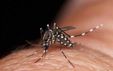
Florida Counties to Issue Mosquito Borne Illness Health Advisories
Florida's summer rainy season is falling in full force. More rain leads to more mosquitoes that only require a bottle cap of standing water to breed and spread. In Florida, mosquito-borne disease activity has already prompted mosquito-control and state health officials in five counties - Orange...

Being Overweight Increases Risk of Developing Cancer
Maintaining a healthy weight is important for general health, but growing evidence reveals obesity’s role in the development of chronic illnesses and even deadly cancers. Obesity is one of the most frequent chronic illnesses in the United States, with one out of every three adults in the nation b...
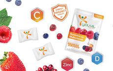
Indonesia’s Top Vitamin Gummy Brand Youvit Enters Malaysia Market
Youvit, formulated in the United States, has become Indonesia’s number-one vitamin gummy brand since 2017 and is now expanding its business to Malaysia. Youvit product range is a complete “one-a-day vitamin gummy" for immunity, hair fall, glowing skin, and active kids that contains natural ingredien...

BayCare and healthPrecision Partner
BayCare Health System and healthPrecision today announced a strategic partnership to bring advanced documentation tools to the hospital bedside to improve accuracy, efficiency, nurse satisfaction and patient outcomes. The co-development of healthPrecision's Medical Brain artificial intelligence ...

Meetup recognized to beneficially impact Mental Health
Meetup, the platform that connects people at events for shared interests, can improve mental health according to new insight from therapists, those that use Meetup, and studies. Unlike most social platforms, Meetup has been recognized to beneficially impact mental health. Embracing this news, the co...

Celebrate World Health Day
World Health Day is celebrated each year on April 7th and helps draw attention to health topics that affect people all over the world. Founded by the World Health Organization (WHO), this important holiday also serves as a reminder to take care of one's health. Actress and health advocate Maggie Q h...

Why Lasik Eye Surgery is Popular in Dubai
Business professionals in Dubai are increasingly turning to LASIK eye surgery to improve their vision and enhance their productivity in the workplace. CEOs and high-level executives often get LASIK eye surgery to improve their vision and eliminate the need for glasses or contact lenses. The reaso...

Global Campaign Empowering Chronic Kidney Disease
Chronic Kidney Disease (CKD) is a growing epidemic and rising global public health concern, with 850 million people affected worldwide. As a progressive disease, very often linked to diabetes, CKD can lead to kidney failure, also known as End Stage Kidney Disease (ESKD), which requires dialysis...

Dealing with Itchy Skin
Are you dealing with itchy skin issues? If so, you're not alone. Itchy skin is a common problem caused by several factors, including skin conditions, allergies, and environmental irritants. There's what you can do to manage and relieve your itchy skin. First, identify the underlying cause of your...
Will 2023 be the Year of the Tripledemic
COVID-19 may have lost its daily news headline status, but the disease rages on with 2700 weekly deaths in the U.S. and millions of Americans chronically disabled from long-COVID, 4 million of which are being kept from work. This winter time, a terrible year for RSV as well as a potentially worse fl...

Dettol sponsors Sporting Cyclothon Event
Reckitt's brand Dettol announces to be Product Sponsor of the 2022 Hong Kong Cyclothon. The partnership aims to enhance hygiene protection of thousands of cyclists at this major sports event held through the diverse cityscape of Hong Kong, featuring a spectacular route. Through this sponsorship, De...

Breast Enhancement Procedures
Doctors have striven to understand the link between breast enhancement and women's health for years. To date, researchers continue to grapple with this issue in attempts to help those who need it. There are lots of reasons why women choose to undergo breast enhancement procedures. Some may feel that...

Four Generations of Workers Are Preparing for Retirement
Seventy-six percent of workers say their life priorities changed as a result of the pandemic, and 56 percent cite saving for retirement as a financial priority, according to Emerging From the COVID-19 Pandemic: Four Generations Prepare for Retirement, a survey report released today by nonprofit Tran...

Health Partnership Awards 2022
Reckitt's brand Dettol is proud to be honoured 'Outstanding Personal Hygiene Marketing Solution' for its success brought by the 'Keep on to Protect Our Future' post-pandemic hygiene protection habit sustaining campaign, in the 'Health Partnership Awards 2022' organised by ET Net, a prominent local m...

GoFit Debuts in Singapore
Gym goers and fitness enthusiasts of all levels can rejoice at the debut of a new high value, low price (HVLP) gym brand in Singapore, GoFit, which is sure to lend more vibrancy to the local fitness scene. No-frills and fuss-free, GoFit is guided by its brand values of Smart, Bold, and Invigorating...